
Abstract
Purpose
The purpose of this study is to construct a guided bone regeneration membrane that is similar to bone components and structurally resembles the native extracellular matrix with sufficient antibacterial properties.
Materials and methods
A novel type of biomimetic and bioactive silver ion-loaded calcium phosphate/chitosan (Ag-CaP/CS) membrane with antibacterial ability was successfully developed by incorporation of silver ion-loaded CaP via a one-step electrospinning method and subsequently crosslinked with vanillin.
Results
Evaluation of the physicochemical properties revealed that the fabricated fibrous membranes mimicked the extracellular matrix structure and the addition of CaP significantly increased the mineralization ability of the membranes. Importantly, the Ag-CaP/CS membranes exhibited a sustainable release of Ag+, which in turn inhibited the adhesion and growth of Staphylococcus mutans. The results of cell adhesion and MTT assay revealed that the Ag-CaP/CS membranes were cytocompatible with bone marrow stromal cells.
Conclusion
The fabricated electrospinning Ag-CaP/CS nanofiber membranes with excellent biocompatibility and strong antimicrobial properties have great potential to be used for guided bone regeneration.
Introduction
Insufficient alveolar bone volume is a common clinical problem that has a direct impact on the survival rate of dental implant treatment. Guided bone regeneration (GBR) is a procedure aiming for the reconstruction of impaired bone tissue, and has become increasingly popular in oral and maxillofacial surgery for augmentation of alveolar bone.Citation1–Citation4 This technique makes use of a membrane barrier to direct the growth of neo-bone tissue while preventing the ingrowth of fibrous tissues to the bone defects.Citation5–Citation7 In the past decades, a variety of barrier membranes, including non-absorbable, for example, polytetrafluoroethylene, and absorbable membranes, such as polyglycolic acid, poly (lactic acid), and collagen membranes, have been developed and applied for GBR techniques.Citation8–Citation10 Although the commercially available GBR membranes showed certain positive outcomes, they are at risk of a biomaterial-centered infection, especially when the membranes are exposed to infected tissue or an area with high accessibility of bacteria, such as the oral cavity. Hence, development of GBR membranes with sufficient antibacterial ability may be a solution for this problem.
In addition to sufficient antibacterial ability, some other essential properties of GBR membranes are required, such as excellent biocompatibility, cell immobility, tissue integration, mechanical stability, optimal porosity, and clinical manageability,Citation11 which may directly influence the outcome of the GBR approach. It is well accepted that a biomaterial mimicking the extracellular matrix (ECM) in terms of both the structure and the components is in favors of cell functionality and accelerates the process of injured tissue regeneration.Citation12 Recently, how to successfully develop a GBR membrane resembling native bone ECM both in composition and structure has become a hot research issue. To manufacture micro-nanoscaled fibers that mimic the structure and morphology of primordial ECM for bone tissue regeneration, electrospinning has been widely used as a proven technique. During electrospinning, polymer solutions are sprayed and spun in a strong electric field and eventually solidified into nanofibers. Some biocompatible and biodegradable polymers are broadly used for electrospinning, including poly (lactic acid), poly (vinyl alcohol), poly (lactic-co-glycolic acid), and poly (epsilon-caprolactone). In addition, electrospinning is a rapid, straightforward, and low-cost method to produce nanofiber-based membranes with high porosity and good pore size distribution.Citation13,Citation14 An optimal porous structure of the GBR membrane is essential for the growth of cells and the exchange of nutrients.Citation4 Therefore, electrospinning was employed to fabricate fibrous membrane for GBR applications.Citation15,Citation16
From a materials point of view, natural human bone is a complex of organic–inorganic composite consisting of calcium phosphate (CaP) nanoparticles and collagen nanofibers. However, electrospinning of collagen denatures collagen to gelatin, and the solvent used in electrospinning will weaken the typical biological properties of collagen and cannot imitate the component of ECM.Citation17 Consequently, other types of natural polymers have been considered, including chitosan (CS). CS is obtained by deacetylation of chitin, which is the second most abundant polysaccharide in the world.Citation18,Citation19 CS has attracted great research interest in the biomedical field because of its excellent performance of biocompatibility, blood compatibility, biosecurity, and microbial degradability.Citation20–Citation23 Silver ions (Ag+) as antibacterial agents have broad-spectrum antibacterial properties, and their resistance to bacteria has rarely been reported relative to traditional antibiotics.Citation24–Citation26 Therefore, Ag+ and silver nanoparticles have been extensively studied in the medical field.Citation27,Citation28 The antimicrobial activity of silver is mainly dependent on the silver cation (Ag+), which can bind strongly to some electron donor groups in biological molecules. In addition, these silver agents are often incorporated in carriers to achieve a sustained release of Ag+.Citation29 CaP materials, such as nano-hydroxyapatite, have been found to provide a large reservoir of silver ions, which could be released gradually resulting in long-term antimicrobial activity. Our previous studies have proved that the silver ion-loaded CaP system possesses strong antibacterial activity, good bio-compatibility, as well as causes slow release of silver ions.Citation30,Citation31 Moreover, chemical composition of CaP is similar to that of natural bone and it has the necessary elements for human metabolism. Importantly, its chemically reactive groups can form chemical bonds with the bone tissue.Citation32 Our goal is to design and fabricate a biomimetic and bioactive silver ion-loaded CaP/CS (Ag-CaP/CS) membrane with antibacterial ability, via electrospinning, which resembles the ECM so as to create conducive living milieu to induce cells to function naturally. It was hypothesized that the Ag-CaP/CS fibrous composite GBR membrane with optimal silver ion content could controllably release silver ions from the fibers to achieve the desired cytocompatibility and sufficient antibacterial effect simultaneously, thus overcoming the drawback of a biomaterial-centered infection with use of the current commercially available GBR membranes. To this end, Ag-CaP/CS fibrous membranes were firstly prepared by electrospinning, and the physicochemical properties, biocompatibility, as well as the antimicrobial activity of the membranes were then studied by in vitro tests in comparison with pure CS and CaP/CS fibrous membrane without silver as the control groups. In general, the higher the content of CaP, the better the osteoconductivity of composite bone repair materials. However, our preliminary experiments showed CaP content higher than 20 wt% made electrospinning of composite membrane difficult. Thus, 20 wt% of CaP was utilized in the present study. In order to determine the optimal concentration of silver ions, we fabricated two types of membranes with different silver content and evaluated the antibacterial activity and cytocompatibility to determine which silver ion concentrations in the composite membrane can show strong antibacterial activity and good cytocompatibility simultaneously.
Materials and methods
Materials
Medical grade CS (degree of deacetylation: 85%; molecular weight: 200–400 kDa) was bought from Heppe Medical Chitosan GmbH (Halle, Germany). Trifluoroacetic acid (TFA) and vanillin were purchased from Sigma-Aldrich Co., Ltd (Gillingham, UK). CaP and silver ion-loaded CaP (Ag-CaP) were prepared using a wet synthesis method as described previously.Citation31
Membrane preparation
CS was dissolved in TFA to form a 5 wt% CS solution at 40°C. Then Ag-CaP or CaP was added and mixed by ultrasonic mixing for 30 minutes. Electrospinning was performed and the spinneret needle was maintained at a voltage of 26 kV by a high voltage power, and an aluminum foil under the needle at a distance of 20 cm was used as the collector. The pump rate was set at 1.0 mL/h. A total of four types of membranes were prepared in this study as shown in , including pure CS, CaP/CS without silver and CaP/CS with different silver content. A scanning electron microscope (SEM, JSM-6510LV; JEOL, Tokyo, Japan) was used to characterize the structure of the fabricated materials. Energy dispersive spectroscopy (EDS, X-MaxN 20; Oxford, UK) was carried out to observe the distribution of the fillers in the fiber matrix. One hundred fibers per sample were used to measure the diameter by Image-Pro Plus.
Table 1 The compositions of the fabricated samples
Crosslinking
Before crosslinking, the membranes were stabilized according to the literature.Citation33 In brief, the membranes were first stabilized by soaking them for 20 minutes in 0.5% sodium hydroxide (NaOH) dissolved in 100% ethanol, followed by five times washing in PBS for 30 seconds. The stabilized membranes were then crosslinked in 5% (w/v) vanillin dissolved in ethanol for 2 hours at 50°C. Subsequently, the crosslinked membranes were rinsed with 100% ethanol for 5 minutes and then with PBS for 30 seconds to remove the unreacted vanillin.
Simulated body fluid (SBF) immersion test
Immersion studies were performed by incubating a membrane (disk shape with a diameter of 15 mm) in 10 mL SBF solution in a 15 mL tube. The pH value of the SBF solution was adjusted to 7.4 before membrane immersion.Citation34 Three samples were used for each condition. All the tubes were placed in a water bath at 37°C under continuous shaking. After immersion periods of 1 and 2 weeks, the membranes were gently washed with deionized water and freeze-dried. The membranes were then examined by X-ray powder diffraction (XRD, DX-2500; Dandong Fangyuan Instrument Co., Ltd., Dandong, China) and observed by SEM after sputter coating with gold.
In vitro Ag+ release
The fibrous membranes with Ag+ (A1 and A2) were cut into square pieces with a length of 10 mm and then immersed in 3 mL PBS. All samples were incubated at room temperature and placed on oscillators at 90 rpm. The supernatant was collected at predetermined time points. After digestion by HNO3 and dilution at a suitable ratio for testing, the Ag concentration of the solution was tested by atomic absorption spectroscopy (SpectrAA 220Z; Varian, Melbourne, VIC, Australia). Meanwhile, A0 membrane was also cut into square pieces with a length of 10 mm and Ag+ was physically absorbed on A0 by immersion of the membrane in AgNO3 aqueous solution for 2 hours, then the Ag+ physically absorbed A0 membrane was dried in a vacuum oven, in which the designed Ag+ content is the same as that in A2 membrane. Thereafter, the release of Ag+ from the Ag+ physically absorbed A0 membrane for 1 day, immersion in PBS was also tested by using the abovementioned methods.
Antimicrobial properties assessment
Antibacterial adhesion assay
The membranes were cut to a size of 5×5 mm and sterilized by ethylene oxide gas prior to the test. Streptococcus mutans (ATCC 25175) in brain–heart infusion (BHI) broth (Oxoid Ltd, Basingstoke, UK) or Porphyromonas gingivalis (ATCC 33277) in tryptic soy broth (Sigma-Aldrich Co., St Louis, MO, USA) were adjusted to a density of 106 colony forming units/mL. Then, each membrane was immersed in 2 mL bacterial suspension at 37°C. After 24 hours, the membranes were gently washed with sterilized PBS to remove the unattached bacterial cells. After being fixed with 2.5% (v/v) glutaraldehyde (Sigma-Aldrich, St Louis, MO, USA) for 2 hours, the samples were dehydrated with a series of ethanol solutions (30%, 50%, 70%, 80%, 90%, 95%, and 100% for 10 minutes each) and dried using the critical point method. At the end of this procedure, the samples were coated with gold and viewed via SEM.
Direct contact test (DCT)
DCT, based on the turbidimetric determination of bacterial growth in 96-well microtiter plates, was used to evaluate the antibacterial activity of root canal sealing materials.Citation35,Citation36 In this study, a modified DCT method was adopted to evaluate the antibacterial ability of the fibrous membranes. In brief, the test membranes (5×5 mm, n=4) were placed separately in 24-well microtiter plates (named as group A well plates). Twenty microliters of bacterial suspension (~1×10⁶ colony forming units/mL) was added to the membranes and incubated at 37°C for 1 hour to facilitate direct contact of bacteria with the membranes. BHI broth (460 µL) was then added to each well and the plates were gently vortex mixed for 2 minutes; 30 µL of the supernatant was then transferred to another well containing fresh BHI broth medium (400 µL) and again mixed for 2 minutes (named as group B well plates). Both groups A (in the presence of the membranes) and B (with the membranes not present) well plates were further incubated at 37°C anaerobically for up to 24 hours. At each predetermined time point, 200 µL of liquid was transferred from each well to a sterilized 96-well microtiter plate and the bacterial growth was measured at 650 nm and 37°C using a microplate spectrophotometer (PerkinElmer 1420 Multilabel Counter; PerkinElmer, Inc., Waltham, MA, USA). After that, the liquid was transferred back to the original wells and the incubation was continued. Empty wells without test materials (n=4) served as controls. All the experiments were carried out under aseptic conditions.
Cytocompatibility
Cell culture
Cell attachment, spread, and proliferation on the different membranes were evaluated using bone marrow stromal cells (BMSCs). Two young Sprague Dawley rats (2 months old and weight about 100 g) obtained from the experimental animal center of Sichuan University were employed for extraction of BMSCs, which was approved by the Ethics Committee of Sichuan University, and all operation procedures and animal care were performed in compliance with the Guide for the Care and Use of Laboratory Animals. BMSCs were obtained from the tibiae and femora of young Sprague Dawley rats and cultured in α-Minimum Essential Medium supplemented with 20% fetal bovine serum, 100 U/mL penicillin, 100 mg/mL streptomycin, 0.219 mg/mL l-glutamine, and 100 mM HEPES buffer (Thermo Fisher Scientific, Waltham, MA, USA) in a humidified incubator with 5% CO2 at 37°C. The culture medium was changed every other day. The third passage of BMSCs was used in the experiments. Prior to seeding, all the membranes of ~1 mm thickness were cut into discs of 10 mm in diameter, sterilized by ethylene oxide gas, and pre-wetted in the culture medium for 24 hours. BMSCs were seeded onto the pre-wetted membranes (2×10⁴ cells per membrane). The seeded membranes were then cultured in a humidified incubator (37°C, 5% CO2) for 11 days with the medium changed every 2 days. Cells cultured without materials were assigned as control.
Attachment and proliferation of the BMSCs
After 4 and 7 days, the samples were collected for SEM observation. The cell membrane constructs were rinsed in PBS, fixed with 3% glutaraldehyde, and dehydrated in graded ethanol concentrations (25%, 50%, 75%, 90%, 95%, and 100% v/v in distilled H2O). After that, the samples were rinsed with isoamyl acetate and then dried in supercritical CO2. Thereafter, the samples were sputter coated with Au and observed by SEM.
The proliferation of BMSC cells cultured on all the membranes was determined by MTT assay (Amresco, Cleveland, OH, USA) according to the instruction of the manufacturer. The absorbance at 490 nm was measured with a microplate reader (PerkinElmer 1420 Multilabel Counter, PerkinElmer, Inc.) after 1, 4, 7, and 11 day (s) of incubation (n=3).
Statistical analysis
For quantitative analysis, Student’s t-tests were used to assess differences between two groups and multiple comparisons were performed via one-way analysis of variance test using SPSS (v. 11.0). P-values <0.05 were considered statistically significant. Data are expressed as mean values±SD.
Results and discussion
Morphology of fibrous membranes
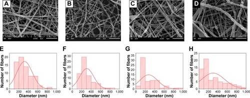
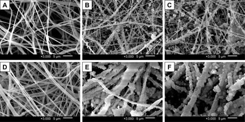
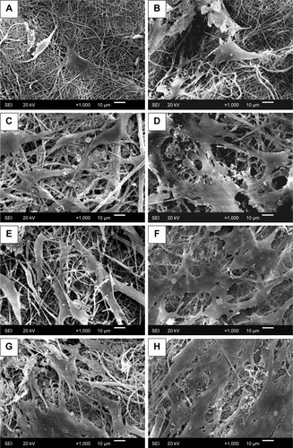
Fibrous membranes were prepared from all the solutions by the same parameters via electrospinning. All the membranes were constructed with fibers with a broad diameter range and they presented a randomly oriented arrangement (). For those groups with CaP, the surface of fibers was rougher than that of pure CS fibers due to the exposure of CaP particles. A porous structure was formed by the randomly arranged fibers, which would facilitate the exchange of nutrients and waste of cell metabolism.
Figure 1 Scanning electron micrographs of the four electrospun fibrous membranes: (A) CS, (B) A0, (C) A1, and (D) A2; (E–H) the corresponding diameter distribution diagrams.
Abbreviation: CS, chitosan.
XRD and Fourier-transform infrared analysis
XRD patterns of hydroxyapatite (HA) and silver-loaded hydroxyapatite (HA-Ag), electrospun membranes (CS, A0 and A1), and A1 immersed in SBF for 2 weeks (A1-SBF) are presented in . The characteristic diffraction peaks of HA-Ag were almost the same as those of HA, indicating that the addition of Ag+ had little effect on the crystal structure of HA. However, the characteristic diffraction peaks of HA were not observed in A0 and A1 samples. Instead, the characteristic diffraction peaks of CaHPO4 were confirmed, indicating the apatitic CaP was dissolved and transformed into CaHPO4 in the acidic solution of TFA during the electrospinning process. HA will dissolve and transform into CaHPO4 in acidic solutions of pH value <4.5.Citation37 Since TFA with low pH value was used as the solvent in this experiment, the electrospinning fibers eventually contain CaHPO4 rather than HA.
Figure 2 (A) XRD patterns of calcium phosphate particles (HA and HA-Ag), electrospun membranes (CS, A0, and A1), A1 immersed in SBF for 2 weeks (A1-SBF), and (B) FTIR spectra of A1, CS, and A1 crosslinked by vanillin (A1-crosslinked).
Abbreviations: CS, chitosan; FTIR, Fourier transform infrared; HA, hydroxyapatite; HA-Ag, silver loaded hydroxyapatite; SBF, simulated body fluid; XRD, X-ray diffraction.
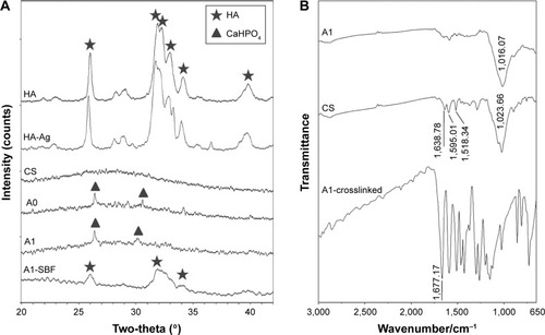
Without crosslinking, the prepared electrospun CS membrane lost integrity in aqueous solution. Therefore, the use of an appropriate crosslinking agent is essential to improve the stability and mechanical property of CS membranes. In this study, we chose vanillin as the crosslinking agent due to its lower cytotoxicity than that of the common crosslinking agents, for example, glutaraldehyde and formaldehyde.Citation38,Citation39 It has been shown that vanillin-crosslinked CS microspheres displayed a better cytocompatibility and much weaker inflammatory reaction than glutaraldehyde-crosslinked CS microspheres.Citation40 Vanillin reacts with CS through the Schiff base reaction between the aldehyde groups of vanillin and the amine groups of CS. The characteristic absorption peak of C=N is seen at 1,677 cm−1 in , which indicates that the Schiff base reaction occurred.
The distribution of CaP and Ag+
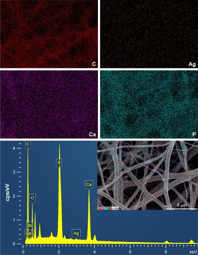
It is widely accepted that the distribution of nanoparticles in fibers directly influences the performance of membrane. EDS mapping was used to characterize the distribution of CaP and Ag+ in the Ag-CaP/CS composite membrane. As shown in , the EDS spectrogram further proved the presence of CaP and Ag+ in fibers. The homogeneous distribution of Ca, P, and Ag elements in EDS mapping indicated that the silver ion-loaded CaP nanoparticles were homogeneously distributed in the fibers.
Figure 3 The distribution of CaP and silver ions in the A2 membrane.
The silver ion loading and release
XPS analysis
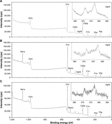
XPS was applied to further confirm the chemical composition and the valence states of the prepared membranes. The XPS spectra of HA-Ag, A1, and A1 crosslinked by vanillin nanofiber membranes are shown in . The presence of the characteristic peak of Ag 3d indicated that silver ions were successfully incorporated into the electro-spun fibrous membranes. The binding energy of Ca and P did not change obviously in the three survey spectra. The characteristic peak of sodium was observed in A1 and A1-crosslinked membranes, which can be explained by the incorporation of sodium in the process of stabilization of the membranes by sodium hydroxide solution. XPS is also a powerful technique to explore the oxidation state of the transition metal compounds with localized valence orbital due to the different energies of the photoelectrons. From the high resolution XPS spectra of Ag 3d (inserted images), the binding energy fits well to a single spin-orbit pair of Ag 3d5/2 at 367.6 eV and Ag 3d3/2 at 373.6 eV, respectively, which is attributed to Ag+.Citation12,Citation41 The spectra of Ag 3d for A1-crosslinked nanofibers illustrated that the valence state of silver ions was stable univalent silver during the electrospinning and crosslinking process, which provides the necessary evidence of the antibacterial action of silver ions in the membrane.
Figure 4 XPS spectra (A) silver-loaded hydroxyapatite, (B) A1, (C) A1 crosslinked by vanillin. (D) Concentration of silver ions released from different silver-loaded electrospun fibrous membranes in deionized, distilled water.
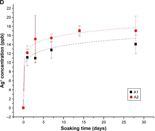
The release of silver ions in vitro
The release behaviors of Ag+ from the Ag-CaP/CS nanofiber membranes was evaluated by in vitro release evaluation (). As the results indicated, apparently, both A1 and A2 exhibited an evident initial burst release within the first day and the amount of the Ag+ released from nanofiber membranes increased slowly with augmentation of soaking time, after 1 day. Although there was no statistical difference between A1 and A2, the amount of Ag+ released from A2 was slightly higher than that released from A1 after 1 day. Ag+ physically absorbed on A0 membrane was used to assess the Ag+ release behaviors as the control. Robust burst release of Ag+ physically absorbed on A0 after 1 day immersion was observed and release amount reached 76.8% of the total absorbed Ag+. Therefore, we did not test the release of Ag+ physically absorbed on A0 for a longer period. Although the burst release of Ag+ from A1 and A2 was also observed, the amount of Ag+ released from A1 and A2 after 1 day immersion was not >30% of the total Ag+ in the membranes. Thus, A1 and A2 showed a relatively slow release for Ag+ in comparison with Ag+ physically absorbed on the fabricated membrane group. The release of Ag+ could be explained by a complex mechanism that includes diffusion, swelling, and corrosion.Citation42 The release of Ag+ should be first separated from the calcium phosphate surface and then be released from the CS nanofiber matrix. Therefore, a considerable part of Ag+ was released within 24 hours, which might achieve a strong antibacterial effect during the early stage after surgery. Another mechanism is that a portion of Ag+ diffused into the solution along with the degradation of CS and CaP.
Immersion in SBF
The bone-linking ability of the different composite nanofiber membranes was assessed by examining the apatitic mineralization ability on the surface of membranes by incubation of the membranes in SBF. shows the SEM image of the surface morphology of the membranes after incubation in SBF for up to 2 weeks. The surface morphology of pure CS did not show any noticeable change after either 1- or 2-week immersion (). In contrast, both A0 and A1 were covered with some spherical minerals and the amount of mineral deposition increased with the immersion time (). However, the crystal structure of the mineral deposition could not be identified by the XRD patterns (, A1-SBF). The deposition of apatite can be explained by the nucleation and growth mechanism. Briefly, the increased local concentration of ions contributed to the formation of nucleation points; thereafter, the CaP nuclei formed and gradually grew into crystals. As a result, the hemispheric apatite formed and accumulated on the surface of fibers over time. The incorporation of CaP into the fibers provided nucleation points for the deposition of apatite.Citation43 The results suggested that the addition of CaP significantly increased the mineralization ability of the membranes, which is beneficial for bone formation.
Figure 5 SEM photos of the electrospun membranes after immersion in SBF: (A) CS, (B) A0, and (C) A1 for 1 week; (D) CS, (E) A0, and (F) A1 for 2 weeks.
Abbreviations: CS, chitosan; SBF, simulated body fluid; SEM, scanning electron micrograph.
Antimicrobial analysis
Anti-adhesion of P. gingivalis
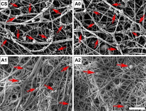
P. gingivalis, the gram-negative and the main pathogenic bacteria of periimplantitis or periodontitis, was used to investigate the antimicrobial activity of various membranes. After immersion of the membranes in the bacterial suspension for 24 hours, the adhesion and growth of P. gingivalis on the fibrous membranes were observed by SEM, and the results are shown in . Notably, the serried bacteria attached to the fibers to form a bead necklace structure of pure CS and CaP/CS (A0). Moreover, a lot of diminutive bacteria were present, which indicated that the P. gingivalis can rapidly proliferate and grow on the surface of membranes. On the contrary, only a few bacteria could be found on the membranes with Ag+ (A1 and A2), and the bacteria on A2 membrane seemed to be slightly less than on A1. The results demonstrated that the prepared membranes with Ag+ had strong antibacterial properties as we expected, which would greatly reduce the risk of postoperative infection.
Figure 6 SEM image of bacterial growth on various membranes after 24 hours of co-culture with Porphyromonas gingivalis (as indicated by the red arrow).
Abbreviations: CS, chitosan; SEM, scanning electron micrograph.
Anti-adhesion of S. mutans
S. mutans, a common oral bacterium, was employed to evaluate the anti-adhesion effect of the prepared membranes. After incubation of the membranes with S. mutans suspension for 24 hours, SEM observation indicated that large numbers of bacteria adhered on the surface of the CS and CaP/CS (A0), whereas few of them could be found on the Ag-CaP/CS membranes (A1 and A2; ). In other words, membranes with Ag+ restrained the adhesion and reproduction of the bacteria on the membrane surface. Low bacterial adhesion would prevent the formation of biofilm, and consequently reduce the risk of a biomaterial-centered infection.Citation44 It is reported that the mechanism of the antibacterial activity of the silver ion is by attaching to the cell wall of bacteria and disturbing the permeability and intermembrane exchange. In addition, the antimicrobial action of silver ions is closely related to their interaction with phosphorous- and sulfur-containing biomolecules, including DNA and proteins, by weakening DNA replication and inactivating proteins.Citation45
Figure 7 SEM images of bacterial adhesion on (A) CS, (B) A0, and (C) A1 and (D) A2 after 24 hours of incubation with Streptococcus mutans (as indicated by the red arrow). DCT results of the four membranes in (E) group A and (F) group B after 24 hours of incubation.
Abbreviations: CS, chitosan; DCT, direct contact test; SEM, scanning electron micrograph.
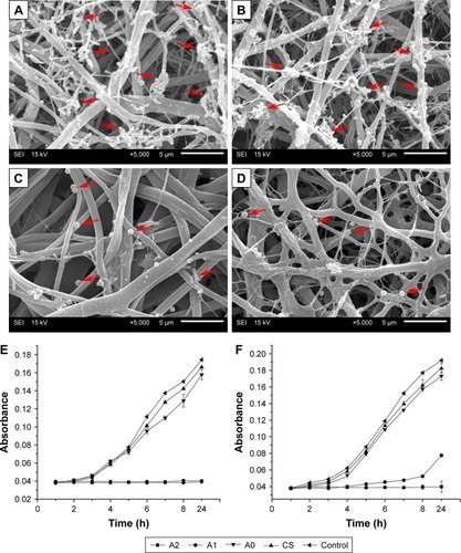
Evaluation of antimicrobial activity by DCT
Furthermore, a modified DCT approach was used to assess the antimicrobial activity of the four fibrous membranes. In the presence of the membranes, that is, the test in group A wells, only the Ag-CaP/CS membranes (A1 and A2) inhibited the proliferation of bacteria within 24 hours, whereas bacteria cultured with A0 and pure CS proliferated at the same rate as the control group (). The situation in vivo was a dynamic system with the exchange of body fluid, resulting in the loss of antibiotic constituents around the implanted materials. Thus, we utilized the group B wells to assess the growth of residual bacteria after just 1 hour of direct contact with the membrane. Unlike the results obtained from group A wells, only A2 exhibited complete inhibition of bacterial proliferation for up to 24 hours (). Bacteria in contact with A1 began to proliferate after 4 hours of incubation. The DCT indicates that the antibacterial property of Ag-CaP/CS composite membranes was associated with the amount of the incorporated silver. We learned that there are, indeed, related studies demonstrating that CS itself has a certain antibacterial effect.Citation46,Citation47 In contrast, research has also shown that CS does not have an antimicrobial effect.Citation26,Citation48 There is a controversial point about the antibacterial properties of CS. We think that whether or not CS exhibits antibacterial properties may be related to the source of its extraction as well as the species of bacteria. In our present study, both gram-positive (S. mutans) and gram-negative (P. gingivalis) bacteria were used to assess the antibacterial properties and the results indicated that CS did not exhibit antibacterial effect.
Cell morphology and proliferation of BMSCs
BMSCs were used to evaluate the attachment, spread and growth of cells on the membranes. As shown in , the morphology of BMSCs on the surface of different membranes after incubation for 4 and 7 days was observed by SEM. All of the membranes facilitated good attachment and growth of cells except for pure CS membrane. This is in accordance with the previous studies which showed that unmodified CS membranes did not perform sufficiently to mediate cell adhesion.Citation49,Citation50 After 4 days of incubation, cells on the membranes were flat and polygonal and the parapodia of BMSCs extended into the fibrous structure of the membranes. At day 7, the cells on the composite membranes were almost confluent. As the fibrous framework of biodegradable polymer membrane, the micro-nanostructure can act as an ECM for cell attachment and adhesion. The difference in the cell growth between the CS and composite membranes is likely due to the addition of CaP, which may increase the surface roughness and protein adsorption to promote cell adhesion.Citation4,Citation51 In addition, Ca and P are necessary elements for human metabolism, which can transduce the chemical signal into the cell, thereby modulating a series of cell behaviors.Citation52
Figure 8 SEM images of BMSCs on the surface of (A, B) CS, (C, D) A0, and (E, F) A1 and (G, H) A2 after (A, C, E, G) 4 days and (B, D, F, H) 7 days of incubation.
Abbreviations: BMSCs, bone marrow stromal cells; CS, chitosan; SEM, scanning electron micrograph.
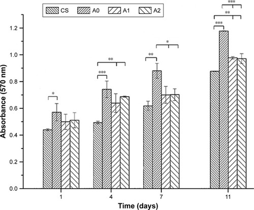
The proliferation of BMSCs on the nanofiber membranes was investigated by MTT assays. shows the cell viability after culturing for 1, 4, 7, and 11 days. With increasing culture time, obviously, the number of cells on the nanofiber membranes significantly increased. The proliferation of BMSCs on A0, A1, and A2 was distinctly higher than on pure CS membrane, which is consistent with the SEM results. The incorporation of CaP facilitated cell adhesion on the surface of membranes, thereby significantly promoting the proliferation of BMSCs.Citation52 However, the cell viability of A0 was higher than those of A1 and A2, suggesting that the addition of silver ions inhibited cell proliferation to some extent, but the silver-loaded membranes were still superior to pure CS membrane for BMSCs proliferation. The Ag-CaP/CS composite membranes with an optimal concentration of Ag+ could have simultaneously good cytocompatibility and antibacterial properties. Bacteria are prokaryotes and human cells are eukaryotic cells with integrated nuclei.Citation53 Therefore, eukaryotic cells are larger than prokaryotic cells and a higher concentration of Ag+ is required to cause cytotoxicity on human cells than on bacterial cells. This difference provides a “therapeutic window” for the membranes to achieve biocompatibility and antibacterial properties at the same time.Citation4 This result also indicates that although a higher content of silver ions in the membrane will achieve stronger antibacterial activity, it will lead to corresponding cytotoxicity.Citation54 Therefore, an optimal concentration of silver in biomaterial is of great importance. The fabricated membrane of A2 achieved good cytocompatibility and strong antibacterial properties simultaneously, so we did not further increase the concentration of silver ions in the membrane in this study.
Figure 9 MTT assay of BMSCs cultured on the membranes.
Notes: Error bars represent SD from the mean (n=3). ***P<0.001; **P<0.01; *P<0.05.
Abbreviations: BMSCs, bone marrow stromal cells; CS, chitosan.
Conclusion
In this study, Ag-CaP/CS bioactive composite fibrous membranes with antibacterial properties were successfully developed by electrospinning method and subsequently crosslinked with vanillin as the potential candidate for GBR application. The physicochemical properties evaluated suggested that the fabricated fibrous membranes mimicked the ECM structure and the addition of CaP significantly increased the apatite mineralization ability of the membranes. Most importantly, the silver ions were successfully incorporated into the fibers, and thereby endowed the membranes with antibacterial function. The incorporation of 0.144% content of silver ions into the nanofibers (A2) endows the fibrous membranes with strong antibacterial properties without induction of adverse cytotoxicity. Hence, the fabricated Ag-CaP/CS fibrous membranes showing a combination of excellent biocompatibility and strong antimicrobial properties have great potential to be used for GBR.
Acknowledgments
The authors acknowledge the support of the National Key Research and Development Program of China (2016YFA0201703/2016YFA0201700) and NFSC fund (31670965).
Disclosure
The authors report no conflicts of interest in this work.
References
- BenicGIHämmerleCHHorizontal bone augmentation by means of guided bone regenerationPeriodontology20146611340
- BottinoMCThomasVSchmidtGRecent advances in the development of GTR/GBR membranes for periodontal regeneration – a materials perspectiveDent Mater201228770372122592164
- JangTSLeeEJJoJHJhJFibrous membrane of nano-hybrid poly-L-lactic acid/silica xerogel for guided bone regenerationJ Biomed Mater Res B Appl Biomater2012100232133022102608
- LiJZuoYManYFabrication and biocompatibility of an antimicrobial composite membrane with an asymmetric porous structureJ Biomater Sci Polym Ed2012231–4819621156105
- SongXLingFMaLYangCChenXElectrospun hydroxyapatite grafted poly(L-lactide)/poly(lactic-co-glycolic acid) nanofibers for guided bone regeneration membraneCompos Sci Technol201379814
- ZhangHWangJMaHHrMBilayered PLGA/wool keratin composite membranes support periodontal regeneration in beagle dogsACS Biomater Sci Eng201621221622175
- ZhangH-LWangJYuNLiuJ-SElectrospun PLGA/multi-walled carbon nanotubes/wool keratin composite membranes: morphological, mechanical, and thermal properties, and their bioactivities in vitroJ Polym Res2014211329
- GentilePChionoVTonda-TuroCFerreiraAMCiardelliGPolymeric membranes for guided bone regenerationBiotechnol J20116101187119721932249
- McallisterBSHaghighatKBone augmentation techniquesJ Periodontol200778337739617335361
- von ArxTBuserDHorizontal ridge augmentation using autogenous block grafts and the guided bone regeneration technique with collagen membranes: a clinical study with 42 patientsClin Oral Implants Res200617435936616907765
- LiJManYZuoYIn vitro and in vivo evaluation of a nHA/PA66 composite membrane for guided bone regenerationJ Biomater Sci Polym Ed2011221–326327520557712
- TangYSubramaniamVPLauTHIn situ formation of large-scale Ag/AgCl nanoparticles on layered titanate honeycomb by gas phase reaction for visible light degradation of phenol solutionAppl Catal B20111063–4577585
- PengHYinZLiuHElectrospun biomimetic scaffold of hydroxyapatite/chitosan supports enhanced osteogenic differentiation of mMSCsNanotechnology2012234848510223128604
- ShenKHuQChenLShenJPreparation of chitosan bicomponent nanofibers filled with hydroxyapatite nanoparticles via electrospinningJ Appl Polym Sci2010115526832690
- RenKWangYSunTYueWZhangHElectrospun PCL/gelatin composite nanofiber structures for effective guided bone regeneration membranesMater Sci Eng C Mater Biol Appl20177832433228575991
- SuHLiuKYKarydisAIn vitro and in vivo evaluations of a novel post-electrospinning treatment to improve the fibrous structure of chitosan membranes for guided bone regenerationBiomed Mater201612101500327910815
- ZeugolisDIKhewSTYewESElectro-spinning of pure collagen nanofibres – just an expensive way to make gelatin?Biomaterials200829152293230518313748
- CelebiHGurbuzMKoparalSDoganADevelopment of antibacterial electrospun chitosan/poly(vinyl alcohol) nanofibers containing silver ion-incorporated HAP nanoparticlesCompos Interfaces2013209799812
- HassibaAJEl ZowalatyMENasrallahGKReview of recent research on biomedical applications of electrospun polymer nanofibers for improved wound healingNanomedicine201611671573726744905
- van Hong ThienDHsiaoSWHoMHLiCHShihJLMhHChLElectrospun chitosan/hydroxyapatite nanofibers for bone tissue engineeringJ Mater Sci201348416401645
- ZhangYVenugopalJREl-TurkiARamakrishnaSSuBLimCTElectrospun biomimetic nanocomposite nanofibers of hydroxyapatite/chitosan for bone tissue engineeringBiomaterials200829324314432218715637
- ZhaoWLiuWLiJLinXWangYPreparation of animal polysaccharides nanofibers by electrospinning and their potential biomedical applicationsJ Biomed Mater Res A2015103280781824733749
- ZhouYYangHLiuXMaoJGuSXuWSjGWlXElectrospinning of carboxyethyl chitosan/poly(vinyl alcohol)/silk fibroin nanoparticles for wound dressingsInt J Biol Macromol201353889223164753
- Bedolla-CázaresFHernández-MarceloPEGómez-HurtadoMASilver nanoparticles from AgNO3–affinin complex synthesized by an ecofriendly route: chitosan-based electrospun composite productionClean Technol Environ Policy2017193897906
- FoudaMMEl-AassarMRAl-DeyabSSAntimicrobial activity of carboxymethyl chitosan/polyethylene oxide nanofibers embedded silver nanoparticlesCarbohydr Polym20139221012101723399122
- SongJRemmersSJShaoJAntibacterial effects of electrospun chitosan/poly(ethylene oxide) nanofibrous membranes loaded with chlorhexidine and silverNanomedicine20161251357136426970025
- López-EsparzaJEspinosa-CristóbalLFDonohue-CornejoAReyes-LópezSYAntimicrobial activity of silver nanoparticles in polycaprolactone nanofibers against gram-positive and gram-negative bacteriaInd Eng Chem Res201655491253212538
- ScocciantiGFrenosFBeltramiGCampanacciDACapannaRLevels of silver ions in body fluids and clinical results in silver-coated mega-prostheses after tumour, trauma or failed arthroplastyInjury201647Suppl 4S11S16
- ZhuangXChengBKangWXuXXlXElectrospun chitosan/gelatin nanofibers containing silver nanoparticlesCarbohydr Polym2010822524527
- MpLJdLQuanZXYsOJiangDMYbLIn vitro evaluation of antibacterial activity and cytotoxicity of novel nanocomposite material for bone fillingMater Res Innov20131512428
- WuXLiJWangLHuangDZuoYLiYJdLYbLThe release properties of silver ions from Ag-nHA/TiO2/PA66 antimicrobial composite scaffoldsBiomed Mater20105404410520683127
- GaoCFengPPengSShuaiCCarbon nanotube, graphene and boron nitride nanotube reinforced bioactive ceramics for bone repairActa Biomater20176112028501710
- FrohberghMEKatsmanABottaGPElectrospun hydroxyapatite-containing chitosan nanofibers crosslinked with genipin for bone tissue engineeringBiomaterials201233369167917823022346
- KokuboTTakadamaHHow useful is SBF in predicting in vivo bone bioactivity?Biomaterials200627152907291516448693
- AnumulaLKumarSKumarVSAn assessment of antibacterial activity of four endodontic sealers on Enterococcus faecalis by a direct contact test: an in vitro studyISRN Dent2012201298978198978522888444
- WangJZuoYZhaoMPhysicochemical and biological properties of a novel injectable polyurethane system for root canal fillingInt J Nanomedicine20151069770925653518
- ShellisRPBarbourMEJonesSBAddyMEffects of pH and acid concentration on erosive dissolution of enamel, dentine, and compressed hydroxyapatiteEur J Oral Sci2010118547548220831581
- BuslovichAHorevBRodovVGedankenAPoverenovEOne-step surface grafting of organic nanoparticles: in situ deposition of antimicrobial agents vanillin and chitosan on polyethylene packaging filmsJ Mater Chem B201751426552661
- YangH-JLeeJ-HLeeK-YSongKBAntimicrobial effect of an Undaria pinnatifida composite film containing vanillin against Escherichia coli and its application in the packaging of smoked chicken breastInt J Food Sci Technol2017522398403
- ZouQLiJLiYJdLYbLPreparation and characterization of vanillin-crosslinked chitosan therapeutic bioactive microcarriersInt J Biol Macromol201579473674726051343
- ZhaoYZhouYWuXWangLXuLWeiSA facile method for electrospinning of Ag nanoparticles/poly (vinyl alcohol)/carboxymethyl-chitosan nanofibersAppl Surf Sci20122582288678873
- ZhengFWangSWenSShenMZhuMShiXCharacterization and antibacterial activity of amoxicillin-loaded electrospun nano-hydroxyapatite/poly(lactic-co-glycolic acid) composite nanofibersBiomaterials20133441402141223168384
- MokabberTLuLQvan RijnPVakisAIPeiYTCrystal growth mechanism of calcium phosphate coatings on titanium by electrochemical depositionSurf Coat Technol2018334526535
- YangWJYangCSHuangCJChenKSLinSFBostrycinLSFBostrycin, a novel coupling agent for protein immobilization and prevention of biomaterial-centered infection produced by Nigrospora sp. No. 407Enzyme Microb Technol2012506–728729222500894
- ZhouYKongYKunduSCirilloJDLiangHAntibacterial activities of gold and silver nanoparticles against Escherichia coli and bacillus Calmette-GuérinJ Nanobiotechnology20121011922559747
- DumontMVilletRGuirandMProcessing and antibacterial properties of chitosan-coated alginate fibersCarbohydr Polym2018190314229628252
- HuangC-YKuoC-HWuC-HKuM-WChenP-WChWMwKExtraction of crude chitosans from squid (Illex argentinus) pen by a compressional puffing-pretreatment process and evaluation of their antibacterial activityFood Chem201825421722329548445
- StroescuMStoica-GuzunAIsopencuGChitosan-vanillin composites with antimicrobial propertiesFood Hydrocoll2015486271
- López-PérezPMda SilvaRMPSerraCPashkulevaIReisRLSurface phosphorylation of chitosan significantly improves osteoblast cell viability, attachment and proliferationJ Mater Chem2010203483491
- SilvaSSLunaSMGomesMEPlasma surface modification of chitosan membranes: characterization and preliminary cell response studiesMacromol Biosci20088656857618350539
- NagasakiTNagataFSakuraiMKatoKEffects of pore distribution of hydroxyapatite particles on their protein adsorption behaviorJournal of Asian Ceramic Societies2017528893
- GaoCPengSFengPShuaiCBone biomaterials and interactions with stem cellsBone Res201751705929285402
- ReidyBHaaseALuchADawsonKLynchIMechanisms of silver nanoparticle release, transformation and toxicity: a critical review of current knowledge and recommendations for future studies and applicationsMaterials2013662295235028809275
- DdsLGullonBCardelle-CobasAChitosan-based silver nanoparticles: a study of the antibacterial, antileishmanial and cytotoxic effectsJ Bioact Compat Poly2016324397410