
Abstract
Introduction
Melanin pigments are produced by melanocytes and are believed to act as antioxidants based on the belief that melanin can suppress electronically stirred states and scavenge the free radicals.
Materials and methods
The study was aimed to verify and prove the toxicity induced by administration of gold nanoparticles (GNPs) and to characterize the role of melanin as an antioxidant against inflammatory liver damage, oxidative stress, and lipid peroxidation induced intraperitoneally by GNPs in vivo.
Results
The findings from this study confirmed that administration of GNPs intraperitoneally caused liver damage in addition to producing oxidative stress and fatty acid peroxidation. The treatment of rats with melanin along with GNPs induced dramatic changes in all the measured biochemical parameters. Our data demonstrated that melanin completely inhibited inflammatory liver damage, oxidative stress, and lipid peroxidation, which was confirmed by the histological investigation of different liver sections stained by H&E.
Conclusion
These results suggest the beneficial use of melanin together with GNPs for alleviating its toxicity. Other studies should be implemented taking into consideration the role of melanin in comparison with other natural antioxidants.
Introduction
Several studies have reported on the production of numerous inflammatory and cytotoxic responses resulting from contact with smaller-sized gold nanoparticles (GNPs) in comparison to contact with larger-sized GNPs with the same mass concentration due to their highly reactive role with biological constituents, and have stressed on the harmful effects produced by a large number of nanoparticles.Citation1–Citation6 The toxic nature of GNPs is related to its variations, surface functional affinity, appearance, and diameter.Citation1–Citation6 The intraperitoneal administration of GNPs induced inflammation, fatty acid change and hypergenesis, hydropic deterioration, and death of most cells.Citation1,Citation3
Exposure to GNPs has been known to cause cardiac tissue destruction, inflammation of the airspaces in the lungs, connective tissue deposition, and chronic inflammatory cell infiltration that solely depends on the amount and length of exposure.Citation3 These changes could be as a result of the damage to the tissues, resulting from poisoning by GNPs, which interfere with the antioxidant defense structure, thereby probably giving rise to reactive oxygen species (ROS) formation, which may ultimately lead to cell death.Citation2,Citation4
Recently, herbal melanin has been extracted from various sources, and the immunoprotective role of melanin has been reported in plants and skin.Citation7–Citation9 Melanin can be found in different organs and tissues and in the blood of living organisms, but its defining role is still unclear. Many diseases are accompanied by an increase or decrease in melanin production. Melanin plays a vital role in preventing, absorbing, and shielding the cell’s DNA in the nucleus against injury due to ultraviolet radiation (UV). Melanin shows various attractive properties, such as being a nonoxidative agent as well as its scavenging activities on free radicals.Citation10–Citation12
Chen et al,Citation13 have reported that toxicity is generated by nanoparticles due to the reduction in their size. Furthermore, as there are many applications for nanoparticles, especially in drug delivery and imaging, it becomes necessary to prevent toxicity of GNPs when used in medical applications. When GNPs are injected into mice, it enters hippocampus and induces cognitive impairment, in addition to other different complications. Pretreatment of tea melanin significantly prevented the deposition of GNPs in mouse brains, especially in the hippocampus.Citation13
The ability of melanin to act as a forager of free radicals in vitro and in vivo is well established.Citation4–Citation16 Previous studies on melanin revealed that melanin combines with the substances containing oxygen such as oxygen,Citation17,Citation18 hydroxyl radical,Citation19 and superoxide negatively charged ion.Citation19,Citation20 Melanin’s ability to function as an antioxidant has been established from its ability to prevent lipid peroxidation.Citation21–Citation23
To make use of GNPs in drug delivery and cancer treatment, knowledge about inhibiting toxicity is essential. Prevention of toxicity stimulated by the injection of small-sized GNPs has not been well confirmed and established in rat liver. Thus, the current study aims to reinforce and prove the toxicity induced by administration GNPs in rats in vivo and also to investigate the role of melanin as an antioxidant against hepatotoxicity, oxidative stress, and lipid peroxidation.
Materials and methods
Preparation of melanin
Melanin was prepared based on calibrated, standardized, and established known techniques.Citation7 The purity of the prepared melanin was almost about 99%.Citation24,Citation25 The botanical name of the plant from which the black seeds were obtained is “Nigella sativa.” N. sativa seed coats were dissolved in NaOH at pH 12.5 for 3 h, giving rise to the formation of a brown solution. The solution formed was dark, clear, and had no suspension. Centrifugation was done to produce a stock solution and filtered, followed by melanin extraction using concentrated hydrochloric acid with a pH of 2.Citation7
To obtain melanin with higher purity, alkali–acid treatment was done 2–3 times. The precipitate was washed with distilled water, filtered, and desiccated at a temperature of 80°C. The dried powder obtained was kept and later used for the preparation of other solutions at pH 7 for biological examination by dissolving the preferred volume of melanin powder (in w/w ratio) in NaOH at pH 12.5 using concentrated hydrochloric acid to correct the pH and to obtain the preferred concentration. To avoid photochemical and photophysical alterations, the herbal melanin was kept refrigerated at a temperature of −20°C.Citation24
GNPs and dosing
Ten-nanometer GNPs with spherical morphology (products MKN-Au-010; M K IMPEX Corp; Divn MK Nano, Mississauga, Canada) were used. Doses of 50 μL of 10 nm GNPs were administered intraperitoneally to the rats. The size and morphology of different gold nanoparticles were calculated from the images taken by the transmission electron microscope. Ten-nanometer GNPs show spherical shape, while the 50 nm GNPs presented hexagonal shape.Citation1–Citation4
Animals
Eighteen (18) male Wistar Kyoto rats, aged 12 weeks, and weighing between 220 and 240 gm were obtained from the College of Pharmacy, King Saud University (KSU) Animal care house. The rats were kept in cages under customary environmental conditions (22°C±5°C temperature, 55%±5% humidity, and 12 h light/dark cycle). The rats had normal supply of water and were fed with pelleted standard rat chew diet. The KSU Animal Use Committee approved the animal experiments, and all experiments were conducted in accordance with the guidelines approved by KSU local animal care.
Experimental design
The acclimatization period for the animals was one week, and then rats were fasted for 24 h before injection and were divided into three sets of 6 animals each. The 1st group (G1) was the normal healthy rats that received no GNPs and served as the control group. The 2nd group (G2) were injected daily with a dose of 50 μL GNPs intraperitoneally for 7 consecutive days. The 3rd group (G3) were GNP-intoxicated rats that were injected with melanin (100 mg/kg BW/d) for 7 successive days.
Blood sampling and tissue preparation
After the final dose, the rats were starved for 12–14 h, and then euthanized; blood samples from each animal were collected into disinfected tubes for serum separation. Serum separation was done by using a centrifuge at 3,000 rpm for 10 min and preserved at a temperature of −80°C for various biochemical assessments. Midline incision method was used to collect and harvest liver and kidney tissues, which were then rinsed with icy isosmotic saline, homogenized, and frozen at a temperature of −80°C for biochemical tissue analysis (such as reduced glutathione [GSH] level estimation).Citation25
Serum liver function markers
To evaluate the liver inflammatory damage, serum liver biomarkers such as γ-glutamyl transferase (GGT), aspartate aminotransferase (AST), total bilirubin (TBIL), alanine ami-notransferase (ALT), total protein, and alkaline phosphatase (ALP) were determined using kits from Salucea (Etten-Leur, the Netherlands).
Oxidative stress biomarkers
Determination of GSH level
To verify the oxidative stress induced by intraperitoneal administration of GNPs, GSH in liver tissues of rats was determined enzymatically by the revised procedure.Citation26
Determination of malondialdehyde (MDA)
To verify the lipid peroxidation induced by GNPs, the level of malondialdehyde (MDA) in rat liver tissues was determined using a spectrophotometer as described by Utley and Hochstein (1967).Citation27
Histological investigation of liver tissues
Samples of liver tissues were collected and fixed in 4% formaldehyde for 24 h, dehydrated in ethyl alcohol, and cleared in xylene. Then it was embedded in paraffin. Paraffin blocks were cut using a microtone at 4 μM and then fixed on glass slides for staining. Sections were stained with hematoxylin to stain the nuclei and then stained with eosin to stain the cytoplasm.
Statistical analysis
Data are presented as mean ± SE. Analysis of data was performed using one-way analysis of variance at a confidence level of *p<0.05 compared with the control group and compared with the GNP group.
Results
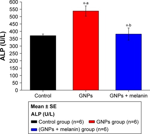
shows a disturbance in the liver ALP activity after the administration of GNPs. A significant elevation (p<0.05) to 538.17±34.71 U/L in the GNPs group was observed compared with 370.67±11.17 U/L in the control group. Rats that were treated with melanin simultaneously for up to 7 days showed significantly decreased activity of liver ALP, to 375.22±11.17 U/L, almost near to that seen in the control group.
Figure 1 Effect of GNPs and melanin on ALP level in rats.
Note: aRelated to GNPs within the control group; bmelanin related to GNPs and control; *P<0.05.
Abbreviations: ALP, alkaline phosphatase; GNP, gold nanoparticle.
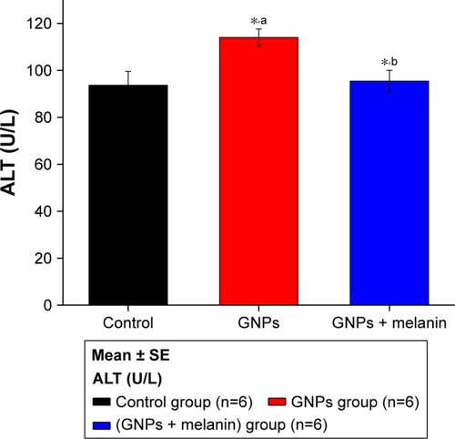
demonstrates the elevation of liver ALT activity, which increased (p<0.05) to 118±3.71 U/L in GNP rats as compared to 97.70±5.88 U/L in control rats. The coadministration of melanin with GNPs significantly reduced this elevation to 98.50±5.88 U/L.
Figure 2 Effect of GNPs and melanin on ALT level in rats.
Note: aRelated to GNPs within the control group; bmelanin related to GNPs and control; *P<0.05.
Abbreviations: ALT, alanine aminotransferase; GNP, gold nanoparticle.
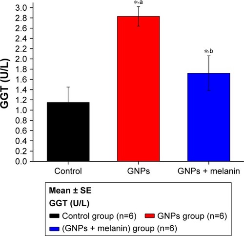
shows a significant elevation in the liver enzyme GGT in the GNP group (2.83±0.18 U/L) in comparison to the control group (1.15±0.30 U/L). Coadministration of melanin with GNPs significantly reduced the GGT level to 1.72±0.30 μ/L.
Figure 3 Effect of GNPs and melanin on GGT level in rats.
Note: aRelated to GNPs within the control group; bmelanin related to GNPs and control; *P<0.05.
Abbreviations: GGT, gamma-glutamyl transferase; GNP, gold nanoparticle.
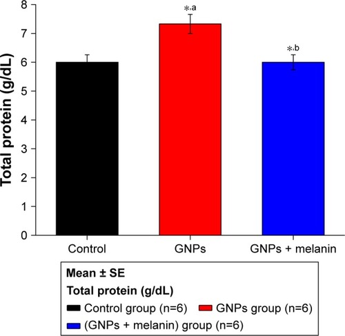
illustrates a notable increase (p<0.05) in the serum level of serum protein in the GNP rats (7.33±0.33 g/dL) compared to the control rats (5.95±0.25 g/dL). The coadministration of melanin with GNPs induced a significant decrease in the serum protein level (6.08±0.25 g/dL) compared to the GNP-administrated rats.
Figure 4 Effect of GNPs and melanin on total protein level in rats.
Abbreviation: GNP, gold nanoparticle.
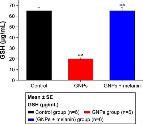
shows a significant reduction in the liver GSH levels in the GNP group; it was 20±0.99 μg/mL compared with the control group, 65±2.89 μg/mL. The coadministration of melanin with GNPs induced a significant elevation in the GSH level to 65.10±2.99 μg/mL compared to the GNPs group.
Figure 5 Effect of GNPs and melanin on liver GSH level in rats.
Note: aRelated to GNPs within the control group; bmelanin related to GNPs and control; *P<0.05.
Abbreviations: GNP, gold nanoparticle; GSH, reduced glutathione.
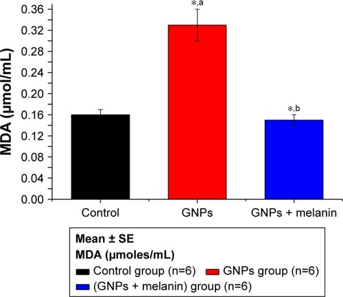
shows an obvious significant elevation on MDA activity in GNP rats (**p<0.01) when compared to control rats (0.33±0.03 vs 0.16±0.01 μmol/mL). The coadministration of melanin with GNPs induced a significant reduction in MDA level to 0.15±0.01 μmol/mL compared to the GNPs group.
Figure 6 Effect of GNPs and melanin on liver MDA level in rats.
Abbreviations: GNP, gold nanoparticle; MDA, malondialdehyde.
Hepatoxicity was confirmed and verified through histological investigation of the liver tissues, stained with H&E.
shows a normal liver section. Each hepatic lobule consists of a central vein lined with simple squamous epithelium, surrounded by polygonal hepatic cells, and contains quite clear blood sinusoids in-between. The cytoplasm of the hepatocytes appeared homogenous and contained various uniformly distributed coarse basophilic granules. The hepatocytes are usually uni-or binucleated. Numerous spindle-shaped Kupffer’s cells arise between the hepatocytes.
Figure 7 Photomicrographs of liver sections stained by H&E, 400×.
Notes: (A) Control rat: showing the typical hepatic architecture of hepatocytes (h), hepatic sinusoids (hs) and Kupffer’s cells (arrows). (B) GNPs-treated rat: showing swollen hepatocytes with vacuolated cytoplasm (arrows), and other hepatocytes with necrosis (stars). (C) GNPs + melanin-treated rat: showing normal hepatocytes (h), hepatic sinusoids (hs), and Kupffer’s cells (arrows).
Abbreviation: GNP, gold nanoparticle.
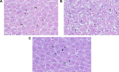
shows liver sections obtained from rats treated with 100 μL of 10 nm GNPs for 7 days revealed different histological lesions compared with the control group; these lesions also showed signs of vacuolar degeneration and necrosis. Necrosis refers to severe morphological changes accompanying the cell death, and it indicates to the presence of potentially irreversible cellular injuries. The necrotic cells showed nuclear alterations including pyknosis, karyorrhexis, and karyolysis.
shows the liver of rats treated with melanin (200 mg/kg/d) along with 100 μL of 10 nm GNPs for 7 days showed protection against hepatocyte vacuolar degeneration and necrosis when compared to the GNP-treated rats, and these cells appeared like those of normal liver cells.
Discussion
This study showed that GNPs caused damage to hepatocytes, as supported by the substantial elevation of serum liver markers in injected rats compared with control rats. Our finding was supported by previous studies,Citation28–Citation31 as these results revealed that excessive zinc administration could lead to liver damage. Coadministration of melanin to GNP-treated rats significantly reduced the levels of all the serum liver markers (p<0.001) when compared with the injected rats. This shows that the melanin could act as a defense against the liver dysfunction triggered by GNPs’ hepatotoxicity.
Some studies have reported that the pathogenic mechanism instigated by in vivo administration of metal oxide particles is dominated by oxidative stress, cell changes and death, and DNA destruction.Citation32,Citation33 Thus, a protective plan geared toward the reduction of the generation of inflammatory precursors could ameliorate liver damage and prevent or improve organ dysfunction. In our study, the ingestion of melanin immediately along with GNPs was beneficial in preventing the GNP-induced inflammatory liver injury.
Due to the recognized relationship between nanoparticles and oxidative stress, fatty peroxidation can be mentioned as one of the probable reasons for DNA destruction triggered by GNPs.Citation34 ROS are known to combine with DNA molecules, leading to the destruction of nitrogenous bases together with DNA support.Citation32
MDA, the main producer of fatty acid peroxidation, is a mutagenic and carcinogenic substance that can combine with DNA to form chemical compounds with adenosine nucleosides (deoxyguanosine and deoxyadenosine) and deoxyribonucleosides (deoxycytidine).Citation35,Citation36 DNA damage may trigger signal transduction pathways that may result in programmed cell death.Citation37
Eumelanin, which is the essential form of melanin found in humans, can protect against UV radiation, and owing to its antioxidant ability, it reduces DNA damage by creating a cap cover round the nucleus.Citation38,Citation39 Our data were consistent with those published earlier that melanin acts as an antioxidant, confirmed by its ability to stop fatty acid peroxidation.Citation22,Citation23 Together with reducing oxidative stress, melanin also reduces the inflammatory reaction of hepatocytes by preventing the creation of proinflammatory cytokines from permeating monocytes, and possibly lymphocytes also.
Our data might suggest or support the ability to use melanin to significantly reduce the inflammatory liver damage caused by administration of GNPs, as observed by the considerable alterations in the liver function markers as well as liver tissues biomarkers observed in this study. Moreover, new experiments should be performed to test the possibility of using melanin as a protective agent against the nephrotoxicity induced by GNPs.
Conclusion
This study underlines the hepatotoxic effects caused by intraperitoneal administration of GNPs that was confirmed by a significant rise in the levels of serum liver function markers such as ALT, ALP, GGT, TBIL, and total protein; this was in addition to the significant elevation in the MDA level and the decrease in the oxidative stress biomarker GSH level. The treatment with melanin successively alleviated ALT, ALP, GGT, TBIL, total protein, and MDA, while it elevated the GSH, which in turn efficiently ameliorated the lipid peroxidation and inflammatory liver damage induced in GNPs intoxicated rats. Our data demonstrated that melanin is a highly potent antioxidant that can protect the hepatocytes from damage produced by oxidative stress and other associated vascular complexities caused by GNP generation, due to the creation of ROS.
Acknowledgments
The authors would like to extend their sincere appreciation to the Deanship of Scientific Research at King Saud University
Disclosure
The authors report no conflicts of interest in this work.
References
- AbdelhalimMAKJarrarBMHistological alterations in the liver of rats induced by different gold nanoparticles size, dose and exposure durationJ Nanobiotechnology201210522276919
- AbdelhalimMAKJarrarBMRenal tissue alterations were size-dependent with smaller ones induced more effects and related with the time exposure of gold nanoparticlesLipids Health Dis20111016321936889
- AbdelhalimMAKJarrarBMGold nanoparticles induced cloudy swelling to hydropic degeneration, cytoplasmic hyaline vacuolation, polymorphism, binucleation, karyopyknosis, karyolysis, karyorrhexis and necrosis in the liverLipids Health Dis20111016621939512
- NelAXiaTMadlerLLiNToxic potential of materials at the nano levelScience2006311576162262716456071
- AbdelhalimMAKExposure to gold nanoparticles produces cardiac tissue damage that depends on the size and duration of exposureLipids Health Dis20111020522073987
- AbdelhalimMAKGold nanoparticles administration induces disarray of heart muscle, hemorrhagic, chronic inflammatory cells infiltrated by small lymphocytes, cytoplasmic vacuolization and congested and dilated blood vesselsLipids Health Dis20111023322151883
- HaseebAElhagHinventorHaseebAElhagHassigneeProcess for producing melanin using cultures of the genus NigellaWIPO patent WO2012125091A12012920 Available from: www.google.com/patents/WO2012125091A1Accessed June 08, 2018
- El-ObeidAAl-HarbiSAl-JomahNHassibAHerbal melanin modulates tumour necrosis factor alpha (TNF-α), interleukin 6 (IL-6) and vascular endothelial growth factor (VEGF) productionPhytomedicine200613532433316635740
- SavaVMHungYCBlagodarskyVAHongMYHuangGSThe liver-protecting activity of melanin-like pigment derived from black teaFood Res Int20036505511
- BridelliMGCiatiACrippaPRBinding of chemicals to melanins re-examined: adsorption of some drugs to the surface of melanin particlesBiophys Chem2006119213714516139945
- MeredithPSarnaTThe physical and chemical properties of melaninPigment Cell Res200619657259417083485
- PernaGFrassanitoMCPalazzoGFluorescence spectroscopy of synthetic melanin in solutionJ Lumin200912914449
- ChenYSHungYCHongMYControl of in vivo transport and toxicity of nanoparticles by tea melaninJ Nanomater201220129
- ProtaGMelanins and MelanogenesisSan Diego, CAAcademic Press1992
- HerrlingTJungKFuchsJThe role of melanin as protector against free radicals in skin and its role as free radical indicator in hairSpectrochim Acta A Mol Biomol Spectrosc20086951429143517988942
- RozanowskaMSarnaTLandEJTruscottTGFree radical scavenging properties of melanin interaction of eu- and pheomelanin models with reducing and oxidising radicalsFree Radic Biol Med1999265–651852510218640
- SarnaTMenonIASealyRCPhotosensitization of melanins: a comparative studyPhotochem Photobiol19854255295324089035
- SealyRCSarnaTWannerEJReszkaKPhotosensitization of melanin: an electron spin resonance study of sensitized radical production and oxygen consumptionPhotochem Photobiol19844044534596095338
- SarnaTPilasBLandEJTruscottTGInteraction of radicals from water radiolysis with melaninBiochim Biophys Acta198688311621673015231
- KorytowskiWKalyanaramanBMenonIASarnaTSealyRCReaction of superoxide anions with melanins: electron spin resonance and spin trapping studiesBiochim Biophys Acta198688221451533011111
- BustamanteJBredestonLMalangaGMordohJRole of melanin as a scavenger of active oxygen speciesPigment Cell Res1993653483538302773
- EzzahirAThe influence of melanins on the photoperoxidation of lipidsJ Photochem Photobiol B1989333413492504900
- KrolESLieblerDCPhotoprotective actions of natural and synthetic melaninsChem Res Toxicol19981112143414409860484
- Al-TayibOAHaseebAMEl-TahirKEIdrissMHThe aqueous extracts of the Nigella sativa melanin: experimental in vivo test and in vitro HEp-2 cell lines cytotoxicity effectsIOSR J Agric Vet Sci2016968490
- LoPachinRMRudyTAYakshTLAn improved method for chronic catheterization of the rat spinal subarachnoid spacePhysiol Behav19812735595617335798
- GurrJRWangASChenCHJanKYUltrafine titanium dioxide particles in the absence of phopactivation can induce oxidative DNA damage to human bronchial epithelial cellsToxicol20052131–26673
- UtleyHGBernheimFHochsteinPEffect of sulfhydryl reagents on peroxidation in microsomesArch Biochem Biophys196711812932
- WilmsLCHollmanPCBootsAWKleinjansJCProtection by quercetin and Quercetin-rich fruit juice against induction of oxidative DNA damage and formation of BPDE-DNA adducts in human lymphocytesMutat Res20055821–215516215781220
- LassASuessenbacherAWolkartGMayerBBrunnerFFunctional and analytical evidence for scavenging of oxygen radicals by L-arginineMol Pharmacol20026151081108811961125
- PiacenzaLPeluffoGRadiRL-arginine-dependent suppression of apoptosis in Trypanosoma cruzi: contribution of the nitric oxide and polyamine path waysProc Natl Acad Sci U S A200198137301730611404465
- ZhangXJChinkesDLWuZHerndonDNEnternal arginine supplementation stimulates DNA synthesis in skin donor woundClin Nutr201130339139621277659
- BormPJRobbinsDHauboldSThe potential risks of nanomaterials: a review carried out for ECETOCPart Fibre Toxicol200631116907977
- GhoshPHanGDeMKimCKRotelloVMGold nanoparticles in delivery applicationsAdv Drug Deliv Rev2011601113071315
- XiongDFangTYuLSimaXZhuWEffects of nano-scale TiO2, ZnO and their bulk counterparts on zebrafish: acute toxicity, oxidative stress and oxidative damageSci Total Environ201140981444145221296382
- MarnettLJOxy radicals, lipid peroxidation and DNA damageToxicology2002181–182219222
- NiedernhoferLJDanielsJSRouzerCAGreeneREMarnettLJMalondialdehyde, a product of lipid peroxidation, is mutagenic in human cellsJ Biol Chem200327833314263143312775726
- SharmaVShuklaRKSaxenaNParmarDDasMDhawanADNA damaging potential of zinc oxide nanoparticles in human epidermal cellsToxicol Lett2009185321121819382294
- KobayashiNNakagawaAMuramatsuTSupranuclear melanin caps reduce ultraviolet induced DNA photoproducts in human epidermisJ Invest Dermatol199811058068109579550
- MontagnaWCarlisleKThe architecture of black and white facial skinJ Am Acad Dermatol1991246 Pt 19299371714469