
 ?Mathematical formulae have been encoded as MathML and are displayed in this HTML version using MathJax in order to improve their display. Uncheck the box to turn MathJax off. This feature requires Javascript. Click on a formula to zoom.
Abstract
Background
Here, electrospun fibers based on a blend of polycaprolactone (PCL), poly(ethylene glycol) (PEG), and gelatin methacryloyl (GelMA) were developed. The careful choice of this polymer combination allowed for the preparation of a biomaterial that preserved the mechanical strength of PCL, while at the same time improving the hydrophilicity of the blended material and human osteoblast maturation.
Methods
The morphology, chemical structure, wettability, and mechanical properties before and after UV photocrosslinking were evaluated. Furthermore, human osteoblasts (hFOB) were cultivated for up to 21 days on the scaffolds, and their potential to upregulate cell proliferation, alkaline phosphatase (ALP) activity, and calcium deposition were investigated.
Results
Contact angle measurement results showed that the developed scaffolds presented hydrophilic properties after PEG and GelMA incorporation before (25°) and after UV photocross-linking (69°) compared to pure PCL (149°). PCL:PEG:GelMA-UV displayed a slight increase in mechanical strength (elastic modulus ~37 MPa) over PCL alone (~33 MPa). Normally, an increase in strength of fibers leads to a decrease in elongation at break, due to the material becoming less deformable and stiffer, thus leading to breaks at low strain. This behavior was observed by comparing PCL (elongation at break ~106%) and PCL:PEG:GelMA-UV (~50%). Moreover, increases in ALP activity (10-fold at day 14) and calcium deposition (1.3-fold at day 21) by hFOBs were detected after PEG and GelMA incorporation after UV photocross-linking compared to pure PCL. Ultrathin and hydrophilic fibers were obtained after PEG and GelMA incorporation after UV photocrosslinking, but the strength of PCL was maintained. Interestingly, those ultrathin fiber characteristics improved hFOB functions.
Conclusion
These findings appear promising for the use of these electrospun scaffolds, based on the combination of polymers used here for numerous orthopedic applications.
Introduction
Ultrathin scaffolds with desirable mechanical and wettability properties for tissue engineering and biomedical applications remain challenging.Citation1 These properties are able to control biological interactions with the scaffolds such as cell adhesion (controlling specific proteins), proliferation, and differentiation.Citation2 In this context, polycaprolactone (PCL) has been largely used in bone tissue engineering applications.Citation3 PCL has favorable mechanical properties compared to other polyesters and has good biocompatibility. PCL has a tensile strength ranging from 10.5 to 16.1 MPa and a tensile yield strength between 8.2 and 10.1 MPa. For electrospun PCL, the mean value of the Young’s modulus has been reported to be 3.5–6 MPa, with an average value of the strain at break of 150–190%.Citation4,Citation5 However, PCL exhibits hydrophobic characteristics and a long degradation time, which may reduce cell adhesion and limit applications where faster erosion of the matrices is needed. For this reason, the development of new strategies for improving its hydrophilicity, degradability, osteogenesis, and controllable mechanical properties for orthopedic applications is needed.Citation6–Citation8
The optimal scaffold for bone regeneration should display sufficient mechanical properties to support bone tissue requirements (Young’s modulus of cortical bone = 15–20 GPa and 0.1–2 GPa for cancellous bone),Citation9 hydrophilicity to improve the infiltration of cells favoring the transportation of water, nutrients, and waste, as well to promote osteogenesis.Citation10
One strategy to design such a biomaterial that fulfills the above requirements is to combine different polymers in a way that synergizes their desirable, unique properties without reducing their efficacy. Such materials can be used for the fabrication of biomaterial fibers (to mimic the extracellular matrix [ECM] and enhance the surface contact) for bone tissue regeneration through the employment of electrospinning.Citation11 There are several advantages with this approach compared to other methods, specifically for preparing polymer blends, such as reproducibility, low cost, and high yield.Citation12 Electrospinning is simple and efficient, since a common solvent can be used to dissolve different polymers, and the solvent can be easily evaporated due to applied high voltage.Citation12
An additional polymer that is of great interest in this context is poly(ethylene glycol) (PEG) that has distinct properties such as biocompatibility, water absorption, hydrophilicity, and the ability to reduce protein adsorption.Citation13 There have been several reports on the combination of PCL and PEG,Citation14,Citation15 PCL and gelatin,Citation4,Citation5,Citation8,Citation16 and PCL/PEG/gelatin for bone tissue regeneration (in these examples noncrosslinkable gelatin was employed).Citation17 These reports have described that electrospun PCL, PEG, and gelatin fibers and their combinations improved the osteogenesis and calcification of the matrix, induced osteoblast maturation and promoted bone regeneration; however, the described combinations resulted in a lower tensile strength (ranging from 2 to 35 MPa) compared to scaffolds for bone regeneration.Citation5,Citation8,Citation16 For example, Tiwari et alCitation18 combined PCL and PEG to produce scaffolds, and obtained an average tensile stress of up to 29 MPa.
The produced scaffold should also promote cell adhesion for better interaction with the host tissue. Gelatin, a natural polymer, is a good candidate for promoting cell adhesion due to a similarity to the ECM.Citation19 A photocrosslinkable gelatin methacryloyl (GelMA) material has been extensively used as a hydrogel scaffold for biomaterial applications.Citation19 GelMA has a chemical similarity to numerous ECMs and has been shown to improve vascularization, water absorption, and permeability of proteins, and possess a fast and controlled degradability.Citation20 However, GelMA has relatively weak mechanical properties for bone tissue engineering applications. To overcome this limitation, GelMA may be combined with other materials for the design of scaffolds suitable for those applications.Citation21,Citation22 The measured Young’s modulus for GelMA fibers alone (GelMA [10 wt%], 70% degree of methacryloyl modification, 2–10 min of UV crosslinking time) ranges from 290 to 350 kPa and its elongation at break is between 51 and 67%.Citation23 Herein, we combined PCL, PEG, and GelMA, followed by an electrospinning process for the design of fibrous blend scaffolds, as a suitable candidate for bone tissue engineering. The produced ultrathin fibers showed hydrophilicity and high mechanical strength while in vitro osteoblast functions were improved. Our study, thus, showcases the applications of this new scaffolding system for bone tissue regeneration, with a potential of extending to engineering other functional tissue types.
Results and discussion
The GelMA was prepared by methacryloyl substitution of gelatin, and the degree of substitution was determined using proton nuclear magnetic resonance (Citation1H NMR) analysis (~70% yield, 66% degree of substitution, ).Citation24 Moreover, it is widely known that low molecular weight PEG alone is challenging to electrospin.Citation25 However, this can be circumvented by a combination with other bio-materials; moreover, low amounts of PEG can be used to introduce hydrophilicity.Citation26 Notably, since PEG is known to be cell-repellent, an optimal concentration between PEG and GelMA is an important balance between hydrophilicity and cell adhesion. However, it has previously been reported that only an addition of 5 w/v% GelMA to PEG (20 w/v%) improved cell adhesion.Citation24
Figure 1 (A) Preparation of GelMA. Gelatin containing primary amino (−NH2) and hydroxyl (−OH) groups was reacted with methacrylic anhydride to add methacryloyl pendant groups. (B) Citation1H-NMR of the prepared GelMA compared to gelatin. (C) The different solutions prepared for the photocrosslinking step after electrospinning. (D) Scheme illustrating the chemistry and possible interactions between the electrospun polymers after photocrosslinking.
Notes: aFor all the solutions hexafluoroisopropan-2-ol (HFIP) was employed and the final volume was 5 mL. bThe photoinitiator lithium phenyl (2,4,6-trimethylbenzoly) phosphinate was added to the mixture to crosslink the prepolymer.
Abbreviations: GelMA, gelatin methacryloyl; Citation1HNMR, proton nuclear magnetic resonance.
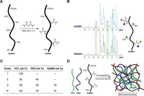
The prepared solutions and their concentrations for electrospinning are depicted in , based on previous reports.Citation18 Electrospinning of a PCL, PEG, and GelMA blend, followed by further crosslinking, provides a covalently-bonded hybrid mat, as illustrated in .
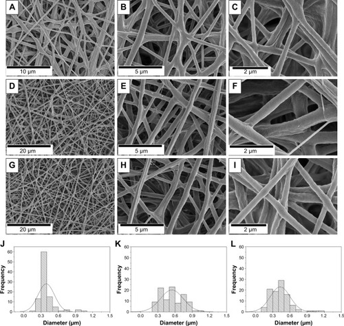
The morphology was investigated through scanning electron microscopy (SEM) analysis (). The micro-fibrous mats made from PCL displayed fiber diameters of 2.63±0.78 µm (). As expected, the electrospinning of pure PEG did not produce fibers, instead only beads were obtained (). However, when PCL and PEG were combined, ultrathin fibrous mats were obtained (), which presented diameter distributions of 0.39±0.14 µm (). The electrospun mats made of pure GelMA provided ultrathin smooth fibers with a diameter of 0.18±0.02 µm () and, by combining PCL, PEG, and GelMA, ultrathin fibers with a similar morphology were obtained () with a fiber diameter of 0.42±0.17 µm (). Importantly, subsequent UV crosslinking of the fiber changed neither its morphology () nor diameter (0.43±0.17 µm, ).
Figure 2 (A–I) Morphology of electrospun fibers from the SEM analysis: (A–C) PCL-PEG, (D–F) PCL-PEG-GelMA, and (G–I) PCL-PEG-GelMA-UV fibers. (J–L) The distribution of the fiber diameters for (J) PCL-PEG, (K) PCL-PEG-GelMA, and (L) PCL:PEG:GelMA-UV fibers.
Abbreviations: GelMA, gelatin methacryloyl; PCL, polycaprolactone; PEG, poly(ethylene glycol); SEM, scanning electron microscopy.
Fourier-transform infrared spectroscopy (FTIR) analysis identified functional groups and interactions of the polymers within the fibers (). The main peaks for the PCL:PEG:GelMA electrospun mats related to each polymer component were clearly identified (). The main peaks referred to PCL were indexed, as follows: 1,724 cm−1 (C=O stretching), 1,342 cm−1 (CH2, bending), 1,240 cm−1 (asymmetric C−O−C stretching), 1,190 cm−1 (O−C−O stretching), 1,170 cm−1 (C−O−C stretching), 1,157 cm−1 (C−C stretching), and 731 cm−1 (−(CH2)n, bending).Citation26,Citation27 The GelMA presence was observed due to the presence of amide I (1,637 cm−1, C=O stretching), 1,634 (methacryloyl group, C=C, stretching), amide II (1,529 cm−1, N−H bending), and amide III (1,448 cm−1, vibrations of C−N and N−H).Citation28 The presence of PEG was also identified: 839 cm−1 (C−H, bending), 951 cm−1 (CH2, rocking), and 1,115 cm−1 (C−O, stretching).Citation29,Citation30
Figure 3 (A, B) FTIR spectra collected of the developed scaffolds: (A) indexed peaks identified by different symbols, (B) C=C bond before and after UV irradiation. (C, D) The mechanical analysis of the electrospun scaffolds: (C) elastic modulus and (D) elongation at break. The values are expressed as means and SDs (***P<0.001, n=3). (E) The contact angles measured and images of the water drops on the PCL, PCL:PEG:GelMA, and PCL:PEG:GelMA-UV. (F, G) The DSC thermograms of the fibers: (F) first heating cycle and (G) first cooling cycle. (H) The thermal analysis of all the fiber components analyzed providing the Tm (crystalline melting temperature), ΔHm (melting enthalpy), ΔHc (crystallization enthalpy) and Xc (fiber crystallinity). All the calculated Xc belongs to the PCL in the mat, thus only the 100% ΔHm = melting enthalpy of a 100% crystalline PCL employed.
Abbreviations: FTIR, Fourier-transform infrared spectroscopy; GelMA, gelatin methacryloyl; PCL, polycaprolactone; DSC, differential scanning calorimetry; PEG, poly(ethylene glycol).
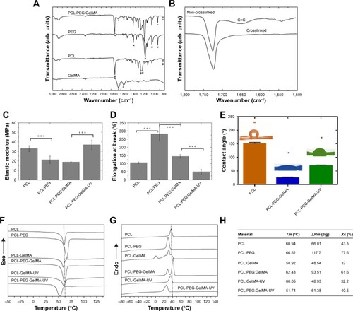
FTIR was also used to examine the potential differences after UV crosslinking (). The band at 1,634 cm−1 could be related to the presence of C=C double bonds in the scaffolds before UV irradiation ().Citation31 Nevertheless, this band disappeared after UV irradiation, indicating the success of the crosslinking procedure (). The effect of the formation of a physical network with increasing subsequent chemical crosslinking efficiency has also been reported previously.Citation21
Additionally, the mechanical properties of the electrospun scaffolds before and after crosslinking were investigated (). The designed PCL:PEG:GelMA scaffold showed a high elastic modulus after UV photocrosslinking compared to the other scaffolds without photocrosslinking, resulting in an elastic modulus of ~37 MPa (PCL:PEG:GelMA-UV), which was in a similar range to that of PCL (~33 MPa). In contrast, the measured elastic moduli for the PCL:PEG scaffold werê21.2 MPa and ~18.85 MPa for the PCL:PEG:GelMA scaffolds without photocrosslinking ().
As expected, the elongation at break for the PCL:PEG: GelMA-UV mats (~50%) was lower than those of all the other samples, probably due to the stiffer polymer obtained after UV crosslinking (). However, the PCL:PEG blends presented a superior elongation property (~283%) than that of pure PCL (~106%) (). However, after incorporation of GelMA, the value decreased by a factor of 2 (~144%), most likely due to the fragility of gelatin compared to PCL and PEG.
Recently, Tiwari et alCitation18 measured similar values between PCL and PCL:PEG mats. However, herein, the mechanical properties measured after the incorporation of GelMA and further UV photocrosslinking (PCL:PEG:GelMA-UV) provided a value 3-fold greater than that already reported for bone tissue engineering applications ().Citation32–Citation38 Scaffolds based on different amounts of PCL, GelMA, and gelatin have been reported by Correia et al.Citation39 They had covered PCL mats with GelMA and gelatin previously, but here our process is more homogenous and easy to reproduce, because PCL and GelMA were electrospun together, followed by photocrosslinking, which greatly improves the mechanical properties of the scaffolds. Clearly, the prepared scaffolds presented a good synergy between PCL and GelMA. Recently, Zhao et alCitation23 electrospun a GelMA solution and obtained ultrathin fibers (700–1,400 nm). The authors also investigated the mechanical properties of GelMA mats before and after UV curing. Herein, we combined PCL, PEG, and GelMA and compared the crosslinking with/without immersion of mats into the photoinitiator solution. The electrospun mats produced here had superior properties compared to other similar studies due to our unique combination of the selected polymers. Specifically, there was a clear improvement of the mechanical strength for our study (our results: 36.95±4.85 MPa; reported results: 400 kPa).Citation39
Moreover, the hydrophilicity of the designed electrospun was improved by the addition of PEG and GelMA before and after UV comparable to PCL (149°) (). In this aspect, the hydrophilicity can be monitored by the comparison of the wettability and surface energy of the material, where a higher surface energy generally corresponds to a lower contact angle.Citation40 The uncrosslinked material provided a water contact angle (CA) of ~25° and a surface energy of 71.4 mJ/m2, while crosslinking resulted in a CA of ~69° and surface energy of 53.3 mJ/m2 ().
It is known that a surface is hydrophobic when the CA>90° and is hydrophilic when CA<90°.Citation41 Correia et alCitation39 coated GelMA, PEG acrylate (PEGA), and PEG diacrylate (PEGDA) onto PCL mats for the preparation of a more hydrophilic material, where they obtained CA that ranged from ~40° to 120°. Different in our approach, we perform a direct incorporation of the various biomaterials, whereas in the presented coating strategy, it required an extra step for the addition of GelMA. Prominently, coating processes can sometimes be limited due to debonding of the coating, mechanically fragility, and instability of the material, thus all avoided by direct incorporation.Citation42
Zhao et alCitation23 obtained a highly porous and water absorbable GelMA and GelMA:PLGA scaffolds suitable for wound healing applications. In our case, we improved the wettability and mechanical properties of the scaffolds favoring the adhesion and growth of osteoblasts.
Differential scanning calorimetry analyzed the thermal parameters, such as crystallization temperature (Tc), melting temperature (Tm), enthalpy of fusion (ΔHm), enthalpy of crystallization (ΔHc), and degree of crystallinity (Xc) of the materials (). The crystallization behavior of PCL after the addition of GelMA slightly shifted to a higher temperature, and Xc, ΔHc, and ΔHm increased significantly from 36.6%, 39.71, and 93.84 J/g to 83.2%, and 69.01–142.9 J/g, respectively. However, the degree of crystallinity of the sample containing both PEG and GelMA was similar to pure PCL (). Noteworthy, after incorporation of PEG, extra peaks were obtained in the cooling cycle corresponding to the PEG moiety. It has previously been reported that a blend between PCL and PEG is highly dependent on the amount of each component in the blend and, in some cases, immiscibility and phase separation between PCL and PEG can occur, confirmed by extra peaks.Citation41
In comparison, after crosslinking, a shift to a lower temperature was immediately observed (), however, all of the parameters (Xc, ΔHc, and ΔHm) increased significantly comparable to the PCL fibers (Xc=73.5% and 85.8%, ΔHc=47.69 and 50.44 J/g, and ΔHm=113 and 126.6 J/g, respectively, ). The result clearly confirms that the crosslinking induces crystallinity in the blend fibers, due to the improved network between the polymers ().
The biological activity (alkaline phosphatase [ALP] activity, calcium deposition, and cell proliferation) of the fabricated electrospun mat was studied for potential applications in bone tissue regeneration (). ALP is a widely used and a classical biomarker to identify osteoblast differentiation (ECM mineralization). Therefore, when high expression is observed, it can be correlated to bone-forming protein expression during osteogenic differentiation, inducing mineralization promoted by a scaffold.Citation43 Interestingly, the groups containing GelMA increased ALP activity, calcium deposition, and osteoblast proliferation to a larger extent compared to pure PCL (). The scaffolds showed an increase in ALP expression compared to PCL (P<0.0001, ). At day 7, PCL:PEG:GelMA scaffolds had higher values compared to pure PCL and PCL:PEG (P<0.0001).
Figure 4 (A) ALP activity showed an increase of calcification of the extracellular matrix after inclusion of GelMA. (B) Calcium deposition demonstrated a further influence of GelMA to enhance the functions of osteoblasts. (C) MTS assay showing that osteoblastic cells were further influenced by hydrophilic properties after inclusion of PEG and GelMA. Data plotted in mean and SD (N=5). Values of P<0.01 were considered significant. Data were normalized by the cells, and the y-axis was multiplied by 104. For the ALP and calcium deposition, the data were compared to control (cells) and between each time. For cellular proliferation assays, the data were compared to pure PCL. N=5. **P<0.01, ***P<0.001, and ****P<0.0001 mean statistical differences. SEM of hFOBs cultivated on scaffolds after 7 days. (D) (i) PCL and (ii) magnified view. (E) (i) PCL-PEG and (ii) magnified view. (F) (i) PCL-PEG-GelMA without UV crosslinking and (ii) magnified view. (G) (i) PCL-PEG-GelMA after UV crosslinking and (ii) magnified view. The cells are spreading on all produced scaffolds presenting filopodium and cytoplasmic extension.
Abbreviations: ALP, alkaline phosphatase; GelMA, gelatin methacryloyl; hFOB, human osteoblasts; MTS, (3-(4,5-dimethylthiazol-2-yl)-5-(3-carboxymethoxyphenyl)-2-(4-sulfophenyl)-2H-tetrazolium); NS, no significance; PCL, polycaprolactone; PEG, poly(ethylene glycol); SEM, scanning electron microscopy.
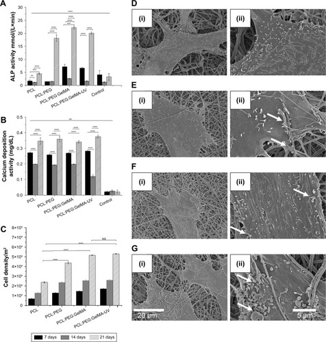
The PCL scaffolds partially induced extracellular calcification at days 14 and 21. On the other hand, when PEG and GelMA were added, the values increased independent of the time points (P<0.01). This highlights the favorable synergistic effect of the –OH groups on PEG and favorable amino acid moieties present in GelMA. At day 21, the ALP activity of the cells on the PCL:PEG scaffolds was 4-fold higher than that of the cells on pure PCL. At the same time (day 21), the ALP activity of cells on the PCL:PEG:GelMA scaffolds were 1.3-fold higher than that of the cells on the PCL:PEG scaffolds, and ~5-fold higher than that of the cells on the pure PCL scaffolds.
Additionally, illustrates the calcium deposition for all the analyzed groups. Calcium deposition is considered the final stage of osteoblast maturation to form bone. This may be because of the hydrophilic behavior associated with the ECM-like components in the GelMA chemical structure. It is also known that lower ALP levels are related to higher calcium deposition due to the last stage of maturation.Citation44 This behavior matched the lowered ALP activity obtained up to 14 days, while the calcium deposition increased at the same time point (). Moreover, the lower value measured for the control (cells in the absence of any scaffold) further supports the hypothesis that our scaffold promotes calcium deposition. After days 7, 14, and 21, all the groups analyzed induced high levels of calcium deposition.
Cell proliferation was measured, and all the groups containing GelMA and PEG were compared to pure PCL nanofibers (). An increase in cell proliferation was observed, independent of the scaffolds. However, an enhancement was observed for groups containing PEG compared to pure PCL for all time points (). After GelMA incorporation, an increase in proliferation of up to 2.5-fold was observed compared to pure PCL for all the time points (P<0.05). Cell proliferation has a strong positive correlation with the hydrophilicity of a surface,Citation45 and therefore the groups containing PEG and GelMA (each greatly hydrophilic) resulted in more cell proliferation compared to pure PCL (hydrophobic).
It has previously been reported that GelMA and PEG induced ECM calcification.Citation46–Citation52 For example, Turkkan et alCitation53 showed that electrospun PCL:PEG with hydroxyapatite induced in vitro osteoblast mineralization. Tiwari et alCitation18 electrospun different combinations of PCL and PEG and evaluated their potential to induce in vitro mineralization. However, the authors only investigated biocompatibility and in vitro mineralization using simulated body fluid.Citation18 Differently, herein we investigated the ability of new scaffolds, fabricated from a unique combination of PCL:PEG:GelMA, to induce in vitro ECM calcification and calcium deposition when cultivated with human osteoblasts (hFOBs).
The cellular adhesion of hFOBs were evaluated by SEM (). The presence of a significant number of filopodium was clearly noticed, independent of the type of scaffolds analyzed (depicted in ). The hydrophobicity of PCL scaffolds did not inhibit hFOB adhesion (). The same behavior was observed after incorporation of PEG (, more hydrophilic). Meanwhile, after inclusion of GelMA, more filopodia were noticed (), indicating monolayer formation, especially after UV crosslinking (). Additionally, the ultrathin scaffold obtained after incorporation of PEG and GelMA could also possibly be another factor promoting cellular adhesion, where microfibrous fibers were obtained with only PCL ( and ).
Conclusion
In summary, we report the preparation of electrospun scaffolds by a blend of PCL:PEG:GelMA polymers. Compared to previous reports, an improvement in efficacy was obtained due to the synergy of the desired properties of each polymer, including enhanced mechanical properties (stiffness) due to the presence of PCL, hydrophilicity due to the presence of PEG, and upregulation of hFOB cell functions from the incorporation of GelMA. The designed biomaterial also resulted in improved mechanical strength comparable to pure PCL. Furthermore, enhanced ALP activity, calcium deposition, and proliferation were obtained for our designed electrospun nanofibers compared to only PCL nanofibers.
Acknowledgments
AOL and FRM would like to thank the Sao Paulo Research Foundation (FAPESP, grants numbers: AOL – 2015/09697-0 and FRM – 2016/00575-1), Coordination for the Improvement of Higher Education Personnel (CAPES, grant numbers AOL – 88881.120138/2016-01 and FRM – 88881.120221/2016-01), Brazilian National Council for Scientific and Technological Development (CNPq, AOL – 303752/2017-3 and FRM – 304133/2017-5), and to the Universidade Brasil for scholarships. SA gratefully acknowledges financial support from the Sweden–America Foundation (The Family Mix Entrepreneur foundation), Olle Engkvist Byggmästare Foundation, and Swedish Chemical Society (Bengt Lundqvist Memory Foundation) for a postdoctoral fellowship. AK acknowledges funding from the National Institutes of Health (AR057837, AR066193, EB022403, EB021148, HL137193, EB021857, AR070647, EB023052, CA214411, and EB024403). YSZ acknowledges funding from the National Institutes of Health (K99CA201603, R21EB025270, R21EB026175).
Supplementary materials
Materials
The polycaprolactone (PCL) (80,000), poly(ethylene glycol) (PEG) (Mw 8,000), gelatin (Type A, 300 bloom from porcine skin), methacrylic anhydride (MA), Alizarin red S, dimethyl sulfoxide, and Irgacure 2959 were purchased from Sigma-Aldrich (St. Louis, MO, USA). Hexafluoroisopropan-2-ol was purchased from Oakhood Chemical (St. Louis, MO, USA). Ethylenediaminetetraacetic acid, Dulbecco’s PBS, and antibiotics were purchased from Sigma-Aldrich. Alpha-MEM was supplied by Invitrogen. HyClone characterized FBS and precleaned microscope slides were obtained from Thermo Fisher Scientific (Waltham, MA, USA). 3-(4,5-dimethylthiazol-2-yl)-5-(3-carboxymethoxyphenyl)-2-(4-sulfophenyl)-2H-tetrazoli solution was provided by Promega (Fitch burg, WI).
Procedure for the synthesis of gelatin methacryloyl (GelMA)
GelMA was prepared in accordance with Nichol et alCitation1 (). PBS (100 mL) was heated at 50°C and 10 g of gelatin (Type A, porcine skin) was dissolved and stirred for up to 1 h. Next, 3 mL of methacrylate anhydride was dripped slowly and stirred in a closed system for 3 h (50°C). Separately, PBS (400 mL) was preheated at 50°C and then mixed (final volume 500 mL). The solution was divided in two portions and dialyzed using DI water for 7 days (dialysis tube, Sigma-Aldrich 12,000–14,000 Da, 40°C). The DI water was changed twice per day. Finally, the solution was tranferred to falcon tubes, frozen at −80°C for 5 days, and then lyophilized for 7 days. Proton nuclear magnetic resonance (Citation1HNMR) detetermined the degree of methacryloyl substitution.
Citation1H NMR analysis
Thirty milligrams of GelMA was dissolved in 1 mL of deuterium oxide (D2O) and run at 37°C. CD LABS 12.0 software analyzed the data. Comparison between methylene lysine protons (2H) around 2.8 ppm (peak b) of gelatin and GelMA were used to determine the degree of methacryloyl substitution (66% by NMR analysis, ).
Preparation of the solutions
PCL, PEG, and GelMA were separately dissolved () for 12 h (using a closed system). A classical electrospinning process was coupled using: a syringe (BD Yale, 3 mL), needle (Inbras, 24G), positive high voltage source (set to 17 kV, Nanospinner Machine, Inovenso), static stainless steel collector (100×100×2 mm) covered by aluminum foil, distance of 10 cm (needle tip to collector), infusion rate of 1 mL/h (Harvard, PHD 2000), temperature at 23°C, and humidity at 40%.
Crosslinking of scaffolds
PCL:PEG:GelMA mats (10×10 mm) (, Entry 4) were immersed in glutaraldehyde (25% in DI water) for 12 h. After, the mats were washed five times in a glycine solution (15 mg/mL of DI water). Next, the mats were immersed in a photoinitiator solution (1.0 g of Irgacure, 10 mL of ethanol, absence of light) for 2 h and photocross-linked for 10 min (365-nm UV light, OminiCure®-2000 Series, 10 cm of working distance). Finally, the cross-linked PCL:PEG:GelMA were washed using the following sequence: ethanol (3×), DI water (3×) and dried overnight (vacuum). summarizes a possible mechanism after photocrosslinking.
Characterization of scaffolds
Micrographs were captured using an FEI Quanta 200, 3 kV, microscope. Previous to the analysis, a thin gold layer was evaporated for 10 min (~10 nm). Attenuated total reflectance Fourier-transform infrared spectrometry (ATR-FTIR, Spotlight-400, Perkin Elmer) analyzed the vibrational chemical groups before and after UV photocrosslinking.
A goniometer (Krüss, Model DSA 100) operating in dynamic mode was used to measure the contact angle between the scaffold surface and air using water and diiodomethane. Two microliters of each liquid was dropped on each scaffold, and images were recorded after 1 min. An N=5 for each sample was used.
The surface energy was calculated using interfacial tension through the Young’s (Equationequation 1(1) )Citation2 and Young-Duprè equations (Equationequation 2
(2) ) using surface tension data from water and diiodomethane liquids ().
Differential scanning calorimetry (TA Q20, TA Instruments) was used to identify the thermal properties of the produced scaffolds. For this, we used the first heating data, collected from −60°C to 150°C, at 10°C/min. The second heating data were also acquired at 10°C/min until reaching 150°C. The glass transition and the crystalline melting temperatures of the samples were obtained from the second heating and used to calculate the degree of the crystallinity () by using Equationequation (4)(4) .
A Dynamic Mechanical Analyzer (DMA Q800, TA Instruments) operating in tension mode (ASTM D 882-12 with some modifications) was used to measure the mechanical properties of the produced scaffolds before and after UV (elastic modulus, MPa; and elongation-at-break, expressed in a percentage) in tension mode. The scaffolds were then cut (30×5×0.1 mm) and preconditioned at 25°C and 50% humidity for 24 h before testing. Measurements were carried out at 25°C at a strain rate of 1 mm/min (N=3). The statistical difference was analyzed using Student’s t-tests.
Biological assays
Cell culture and sample sterilization
Totally 5,000 cells/cm2 of human osteoblast cells line (hFOB 1.19, bone-forming cells; Lonza, CRL-11372, second passage) were cultured using C27015 media (Osteoblast Basal Medium, Promocell GmbH), 1% penicillin/streptomycin (P/S; Hyclone), and an Osteoblast Growth Medium Supplement Mix under standard cell culture conditions (37°C, 5% CO2, and 95% air). UV irradiation (30 min) was used to sterilize the scaffolds (10 mm2).
Cell Proliferation assay
An MTS CellTiter 96® ((3-(4,5-dimethylthiazol-2-yl)-5-(3-carboxymethoxyphenyl)-2-(4-sulfophenyl)-2H-tetrazolium), G3581; Promega Corporation) assay was used to determine cell density up to 21 days. The scaffolds were first immersed into 24-wells plates; 5,000 cells/cm2 were placed onto scaffolds and cultivated for 14 days. The culture media was changed every 2 days. After 14 days, an MTS reagent (1:5 ratio with cell culture medium) was added to each well and incubated for 4 h. Absorbance from each well was measured by a SpectraMax M3(MT05412) at 490 nm, and a color change from pink to dark brown was seen. A standard curve was created with known numbers of cells to correlate absorbance to cell numbers.
ALP assay
A BioAssay QuantiChrom™ ALP Assay Kit (DALP-250) was used. After each incubation time (7, 14, and 21 days), 200 µL of the assay buffer, 5 µL of an acetate solution (final 5 mM), and 2 µL of the p-nitrophenylphosphate liquid substrate (10 mM) were mixed and added to each 96-well assay. Two hundred microliters of DI water and the same volume of a calibration solution was transferred into separate wells of a clear bottom 96-well plate. Then, 50 µL samples were carefully transferred into other wells, and 150 µL of the working solution was pipetted into sample wells. The final reaction volume in the sample wells was 200 µL. The optical density was measured at 405 nm (t=0) and again after 4 min (t=4 min) on a plate reader using the supernatant.
Calcium deposition
Totally 5,000 cells/cm2 were cultivated on the scaffolds for 7, 14, and 21 days. After each time, the scaffolds were washed three times using DI water. After, 1 mL of 0.6 M hydrochloric acid was added to each well for 4 h while in a shaken incubator. Next, the resulting samples were collected after being centrifuged at 12,000 rpm at 4°C for 3 min, and the supernatants were collected. Next, the aliquots of the Quanti-Chrom Calcium Assay Kit (BioAssay Systems) were added. Finally, the contents were transferred to 96-well plates and placed into a SpectraMax M3 microplate reader (Molecular Device), and the absorbance was measured at 612 nm. The values were obtained in absorbance according to the manufacturer’s instructions. The absorbance was measured (612 nm) and compared to a standard curve constructed at the beginning of each trial.
Cellular adhesion
Totally 5,000 cells/cm2 were cultivated on each scaffold for 7 days. After time, the scaffolds were washed (3× using PBS) and fixed in paraformaldehyde (4%) at room temperature (20 min). Afterward, an ascending series of ethanol was used to dehydrate the samples. Finally, a thin gold layer was sputtered (10 nm) and micrographs were collected using a field emission gun scanning electron microscope (FEI Quanta 200, 3 kV).
Statistical analysis
The experiments were analyzed in triplicate and repeated three times. ANOVA and Student’s t-tests were used to determine the significance and statistical differences. The populations were obtained from a normal distribution.
Figure S1 (A–I) Morphology of electrospun (A–C) PCL fibers, (D–F) GelMA fibers and (G–I) PEG beads. (A, D, G) The distribution of diameters for (J) PCL and (K) GelMA fibers.
Abbreviations: GelMA, gelatin methacryloyl; PCL, polycaprolactone; PEG, poly(ethylene glycol).
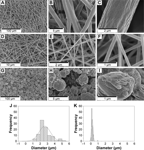
Table S1 Liquids and surface tension components
Table S2 Contact angle and surface free energy calculated for the PCL:PEG:GelMA and PCL:PEG:GelMA-UV scaffoldsTable Footnotea
References
- NicholJWKoshySTBaeHHwangCMYamanlarSKhademhosseiniACell-laden microengineered gelatin methacrylate hydrogelsBiomaterials201031215536554420417964
- YoungTIII. An essay of cohesion of fluidsPhilos Trans R Soc London18099565
- Van OssCJGoodRJChaudhuryMKThe role of van der Waals forces and hydrogen bonds in “hydrophobic interactions” between biopolymers and low energy surfacesJ Colloid Interface Sci1986111378390
- FlahautEDurrieuMCRemy-ZolghadriMBareilleRBaqueyCInvestigation of the cytotoxicity of CVVD carbon nanotubes towards human umblical vein endothelial cellsCarbon20064410931099
- ZhangYOuyangHLimCTRamakrishnaSHuangZ-MElectrospinning of gelatin fibers and gelatin/PCL composite fibrous scaffoldsJ Biomed Mater Res B Appl Biomater200572B1156165
- Navarro-BaenaVSessiniVDominiciFTorreLKennyJMPeponiLDesign and biodegradable blends based on PLA and PCL: from morphological, thermal and mechanical studies to shape memory behaviorPolym Degrad Stab201613297108
Disclosure
The authors report no conflicts of interest in this work.
References
- YoshimotoHShinYMTeraiHVacantiJPA biodegradable nanofiber scaffold by electrospinning and its potential for bone tissue engineeringBiomaterials200324122077208212628828
- Schaap-OziemlakAMKühnPTvan KootenTGvan RijnPBiomaterial–stem cell interactions and their impact on stem cell responseRSC Adv20144955330753320
- WilliamsJMAdewunmiASchekRMBone tissue engineering using polycaprolactone scaffolds fabricated via selective laser sinteringBiomaterials200526234817482715763261
- BinulalNSNatarajanAMenonDBhaskaranVKMonyUNairSVPCL-gelatin composite nanofibers electrospun using diluted acetic acid-ethyl acetate solvent system for stem cell-based bone tissue engineeringJ Biomater Sci Polym Ed201425432534024274102
- ZhangYOuyangHLimCTRamakrishnaSHuangZMElectro-spinning of gelatin fibers and gelatin/PCL composite fibrous scaffoldsJ Biomed Mater Res B Appl Biomater200572115616515389493
- FujiharaKKotakiMRamakrishnaSGuided bone regeneration membrane made of polycaprolactone/calcium carbonate composite nano-fibersBiomaterials200526194139414715664641
- CausaFNettiPAAmbrosioLPoly-epsilon-caprolactone/hydroxyapatite composites for bone regeneration: in vitro characterization and human osteoblast responseJ Biomed Mater Res A200676115116216258959
- RenKWangYSunTYueWZhangHElectrospun PCL/gelatin composite nanofiber structures for effective guided bone regeneration membranesMater Sci Eng C Mater Biol Appl20177832433228575991
- BoseSRoyMBandyopadhyayARecent advances in bone tissue engineering scaffoldsTrends Biotechnol2012301054655422939815
- YiHUr RehmanFZhaoCLiuBHeNRecent advances in nano scaffolds for bone repairBone Res201641605028018707
- JangJHCastanoOKimHWElectrospun materials as potential platforms for bone tissue engineeringAdv Drug Deliv Rev200961121065108319646493
- PramanikSPingguan-MurphyBAbu OsmanNAProgress of key strategies in development of electrospun scaffolds: bone tissueSci Technol Adv Mater201213404300227877500
- ZhuJBioactive modification of poly(ethylene glycol) hydrogels for tissue engineeringBiomaterials201031174639465620303169
- PazarçevirenEErdemliÖKeskinDTezcanerAClinoptilolite/PCL-PEG-PCL composite scaffolds for bone tissue engineering applicationsJ Biomater Appl20173181148116827881642
- KoupaeiNKarkhanehADaliri JoupariMPreparation and characterization of (PCL-crosslinked-PEG)/hydroxyapatite as bone tissue engineering scaffoldsJ Biomed Mater Res A2015103123919392626015080
- YaoRHeJMengGJiangBWuFElectrospun PCL/Gelatin composite fibrous scaffolds: mechanical properties and cellular responsesJ Biomater Sci Polym Ed201627982483827044505
- YangGWangJWangYLiLGuoXZhouSAn implantable active-targeting micelle-in-nanofiber device for efficient and safe cancer therapyACS Nano2015921161117425602381
- TiwariAPJoshiMKLeeJHeterogeneous electrospun polycaprolactone/polyethylene glycol membranes with improved wettability, biocompatibility, and mineralizationColloids Surf A Physicochem Eng Asp2017520105113
- ShuXZLiuYPalumboFPrestwichGDDisulfide-crosslinked hyaluronan-gelatin hydrogel films: a covalent mimic of the extracellular matrix for in vitro cell growthBiomaterials200324213825383412818555
- YueKTrujillo-de SantiagoGAlvarezMMTamayolAAnnabiNKhademhosseiniASynthesis, properties, and biomedical applications of gelatin methacryloyl (GelMA) hydrogelsBiomaterials20157325427126414409
- ThakurTXavierJRCrossLPhotocrosslinkable and elastomeric hydrogels for bone regenerationJ Biomed Mater Res A2016104487988826650507
- EchaveMCSánchezPPedrazJLOriveGProgress of gelatin-based 3D approaches for bone regenerationJ Drug Deliv Sci Technol2017426374
- ZhaoXSunXYildirimerLCell infiltrative hydrogel fibrous scaffolds for accelerated wound healingActa Biomater201749667727826004
- NicholJWKoshySTBaeHHwangCMYamanlarSKhademhosseiniACell-laden microengineered gelatin methacrylate hydrogelsBiomaterials201031215536554420417964
- KlossnerRRQueenHACoughlinAJKrauseWECorrelation of chitosan’s rheological properties and its ability to electrospinBiomacromolecules20089102947295318785774
- ChakrapaniVYGnanamaniAGiridevVRMadhusoothananMSekaranGElectrospinning of type I collagen and PCL nanofibers using acetic acidJ Appl Polym Sci2012125432213227
- FukushimaKFeijooJLYangM-CComparison of abiotic and biotic degradation of PDLLA, PCL and partially miscible PDLLA/PCL blendEur Polym J2013493706717
- DallasPNiarchosDVrbanicDInterfacial polymerization of pyrrole and in situ synthesis of polypyrrole/silver nanocompositesPolymer200748720072013
- SeifpoorMNouriMMokhtariJThermo-regulating nanofibers based on nylon 6,6/polyethylene glycol blendFibers and Polymers2011126706714
- ChenHMaQWangSLiuHWangKMorphologyWKMorphology, compatibility, physical and thermo-regulated properties of the electrospinning polyamide 6 and polyethylene glycol blended nanofibersJournal of Industrial Textiles201645614901503
- ShinHOlsenBDKhademhosseiniAThe mechanical properties and cytotoxicity of cell-laden double-network hydrogels based on photocrosslinkable gelatin and gellan gum biomacromoleculesBiomaterials201233113143315222265786
- LiYFRubertMAslanHUltraporous interweaving electrospun microfibers from PCL-PEO binary blends and their inflammatory responsesNanoscale2014663392340224531205
- XiaoKZhaiYYuJDingBPoly N-StructuredNanonet-structured poly(m-phenylene isophthalamide)–polyurethane membranes with enhanced thermostability and wettability for high power lithium ion batteriesRSC Adv20155685547855485
- GuanJWagnerWRSynthesisWWRSynthesis, characterization and cytocompatibility of polyurethaneurea elastomers with designed elastase sensitivityBiomacromolecules2005652833284216153125
- KwonIKKidoakiSMatsudaTElectrospun nano- to microfiber fabrics made of biodegradable copolyesters: structural characteristics, mechanical properties and cell adhesion potentialBiomaterials200526183929393915626440
- PantHRBajgaiMPNamKTChuKHParkS-JKimHYFormation of electrospun nylon-6/methoxy poly(ethylene glycol) oligomer spider-wave nanofibersMater Lett2010641920872090
- PantHRNamKTOhHJEffect of polymer molecular weight on the fiber morphology of electrospun matsJ Colloid Interface Sci2011364110711121889156
- OlsztaMJChengXJeeSSBone structure and formation: A new perspectiveMater Sci Eng: R: Rep2007583–577116
- CorreiaTRFerreiraPVazRDevelopment of UV cross-linked gelatin coated electrospun poly(caprolactone) fibrous scaffolds for tissue engineeringInt J Biol Macromol201693Pt B1539154827185071
- MorouçoPBiscaiaSVianaTFabrication of poly(ε-caprolactone) scaffolds reinforced with cellulose nanofibers, with and without the addition of hydroxyapatite nanoparticlesBiomed Res Int20162016159615727872844
- VoglerEAStructure and reactivity of water at biomaterial surfacesAdv Colloid Interface Sci1998741691179561719
- YukHZhangTLinSParadaGAZhaoXTough bonding of hydrogels to diverse non-porous surfacesNat Mater201615219019626552058
- MizunoMKubokiYOsteoblast-related gene expression of bone marrow cells during the osteoblastic differentiation induced by type I collagenJ Biochem2001129113313811134967
- KorolevaADeiwickANguyenAOsteogenic differentiation of human mesenchymal stem cells in 3-D Zr-Si organic-inorganic scaffolds produced by two-photon polymerization techniquePLoS One2015102e011816425706270
- LampinMWarocquier-CléroutLegrisCDegrangeMSigot-LuizardMFCorrelation between substratum roughness and wettability, cell adhesion, and cell migrationJ Biomed Mater Res1997361991089212394
- ByambaaBAnnabiNYueKBioprinted osteogenic and vasculogenic patterns for engineering 3D bone tissueAdv Healthc Mater20176161700015
- KhayatAMonteiroNSmithEEGelMA-Encapsulated hDPSCs and HUVECs for dental pulp regenerationJ Dent Res201796219219928106508
- ChenXBaiSLiBFabrication of gelatin methacrylate/nanohydroxyapatite microgel arrays for periodontal tissue regenerationInt J Nanomedicine2016114707471827695327
- BaratiDShariatiSRPMoeinzadehSMelero-MartinJMKhademhosseiniAJabbariESpatiotemporal release of BMP-2 and VEGF enhances osteogenic and vasculogenic differentiation of human mesenchymal stem cells and endothelial colony-forming cells co-encapsulated in a patterned hydrogelJ Control Release201622312613626721447
- AparnathiMKPatelJSBiodegradable gelatin methacrylate gel as a potential scaffold for bone tissue engineering of canine adipose-derived stem cellsJ Stem Cells201611311111928296875
- JiangPMaoZGaoCCombinational effect of matrix elasticity and alendronate density on differentiation of rat mesenchymal stem cellsActa Biomater201519768425805109
- VisserJGawlittaDBendersKEEndochondral bone formation in gelatin methacrylamide hydrogel with embedded cartilage-derived matrix particlesBiomaterials20153717418225453948
- TürkkanSPazarçevirenAEKeskinDMachinNEDuyguluÖTezcanerANanosized CaP-silk fibroin-PCL-PEG-PCL/PCL based bilayer membranes for guided bone regenerationMater Sci Eng C Mater Biol Appl20178048449328866191