
Abstract
Purpose
Targeted nanobubbles can penetrate the tumor vasculature and achieve ultrasound molecular imaging (USMI) of tumor parenchymal cells. However, most targeted nanobubbles only achieve USMI of tumor parenchymal cells from one organ, and their distribution, loading ability, and binding ability in tumors are not clear. Therefore, targeted nanobubbles loaded with carbonic anhydrase IX (CAIX) aptamer were fabricated for USMI of various tumors, and the morphological basis of USMI with targeted nanobubbles was investigated.
Materials and methods
The specificity of CAIX aptamer at the cellular level was measured by immunofluorescence and flow cytometry. Targeted nanobubbles loaded with CAIX aptamer were prepared by a maleimidethiol coupling reaction, and their binding ability to CAIX-positive tumor cells was analyzed in vitro. USMI of targeted and non-targeted nanobubbles was performed in tumor-bearing nude mice. The distribution, loading ability, and binding ability of targeted nanobubbles in xenograft tumor tissues were demonstrated by immunofluorescence.
Results
CAIX aptamer could specifically bind to CAIX-positive 786-O and Hela cells, rather than CAIX-negative BxPC-3 cells. Targeted nanobubbles loaded with CAIX aptamer had the advantages of small size, uniform distribution, regular shape, and high safety, and they could specifically accumulate around 786-O and Hela cells, while not binding to BxPC-3 cells in vitro. Targeted nanobubbles had significantly higher peak intensity and larger area under the curve than non-targeted nanobubbles in 786-O and Hela xenograft tumor tissues, while there was no significant difference in the imaging effects of targeted and non-targeted nanobubbles in BxPC-3 xenograft tumor tissues. Immunofluorescence demonstrated targeted nanobubbles could still load CAIX aptamer after penetrating the tumor vasculature and specifically binding to CAIX-positive tumor cells in xenograft tumor tissues.
Conclusion
Targeted nanobubbles loaded with CAIX aptamer have a good imaging effect in USMI of tumor parenchymal cells, and can improve the accuracy of early diagnosis of malignant tumors from various organs.
Introduction
The ultrasound contrast agents (UCAs) that are currently used in the clinic can only achieve perfusion imaging of tumor vasculature. Due to the lack of tumor specificity, they cannot specifically accumulate in tumor tissues and thus are not able to achieve ultrasound molecular imaging (USMI) of malignant tumors.Citation1,Citation2 Targeted UCAs can be constructed by coupling specific ligands against tumor cells with the surfaces of UCAs, which can specifically accumulate in tumor tissues and be used for USMI of malignant tumors. Targeted UCAs are mostly on the micron scale, and their binding targets are mainly membrane proteins of tumor vascular endothelial cells, such as vascular endothelial growth factor receptor-2, endothelin, nucleolin, and so on.Citation3–Citation6 Therefore, they only specifically bind to tumor vasculature and achieve USMI of tumor blood pool. It is a challenge for targeted UCAs to penetrate the tumor vasculature and achieve extravascular USMI of malignant tumors.
Nanoscale UCAs with small particle size, strong penetration, and long imaging time can aggregate in the extravascular spaces via the enhanced permeability and retention (EPR) effect.Citation7 Extravascular targeted ligands on the surfaces of targeted nanoscale UCAs can further enhance the accumulation of nanoscale UCAs in tumor tissues. After intravenous injection, targeted nanoscale UCAs can specifically bind to tumor parenchyma cells, achieve extravascular USMI of malignant tumors, and find the changes in protein expression on the cell membranes of tumor parenchyma cells in vivo.Citation8,Citation9 Nanobubbles with a lipid outer shell are a novel type of nanoscale UCAs.Citation10 Compared with other shell materials, nanobubbles with a lipid shell have a good imaging effect and high biocompatibility, and have been widely applied in USMI.Citation11–Citation14 However, current studies about USMI of targeted lipid nanobubbles have two challenges: 1) targeted nanobubbles are mainly used for USMI of tumor parenchyma cells from one organ, such as targeted nanobubbles carrying anti-prostate specific membrane antigen aptamer mainly achieve USMI of tumor parenchymal cells in prostate cancer.Citation15 Although they can achieve intravascular USMI in non-prostatic tumors, USMI of tumor parenchymal cells in non-prostatic tumors cannot be achieved.Citation16,Citation17 2) Most studies only focus on the imaging effect of targeted nanobubbles and have not yet studied their distribution, loading ability, and specific binding ability in vivo.
Carbonic anhydrase IX (CAIX) plays an important role in the growth and metastasis of numerous tumors (including renal cancer, cervical cancer, colon cancer, prostate cancer, breast cancer, head and neck tumors, and so on), and has been used for the diagnosis, treatment, and prognosis of malignant tumors.Citation18–Citation23 Targeted ligands against CAIX include monoclonal antibodies, nanobodies, and affibodies, as well as polypeptides that we studied earlier.Citation18,Citation22,Citation24,Citation25 Aptamers are single-stranded DNA or RNA oligonucleotides that specifically bind to biological macromolecules or cells through a specific spatial structure. Compared with other ligands, aptamers have several attractive features, including diverse structure, high specificity, high stability, rapid tissue penetration, no immunogenicity, and easy synthesis and modification.Citation26–Citation29 Molecular probes loaded with specific aptamers can specifically gather in target tissues and achieve molecular imaging of target tissues.Citation15,Citation30
This study prepared targeted lipid nanobubbles loaded with CAIX aptamer, explored their specific binding ability, and targeted imaging ability in various tumors, and further investigated the morphological basis of USMI in vivo. The aim was not only to provide a novel UCA with small size, safety, and high specificity for USMI, but also to provide the experimental basis and methods for targeted nanobubbles to achieve USMI of tumor parenchyma cells from various organs.
Materials and methods
Screening and sequencing of CAIX aptamer
A single-stranded DNA strand against CAIX was screened by the systematic evolution of ligands by exponential enrichment. Magnetic beads (Thermo Fisher Scientific, Waltham, MA, USA) were incubated with CAIX (Sino Biological Inc., Beijing, China) for 30 minutes to obtain the magnetic beads functionalized with CAIX. An initial single-stranded DNA library (20 μM) was heated at 95°C for 10 minutes and cooled immediately. Magnetic beads were incubated with the renatured DNA library for 30 minutes, and the single-stranded DNA strands unbound to magnetic beads were obtained. The single-stranded DNA strands were incubated with the magnetic beads functionalized with CAIX for 2 hours, and the single-stranded DNA strands bound to CAIX were obtained. The single-stranded DNA strands were PCR-amplified using phosphorylation-modified primers and then used as the new library for the next round of screening. After 10 rounds of screening, a single-stranded DNA strand specifically bound to CAIX was obtained and sequenced.
Cell culture
Human renal cell carcinoma 786-O cells, human cervical cancer Hela cells and human pancreatic cancer BxPC-3 cells used in the experiment were all from American Type Culture Collection (Manassas, VA, USA). All tumor cells were cultured in Roswell Park Memorial Institute 1640 supplemented with 10% FBS and 100 IU/mL penicillin-streptomycin (Thermo Fisher Scientific), and maintained at 37°C in an incubator containing 5% CO2.
Synthesis and evaluation of CAIX aptamer
CAIX aptamer modified with a thiol group at the 3′ terminus and 6-carboxyfluorescein (6-FAM) at the 5′ terminus was synthesized by chemical synthesis. A nonsense aptamer of the same sequence length was synthesized and used as a negative control (Sangon Biotech (Shanghai) Co., Ltd., Shanghai, China). The binding ability of CAIX aptamer to CAIX-positive 786-O and Hela cells was measured by immunofluorescence, and CAIX-negative BxPC-3 cells were used as a control. The tumor cells were incubated with 2.5 μM CAIX aptamer or nonsense aptamer at 37°C for 60 minutes and stained with DAPI (Beyotime Institute of Biotechnology, Shanghai, China). The binding ability of these two aptamers to the tumor cells was observed using a Zeiss 780 confocal laser scanning microscopy (CLSM; Carl Zeiss AG, Oberkochen, Germany). In addition, the tumor cells (1×10⁵/mL) were incubated with 2.5 μM CAIX aptamer, nonsense aptamer, or PBS for 60 minutes, and the number of fluorescence-labeled tumor cells was measured using a FACSCalibur flow cytometer (BD Biosciences, San Jose, CA, USA) to quantitatively analyze the binding ability of these two aptamers to the tumor cells.
Preparation of targeted nanobubbles
The lipid components, including 1,2-dipalmitoyl-sn-glycero-3-phosphoethanolamine (Corden Pharma, Liestal, Switzerland), maleimide-modified 1,2-distearoyl-sn-glycero- 3-phosphoethanolamine (NANOCS, Boston, MA, USA), and 1,2-dipalmitoyl-sn-glycero-3-phosphocholine (Avanti Polar Lipids, Alabaster, AL, USA), were fully dissolved in a solution containing glycerin and PBS. Perfluoropropane gas (Research Institute of Physical and Chemical Engineering of Nuclear Industry, Tianjin, China) replaced the air in the bottle, and the bottle was shaken for 90 seconds in an ST-B series amalgamator (AT&M Biomaterials Co., Beijing, China). The obtained suspension was centrifuged at 1,300 rpm for 3 minutes to remove the bottom lipid material that did not form bubbles, and the supernatant mixture including nanobubbles and microbubbles was obtained. The supernatant mixture was continuously centrifuged at 300 rpm for 3 minutes to remove supernatant microbubbles and to obtain blank nanobubbles. A concentration of 10 mM EDTA (Beyotime Institute of Biotechnology), 10 mM Tris (2-carboxyethyl) phosphine hydrochloride (Thermo Fisher Scientific), and 2 mM CAIX aptamer were reacted at 37°C for 1 hour. Blank nanobubbles were then added and incubated for 2 hours.Citation31 After the completion of coupling reaction, targeted nanobubbles loaded with CAIX aptamer were obtained and washed three times with cold PBS by centrifugation flotation at 300 rpm for 3 minutes. Targeted nanobubbles floated on the top of the suspension after centrifugation, and the bottom solution was removed. Non-targeted nanobubbles loaded with nonsense aptamer were prepared by the same method and used as a control. To further evaluate the ability of targeted nanobubbles to load CAIX aptamer, CAIX aptamer modified with 6-FAM was loaded on targeted nanobubbles and the lipid membranes of targeted nanobubbles were labeled with 1,1′-dioctadecyl-3,3,3′,3′-tetramethylindocarbocyanine perchlorate (DiI; Beyotime Institute of Biotechnology) dye, and the fluorescence distribution on the surfaces of targeted nanobubbles was observed under CLSM.
Characteristics of targeted nanobubbles
A hemocytometer was used to measure the concentration of targeted and non-targeted nanobubbles. The particle size and dispersion degree of targeted and non-targeted nanobubbles were compared using a Malvern Zetasizer Nano ZS90 detector (Malvern Instruments Inc., Worcestershire, UK). The distribution and morphology of targeted nanobubbles were observed using a JEM-1400 transmission electron microscope (JEOL, Tokyo, Japan) after negative staining with phosphotungstic acid. A Cell Counting Kit-8 (CCK-8; Beyotime Institute of Biotechnology) assay was used to assess the cytotoxicity of lipid material to 786-O cells. A 50 mm linear broadband probe and an IU22 ultrasound diagnosis apparatus (Philips, Amsterdam, the Netherlands) were used to perform ultrasound imaging of targeted and non-targeted nanobubbles at various concentrations (1×10⁸/mL, 5×10⁷/mL, 2×10⁷/mL, 1×10⁷/mL, and 5×10⁶/mL) in vitro. Ultrasound images were collected under contrast-enhanced ultrasound (CEUS) mode (the mechanical index was 0.12, and the frequency was 5–12 MHz). The imaging intensity of targeted and non-targeted nanobubbles in vitro was analyzed using Qlab 8.1 quantitative analysis software (Philips).
Binding ability of targeted nanobubbles in vitro
After culturing overnight, 786-O, Hela and BxPC-3 cells were each separated into three groups and incubated with DiI-labeled nanobubbles. One group was incubated with targeted nanobubbles (1.5×10⁸/mL), one group was incubated with non-targeted nanobubbles (1.5×10⁸/mL), and another group was reacted with 100 μM CAIX aptamer for 60 minutes before incubation with targeted nanobubbles (1.5×10⁸/mL). After incubation at 37°C for 1 hour, unbound nanobubbles were removed, and the binding ability of targeted and non-targeted nanobubbles to the tumor cells in vitro was observed under CLSM.
Imaging effect of targeted nanobubbles in vivo
The animal protocol in this study was approved by the Laboratory Animal Welfare and Ethics Committee of the Third Military Medical University, and the animals were cared for according to the guidelines of the Third Military Medical University for Animal Welfare. 786-O, Hela and BxPC-3 cells (1×10⁷/mL) were subcutaneously injected into the back of 4- to 6-week-old BALB/c-nu nude mice (HFK Bioscience Co., Beijing, China). After the length of xenograft tumor tissues reached 0.8–1.2 cm, eight tumor-bearing nude mice of each tumor cell type were used for ultrasound imaging. After intraperitoneal injection of 1% pentobarbital sodium, a 50 mm linear broadband probe and an IU22 ultrasound diagnosis apparatus were used to display the maximum section of xenograft tumor tissues, and then the probe was fixed. Targeted or non-targeted nanobubbles (1.5×10⁸/mL) were randomly injected into each tumor-bearing nude mouse through the retroorbital venous sinus, and dynamic images were collected under CEUS mode (the mechanical index was 0.12, and the frequency was 5–12 MHz). The “manual flash” technique was used to destruct residual nanobubbles before injection of another type of nanobubble. The injection interval between targeted and non-targeted nanobubbles was longer than an hour, which guaranteed the imaging effects would not interfere with each other. The imaging intensity of nanobubbles at different times was obtained using Qlab 8.1 quantitative analysis software, and peak time, peak intensity, and area under the curve of targeted and non-targeted nano-bubbles in xenograft tumor tissues were compared.
To further evaluate the aggregation ability of targeted nanobubbles in tumor-bearing nude mice, fluorescence imaging of live small animals was performed. After anesthesia with isoflurane, 1,1′-dioctadecyl-3,3,3′,3′–tetramethylindotricarbocyanine iodide (DiR; Thermo Fisher Scientific)-labeled targeted nanobubbles (5×10⁸/mL) were injected into each tumor-bearing nude mouse through the ret-roorbital venous sinus. The variations in fluorescence intensity of xenograft tumor tissues at different times (0 minute, 1 minute, 5 minutes, and 10 minutes) were compared using an IVIS Spectrum living animal imaging system (PerkinElmer Inc., Waltham, MA, USA).
Binding ability of targeted nanobubbles in vivo
Tumor-bearing nude mice were sacrificed at 8 minutes after injection of fluorescence-labeled nanobubbles (5×10⁸/mL), and xenograft tumor tissues were collected. After injection of DiI-labeled targeted nanobubbles, the sections of xenograft tumor tissues were incubated with rat anti-mouse CD31 monoclonal antibody (1:300; Abcam, Cambridge, UK) at 4°C overnight and Alexa Fluor® 488-labeled rabbit anti-rat antibody (1:300; Abcam) for 1 hour. After being counterstained with DAPI, the sections were observed under CLSM to evaluate the distribution of targeted nanobubbles. To confirm the ability of targeted nanobubbles to load CAIX aptamer in vivo, the sections of xenograft tumor tissues after injection of 6-FAM and DiI-labeled targeted nanobubbles were stained with DAPI and observed under CLSM. The cryosections of xenograft tumor tissues after injection of DiI-labeled targeted or non-targeted nanobubbles were used to evaluate the binding ability of nanobubbles in xenograft tumor tissues. The sections were incubated with rabbit anti-human CAIX polyclonal antibody (1:200; Abcam) at 4°C overnight, then incubated with Alexa Fluor® 488-labeled goat anti-rabbit antibody (1:300; Abcam) for 1 hour, and observed under CLSM.
H&E staining and CAIX expression
The sections of xenograft tumor tissues (n=9) were stained with H&E, and the histologic features were described using a DP26 optical microscope (Olympus Optical Co., Ltd., Kyoto, Japan). Xenograft tumor tissues (n=9) were homogenized, and the supernatant was collected. The proteins were separated by 12% SDS-PAGE and transferred to a polyvinylidene fluoride (PVDF) membrane. The membrane was blocked with 5% non-fat milk, incubated with rabbit anti-human CAIX monoclonal antibody (1:1,000; Abcam) at 4°C overnight and horseradish peroxidase-labeled goat anti-rabbit antibody (1:1,000; Beyotime Institute of Biotechnology) for 1 hour. After incubation with enhanced chemiluminescence reagents, the bands were analyzed using Image Lab™ software version 5.2.1 (Bio-Rad Laboratories Inc., Hercules, CA, USA).
Statistical analysis
Statistical analysis was performed with SPSS 22.0 software (IBM Corporation, Armonk, NY, USA). All quantitative data are presented as the mean ± SD. Paired-sample t-tests and one-way ANOVA were used to compare all parameter indicators. P<0.05 was considered statistically significant. GraphPad Prism 6.0 (GraphPad Software, Inc., La Jolla, CA, USA) was used to draw histograms and line graphs.
Results
Specificity of CAIX aptamer
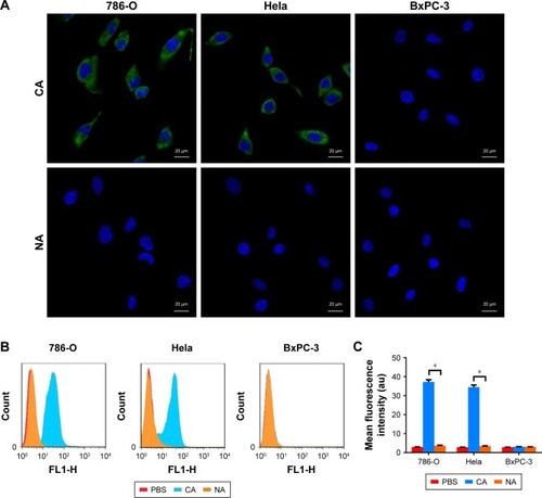
A single-stranded DNA strand (CAIX aptamer) that bound to CAIX was sequenced by high-throughput sequencing, and the sequence was: AGCAGCACAGAGGTCAGATGT GGT-GCGCAGTGATGTGGTTGGTCCTATGCGTGCTAC-CGTCCTATGCGTGCTACCGTGAA. CLSM showed CAIX aptamer could bind to the cell membranes of 786-O and Hela cells, but could not bind to the cell membranes of BxPC-3 cells, and nonsense aptamer could not bind to the cell membranes of any of the three types of tumor cells (). Flow cytometry found the mean fluorescence intensity of 786–O and Hela cells (37.17±1.20 au and 34.47±2.08 au, respectively) after incubation with CAIX aptamer was significantly stronger than that of 786–O and Hela cells (3.66±0.22 au and 3.44±0.10 au, respectively) after incubation with nonsense aptamer (P<0.05) (). However, the mean fluorescence intensity of BxPC-3 cells (3.09±0.25 au and 3.00±0.14 au) was not significantly different after incubation with CAIX aptamer and nonsense aptamer (P>0.05) ().
Figure 1 The specificity of aptamers in cells.
Notes: (A) CLSM images of 786-O cells, Hela cells, and BxPC-3 cells after incubation with CAIX aptamer and nonsense aptamer. (B) Flow cytometry analysis of the binding ability of the two aptamers to 786-O cells, Hela cells, and BxPC-3 cells. (C) Quantification of the binding ability of the two aptamers to the tumor cells (*P<0.05).
Abbreviations: CLSM, confocal laser scanning microscopy; CA, CAIX aptamer; CAIX, carbonic anhydrase IX; NA, nonsense aptamer.
Loading ability of targeted nanobubbles
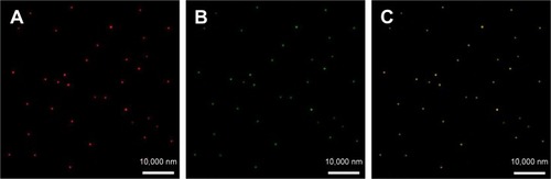
The coupling between the surfaces of targeted nanobubbles and CAIX aptamer was the prerequisite for targeted imaging. Under CLSM, the lipid membranes of DiI-labeled targeted nanobubbles presented red fluorescence and 6-FAM-modified CAIX aptamer on targeted nanobubbles presented green fluorescence (). The merged image revealed the green fluorescence of 6-FAM-modified CAIX aptamer could overlap with the red fluorescence of the DiI-labeled lipid membranes (). However, nanobubbles unloaded with CAIX aptamer or loaded with no fluorescence-modified CAIX aptamer only presented red fluorescence.
Figure 2 Loading ability of targeted nanobubbles.
Notes: (A) Red fluorescence of DiI-labeled lipid membranes. (B) Green fluorescence of 6-FAM modified CAIX aptamer. (C) The merged image confirms targeted nanobubbles were loaded with CAIX aptamer.
Abbreviations: DiI, 1,1′-dioctadecyl-3,3,3′,3′-tetramethylindocarbocyanine perchlorate; 6-FAM, 6-carboxyfluorescein; CAIX, carbonic anhydrase IX.
Characteristics of targeted nanobubbles
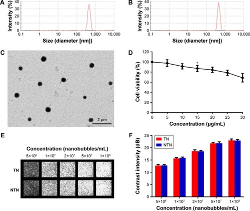
A hemocytometer showed the concentration of targeted nanobubbles (3.07×10⁹ ± 0.29×10⁹/mL) was not significantly different from that of non-targeted nanobubbles loaded with nonsense aptamer (2.99×10⁹ ± 0.34×10⁹/mL) (P>0.05). The particle size of targeted nanobubbles was 478.43±68.46 nm, and the polydispersity index was 0.148 (). The particle size of non-targeted nanobubbles was 483.97±62.08 nm, and the polydispersity index was 0.251 (). Under transmission electron microscope, targeted nanobubbles with uniform distribution and homogeneous size exhibited a black round area encapsulated in a gray ring, whose particle size was about 500 nm (). The CCK-8 assay demonstrated when the concentration of lipid material reached 15 μg/mL, the cell viability of 786-O cells (87.25±6.06%) significantly decreased compared with that of 786-O cells incubated with no lipid material (P<0.05), but when the concentration of lipid material was 10 μg/mL, the cell viability of 786-O cells (91.36±5.14%) did not significantly change (P>0.05) (). The imaging effect was not significantly different between targeted and non-targeted nanobubbles at the same concentration in vitro (P>0.05), and the imaging intensity of targeted and non-targeted nanobubbles increased with the increase of concentration ().
Figure 3 Characteristics of nanobubbles.
Notes: (A) Size-distribution curve of targeted nanobubbles. (B) Size-distribution curve of non-targeted nanobubbles. (C) Observation of targeted nanobubbles under transmission electron microscope. (D) Cell viability of 786-O cells after incubation with lipid material (*P<0.05, a significant difference in comparison with the cell viability of 786-O cells incubated with no lipid material). (E) Ultrasound images of nanobubbles in vitro. (F) Quantification of ultrasound images of nanobubbles in vitro.
Abbreviations: TN, targeted nanobubbles; NTN, non-targeted nanobubbles.
Binding ability of targeted nanobubbles in vitro
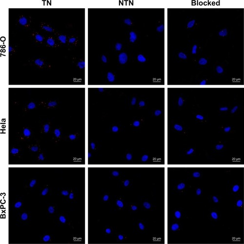
The binding ability of targeted and non-targeted nanobubbles to the three types of tumor cells was observed under CLSM. A large number of nanobubbles gathered around 786-O and Hela cells after incubation with targeted nanobubbles, while only few nanobubbles could bind to 786-O and Hela cells after incubation with non-targeted nanobubbles, and only few nanobubbles could bind to BxPC-3 cells after incubation with targeted or non-targeted nanobubbles (). After the three types of tumor cells were pre-blocked with CAIX aptamer, only a small amount of targeted nanobubbles could bind to the tumor cells ().
Figure 4 Binding ability of nanobubbles in vitro.
Notes: The binding ability of nanobubbles to 786-O cells (upper), the binding ability of nanobubbles to Hela cells (middle), the binding ability of nanobubbles to BxPC-3 cells (lower). Blocked indicates the tumor cells were pre-blocked with CAIX aptamer. Blue fluorescence indicates cell nuclei, and red fluorescence indicates nanobubbles.
Abbreviations: TN, targeted nanobubbles; NTN, non-targeted nanobubbles; CAIX, carbonic anhydrase IX.
Imaging effect of targeted nanobubbles in vivo
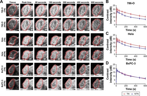
Ultrasound images of targeted and non-targeted nanobubbles in 786-O, Hela, and BxPC-3 xenograft tumor tissues are shown in . Qlab 8.1 quantitative analysis software was used to quantitatively analyze the imaging intensity of targeted and non-targeted nanobubbles. The time-intensity curves exhibited that the imaging intensity was always significantly different between targeted and non-targeted nanobubbles in 786-O and Hela xenograft tumor tissues (P<0.05) (). However, there was no significant difference in the imaging intensity of targeted and non-targeted nanobubbles in BxPC-3 xenograft tumor tissues (P>0.05) (). Peak time of targeted and non-targeted nanobubbles was not significantly different in the three types of xenograft tumor tissues (P>0.05) (). Peak intensity and area under the curve were significantly different between targeted and non-targeted nanobubbles in 786-O and Hela xenograft tumor tissues (P<0.05), whereas there was no significant difference in BxPC-3 xenograft tumor tissues (P>0.05) ().
Table 1 Ultrasound imaging parameters of targeted and non-targeted nanobubbles in xenografts
Figure 5 Ultrasound imaging of nanobubbles in vivo.
Notes: (A) Ultrasound images of nanobubbles in xenograft tumor tissues. Red dotted circles indicate the regions of xenograft tumor tissues. (B–D) Time-intensity curves of nanobubbles in 786-O, Hela and BxPC-3 xenograft tumor tissues.
Abbreviations: TN, targeted nanobubbles; NTN, non-targeted nanobubbles.
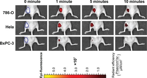
Fluorescence imaging of live small animals revealed a large number of fluorescent signals were produced in 786-O and Hela xenograft tumor tissues after injection with DiR-labeled targeted nanobubbles, and the fluorescence intensity decreased with time (). However, only a few fluorescent signals were presented in BxPC-3 xenograft tumor tissues, and duration time of fluorescent signals was shorter than that in 786-O and Hela xenograft tumor tissues ().
Figure 6 Fluorescence imaging of xenograft tumor tissues of live small animals.
Notes: Fluorescence imaging of live small animals was performed in 786-O, Hela, and BxPC-3 tumor-bearing nude mice over time (0 minute, 1 minute, 5 minutes, and 10 minutes). Blue dotted circles indicate the regions of xenograft tumor tissues.
Binding ability of targeted nanobubbles in vivo
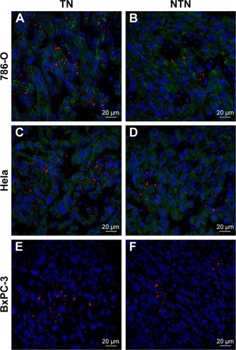
The ability of targeted nanobubbles to be distributed in the extravascular spaces and bind to tumor parenchyma cells in xenograft tumor tissues was demonstrated by immuno-fluorescence. As shown in , DiI-labeled targeted nanobubbles were distributed not only in the tumor vascula-ture, but also in the extravascular spaces of xenograft tumor tissues. As expected, CLSM found the green fluorescence of 6-FAM-modified CAIX aptamer could overlap with the red fluorescence of the lipid membranes of DiI-labeled targeted nanobubbles in xenograft tumor tissues (). The specific binding of targeted nanobubbles to tumor parenchymal cells in xenograft tumor tissues was the key process of USMI. Immunofluorescence described CAIX was expressed in 786-O and Hela xenograft tumor tissues, while not in BxPC-3 xenograft tumor tissues (). In 786-O and Hela xenograft tumor tissues, the number was significantly different between targeted and non-targeted nanobubbles (P<0.05), and targeted nanobubbles could gather around CAIX-positive tumor cells (). However, the number was not significantly different between targeted and non-targeted nanobubbles in BxPC-3 xenograft tumor tissues (P>0.05) ().
Figure 7 Distribution of targeted nanobubbles in xenograft tumor tissues.
Notes: The distribution of targeted nanobubbles in 786-O xenograft tumor tissues (upper), in Hela xenograft tumor tissues (middle), and in BxPC-3 xenograft tumor tissues (lower). Blue fluorescence indicates cell nuclei, green fluorescence indicates tumor vasculature, and red fluorescence indicates nanobubbles.
Abbreviation: TN, targeted nanobubbles.
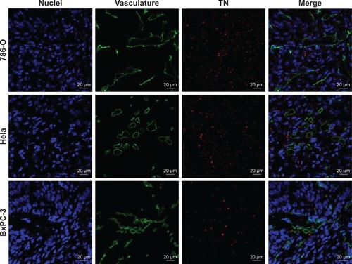
Figure 8 Ability of targeted nanobubbles to load CAIX aptamer in xenograft tumor tissues.
Notes: The ability of targeted nanobubbles to load CAIX aptamer in 786-O xenograft tumor tissues (upper), in Hela xenograft tumor tissues (middle), and in BxPC-3 xenograft tumor tissues (lower). Blue fluorescence indicates cell nuclei, red fluorescence indicates the lipid membranes of targeted nanobubbles, and green fluorescence indicates CAIX aptamer.
Abbreviations: TN, targeted nanobubbles; CAIX, carbonic anhydrase IX.
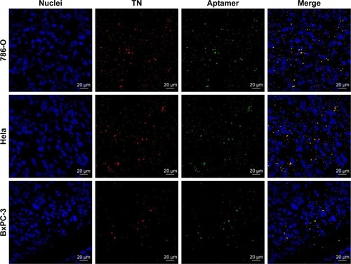
Figure 9 Binding ability of nanobubbles in xenograft tumor tissues.
Notes: (A, B) The binding ability of nanobubbles to tumor cells in 786-O xenograft tumor tissues, (C, D) in Hela xenograft tumor tissues, and (E, F) in BxPC-3 xenograft tumor tissues. Blue fluorescence indicates cell nuclei, green fluorescence indicates the cell membranes of CAIX-positive cells, and red fluorescence indicates nanobubbles.
Abbreviations: TN, targeted nanobubbles; NTN, non-targeted nanobubbles; CAIX, carbonic anhydrase IX.
H&E staining and CAIX expression
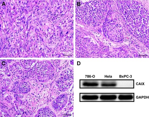
H&E staining showed 786-O xenograft tumor tissues exhibited a cord-like arrangement, and tumor stroma was diffusely distributed in tumor parenchyma (). Tumor cells were obviously heterogeneous, and cell nuclei were large and hyperchromatic (). Hela and BxPC-3 xenograft tumor tissues displayed a sheet or bulk arrangement, and tumor parenchyma was wrapped in tumor stroma (). Tumor cells were of different sizes, and cell nuclei were large and had no polarity (). Western blot (WB) confirmed the high expression of CAIX in 786-O and Hela xenograft tumor tissues, whereas not in BxPC-3 xenograft tumor tissues (), which was consistent with immunofluorescence.
Figure 10 H&E staining and CAIX expression of xenograft tumor tissues.
Notes: (A) 786-O xenograft tumor tissues stained with H&E. (B) Hela xenograft tumor tissues stained with H&E. (C) BxPC-3 xenograft tumor tissues stained with H&E. (D) Expression of CAIX in xenograft tumor tissues.
Abbreviations: CAIX, carbonic anhydrase IX; GAPDH, glyceraldehyde-3-phosphate dehydrogenase.
Discussion
The early diagnosis of malignant tumors is the key to improving the survival rate and reducing the mortality.Citation32–Citation34 Molecular imaging (such as USMI, fluorescent imaging, positron emission tomography, and multimodal imaging) can monitor the biological process in vivo at the cellular and molecular level, which is beneficial to the early diagnosis and treatment evaluation of malignant tumors.Citation35–Citation41 The construction of targeted UCAs that can significantly enhance ultrasound imaging is the key link of USMI.Citation7,Citation42,Citation43 BR55 is currently the most successful targeted UCAs, which can effectively enhance ultrasound imaging of tumor tissues and has been reported in the early diagnosis of malignant tumors.Citation44–Citation47 However, because the particle size is on the micron scale, BR55 can only achieve intravascular USMI. Nanobubbles with a particle size of less than 700 nm can penetrate the tumor vasculature to achieve USMI of tumor parenchymal cells and targeted delivery of drugs or genes to tumor parenchymal cells.Citation8,Citation48–Citation51
CAIX is a transmembrane protein expressed in numerous tumors, which can be used as a specific target for USMI of malignant tumors.Citation18 We have prepared targeted nanobubbles loaded with CAIX polypeptides in our previous research, which could be used for USMI of CAIX-positive xenograft tumor tissues.Citation23 However, polypeptides are composed of amino acid sequences, which can cause an immune response after repeated injection, thus restricting their clinical application.Citation52 Aptamers are composed of nucleic acid sequences and have been widely applied in molecular diagnosis and disease treatment, including USMI of malignant tumors.Citation15,Citation50
Therefore, this study combined lipid nanobubbles with small particle size, CAIX expressed in numerous tumors, and safe and stable aptamers to construct targeted nanobubbles loaded with CAIX aptamer. CAIX aptamer could specifically bind to CAIX-positive 786-O and Hela cells, rather than CAIX-negative BxPC-3 cells, which laid the foundation for the specific binding of targeted nanobubbles loaded with CAIX aptamer to CAIX-positive tumor cells. Compared with the traditional biotin–avidin system, the maleimide-thiol coupling reaction not only loaded specific ligands on targeted nanobubbles, but also did not cause an immune response after intravenous injection of targeted nanobubbles.Citation31,Citation53 Therefore, targeted nanobubbles loaded with CAIX aptamer were prepared by a maleimidethiol coupling reaction in this study, and fluorescence labeling confirmed CAIX aptamer was distributed uniformly on targeted nanobubbles. Targeted nanobubbles were round and distributed uniformly, and their particle size was 478.43±68.46 nm. In in vitro experiments, targeted nanobubbles could specifically bind to 786-O and Hela cells, and the binding ability of targeted nanobubbles to CAIX-positive tumor cells could be blocked by CAIX aptamer. It was considered that targeted nanobubbles were specific to CAIX-positive tumor cells.
The essence of USMI is to specifically enhance ultrasound imaging of tumor tissues with UCAs. Targeted nanobubbles had a better imaging effect in 786-O and Hela xenograft tumor tissues compared with non-targeted nanobubbles loaded with nonsense aptamer, which displayed higher peak intensity and larger area under the curve. However, there was no significant difference in the imaging effects of targeted and non-targeted nanobubbles in BxPC-3 xenograft tumor tissues. These attributes suggested targeted nanobubbles loaded with CAIX aptamer could specifically gather in CAIX-positive xenograft tumor tissues compared with non-targeted nanobubbles, whereas the aggregation ability of targeted and non-targeted nanobubbles was not significantly different in CAIX-negative xenograft tumor tissues. Fluorescence imaging of live small animals showed only a few fluorescent signals were seen in BxPC-3 xenograft tumor tissues, and duration time was short. The reason was that only a few targeted nanobubbles gathered in CAIX-negative xenograft tumor tissues, and DiR dye on targeted nanobubbles was not enough for fluorescence imaging in BxPC-3 xenograft tumor tissues over time.
The morphological basis of USMI of tumor parenchyma cells includes targeted nanobubbles penetrating the tumor vasculature and binding to tumor cells by specific ligands on their surfaces. Because the imaging intensity of tumor tissues mainly came from bound targeted UCAs at 8 minutes after injection of targeted UCAs, we observed the sections at 8 minutes after injection of nanobubbles to investigate the morphological basis of USMI.Citation5,Citation52 Targeted nanobubbles were seen in the vascular and extravascular spaces of 786-O, Hela, and BxPC-3 xenograft tumor tissues, which meant targeted nanobubbles could penetrate the tumor vasculature of xenograft tumor tissues. Fluorescence labeling found targeted nanobubbles distributed in the extravascular spaces of xenograft tumor tissues could still load CAIX aptamer. In addition, immunofluorescence not only displayed the distribution differences of targeted and non-targeted nanobubbles in 786-O and Hela xenograft tumor tissues, but also demonstrated targeted nanobubbles could specifically bind to CAIX-positive tumor cells in 786-O and Hela xenograft tumor tissues, whereas not to CAIX-negative tumor cells in BxPC-3 xenograft tumor tissues. WB further showed the high expression of CAIX in 786-O and Hela xenograft tumor tissues, while not in BxPC-3 xenograft tumor tissues. Therefore, the morphological basis of USMI of malignant tumors after intravenous injection of targeted nanobubbles had been illustrated, namely, targeted nanobubbles in circulation could passively diffuse into the extravascular spaces via EPR effect, actively bind to CAIX-positive tumor cells under the action of CAIX aptamer, significantly enhance ultrasound imaging of CAIX-positive xenograft tumor tissues, and thus achieve USMI of tumor parenchyma cells.
This study had the following characteristics: 1) aptamer-mediated targeted nanobubbles were constructed, which could specifically bind to tumor cells and safely and effectively enhance ultrasound imaging of tumor tissues; 2) targeted nanobubbles were constructed by a maleimidethiol coupling reaction, which could not only load CAIX aptamer on targeted nanobubbles stably and effectively, but also avoid an immune response in vivo; 3) because numerous tumors express CAIX, targeted nanobubbles loaded with CAIX aptamer can be used for USMI of various tumors; 4) the distribution, loading ability, and binding ability of targeted nanobubbles in xenograft tumor tissues were investigated in depth, which fully clarified the morphological basis of USMI. Though targeted nanobubbles could load CAIX aptamer and specifically bind to CAIX-positive tumor cells in vitro and in vivo, future experiments need to investigate the relationship between the number of CAIX aptamer molecules on targeted nanobubbles and the binding ability of targeted nanobubbles to CAIX.
Conclusion
Combining the advantages of nanobubbles and aptamers, this study successfully constructed targeted nanobubbles loaded with CAIX aptamer by a maleimidethiol coupling reaction, which possessed the attractive features of small size, strong penetration, high safety, high specificity, and excellent imaging effect. Furthermore, the distribution of targeted nanobubbles in the extravascular spaces and the ability to load CAIX aptamer and bind to CAIX-positive tumor cells in xenograft tumor tissues were confirmed by immunofluorescence. This study not only introduces targeted nanobubbles carrying aptamers for USMI of malignant tumors from various organs, but also further determines the morphological basis of USMI of malignant tumors after intravenous injection of targeted nanobubbles and lays the foundation for the application of targeted nanobubbles in the early diagnosis of malignant tumors.
Acknowledgments
This work was supported by the International Science & Technology Cooperation Program of China (No 2015DFA30920), the National Natural Science Foundation of China (No 81771856), and the Science & Technology (International Science & Technology Cooperation) Research Base Construction Program of Chongqing (No cstc2014gjhz110004).
Disclosure
The authors report no conflicts of interest in this work.
References
- TerziEIavaroneMPompiliMContrast ultrasound LI-RADS LR-5 identifies hepatocellular carcinoma in cirrhosis in a multicenter restropective study of 1,006 nodulesJ Hepatol201868348549229133247
- Abou-ElkacemLWilsonKEJohnsonSMUltrasound Molecular Imaging of the Breast Cancer Neovasculature using Engineered Fibronectin Scaffold Ligands: A Novel Class of Targeted Contrast Ultrasound AgentTheranostics20166111740175227570547
- BzylJPalmowskiMRixAThe high angiogenic activity in very early breast cancer enables reliable imaging with VEGFR2-targeted microbubbles (BR55)Eur Radiol201323246847522878592
- ZhouYGuHXuYTargeted antiangiogenesis gene therapy using targeted cationic microbubbles conjugated with CD105 antibody compared with untargeted cationic and neutral microbubblesTheranostics20155439941725699099
- ZhangHInghamESGagnonMKInvitro characterization and invivo ultrasound molecular imaging of nucleolin-targeted microbubblesBiomaterials2017118637327940383
- BachawalSVJensenKCWilsonKETianLLutzAMWillmannJKBreast Cancer Detection by B7-H3-Targeted Ultrasound Molecular ImagingCancer Res201575122501250925899053
- CaiWBYangHLZhangJThe Optimized Fabrication of Nanobubbles as Ultrasound Contrast Agents for Tumor ImagingSci Rep201551372526333917
- LiJTianYShanDNeuropeptide Y Y1 receptor-mediated biodegradable photoluminescent nanobubbles as ultrasound contrast agents for targeted breast cancer imagingBiomaterials201711610611727914983
- YangHCaiWXuLNanobubble-Affibody: Novel ultrasound contrast agents for targeted molecular ultrasound imaging of tumorBiomaterials20153727928825453958
- FanXWangLGuoYXiongXZhuLFangKInhibition of prostate cancer growth using doxorubicin assisted by ultrasound-targeted nanobubble destructionInt J Nanomedicine2016113585359627536100
- SegersTde RondLde JongNBordenMVersluisMStability of Monodisperse Phospholipid-Coated Microbubbles Formed by Flow-Focusing at High Production RatesLangmuir201632163937394427006083
- PereraRHWuHPeirisPImproving performance of nanoscale ultrasound contrast agents using N,N-diethylacrylamide stabilizationNanomedicine2017131596727565686
- DuanSGuoLShiDShangMMengDLiJDevelopment of a novel folate-modified nanobubbles with improved targeting ability to tumor cellsUltrason Sonochem20173723524328427629
- GaoYHernandezCYuanHXUltrasound molecular imaging of ovarian cancer with CA-125 targeted nanobubble contrast agentsNanomedicine20171372159216828603079
- FanXGuoYWangLXiongXZhuLFangKDiagnosis of prostate cancer using anti-PSMA aptamer A10-3.2-oriented lipid nanobubblesInt J Nanomedicine2016113939395027574424
- ChangSSOverview of prostate-specific membrane antigenRev Urol20046Suppl 10S13S18
- TangLTongRCoyleVJTargeting tumor vasculature with aptamer-functionalized doxorubicin-polylactide nanoconjugates for enhanced cancer therapyACS Nano2015955072508125938427
- GarousiJHonarvarHAnderssonKGComparative Evaluation of Affibody Molecules for Radionuclide Imaging of in Vivo Expression of Carbonic Anhydrase IXMol Pharm201613113676368727529191
- EllingsenCAndersenLMGalappathiKRofstadEKHypoxia bio-markers in squamous cell carcinoma of the uterine cervixBMC Cancer20151580526502718
- CovielloVMarchiBSartiniS1,2-Benzisothiazole Derivatives Bearing 4-, 5-, or 6-Alkyl/arylcarboxamide Moieties Inhibit Carbonic Anhydrase Isoform IX (CAIX) and Cell Proliferation under Hypoxic ConditionsJ Med Chem201659136547655227305384
- Viola-VillegasNTCarlinSDAckerstaffEUnderstanding the pharmacological properties of a metabolic PET tracer in prostate cancerProc Natl Acad Sci U S A2014111207254725924785505
- van BrusselASAdamsAOliveiraSHypoxia-Targeting Fluorescent Nanobodies for Optical Molecular Imaging of Pre-Invasive Breast CancerMol Imaging Biol201618453554426589824
- ZegersCMHoebersFJvan ElmptWEvaluation of tumour hypoxia during radiotherapy using [18F]HX4 PET imaging and blood biomarkers in patients with head and neck cancerEur J Nucl Med Mol Imaging201643122139214627251643
- MuselaersCHRijpkemaMBosDLRadionuclide and Fluorescence Imaging of Clear Cell Renal Cell Carcinoma Using Dual Labeled Anti-Carbonic Anhydrase IX Antibody G250J Urol2015194253253825686542
- ZhuLGuoYWangLConstruction of ultrasonic nanobubbles carrying CAIX polypeptides to target carcinoma cells derived from various organsJ Nanobiotechnology20171516328962657
- WuQWuLWangYEvolution of DNA aptamers for malignant brain tumor gliosarcoma cell recognition and clinical tissue imagingBiosens Bioelectron2016801826802746
- KangSATsolmonBMannAPSafety evaluation of intravenously administered mono-thioated aptamer against E-selectin in miceToxicol Appl Pharmacol20152871869226048585
- SongYZhuZAnYSelection of DNA aptamers against epithelial cell adhesion molecule for cancer cell imaging and circulating tumor cell captureAnal Chem20138584141414923480100
- AlibolandiMRamezaniMAbnousKIn vitro and in vivo evaluation of therapy targeting epithelial-cell adhesion-molecule aptamers for non-small cell lung cancerJ Control Release20152098810025912964
- JacobsonOWeissIDWangL18F-Labeled Single-Stranded DNA Aptamer for PET Imaging of Protein Tyrosine Kinase-7 ExpressionJ Nucl Med201556111780178526315836
- GaoZHouYZengJTumor Microenvironment-Triggered Aggregation of Antiphagocytosis 99m Tc-Labeled Fe3 O4 Nanoprobes for Enhanced Tumor Imaging In VivoAdv Mater201729241701095
- ChenWZhengRBaadePDCancer statistics in China, 2015CA Cancer J Clin201666211513226808342
- ChenWZhengRZhangSCancer incidence and mortality in China, 2013Cancer Lett2017401637128476483
- DoroudiMSchoenREPinskyPFEarly detection versus primary prevention in the PLCO flexible sigmoidoscopy screening trial: Which has the greatest impact on mortality?Cancer2017123244815482228976536
- LiuJChenYWangGUltrasound molecular imaging of acute cardiac transplantation rejection using nanobubbles targeted to T lymphocytesBiomaterials201816220020729453053
- ZhouTCaiWYangHAnnexin V conjugated nanobubbles: A novel ultrasound contrast agent for in vivo assessment of the apoptotic response in cancer therapyJ Control Release201827611312429522835
- WangSHerbstEBMauldinFWDiakovaGBKlibanovALHossackJAUltra-Low-Dose Ultrasound Molecular Imaging for the Detection of Angiogenesis in a Mouse Murine Tumor Model: How Little Can We See?Invest Radiol2016511275876627654582
- SunYMaXChengKStrained cyclooctyne as a molecular platform for construction of multimodal imaging probesAngew Chem Int Ed Engl201554205981598425800807
- DingFChenSZhangWTuYSunYUPAR targeted molecular imaging of cancers with small molecule-based probesBioorg Med Chem201725205179518428869084
- DingFZhanYLuXSunYRecent advances in near-infrared II fluorophores for multifunctional biomedical imagingChem Sci20189194370438029896378
- SunYZengXXiaoYNovel dual-function near-infrared II fluorescence and PET probe for tumor delineation and image-guided surgeryChem Sci2018982092209729675250
- LutzAMBachawalSVDrescherCWPyszMAWillmannJKGambhirSSUltrasound molecular imaging in a human CD276 expression-modulated murine ovarian cancer modelClin Cancer Res20142051313132224389327
- HoytKWarramJMWangDRatnayakaSTraylorAAgarwalAMolecular Ultrasound Imaging of Tissue Inflammation Using an Animal Model of Acute Kidney InjuryMol Imaging Biol201517678679225905474
- Baron ToaldoMSalvatoreVMarinelliSUse of VEGFR-2 targeted ultrasound contrast agent for the early evaluation of response to sorafenib in a mouse model of hepatocellular carcinomaMol Imaging Biol2015171293725082536
- HacklCSchachererDAndersMImproved Detection of preclinical Colorectal Liver Metastases by High Resolution Ultrasound including Molecular Ultrasound Imaging using the targeted Contrast Agent BR55Ultraschall Med201637329029627112624
- WillmannJKBonomoLCarla TestaAUltrasound Molecular Imaging With BR55 in Patients With Breast and Ovarian Lesions: First-in-Human ResultsJ Clin Oncol201735192133214028291391
- SmeengeMTranquartFMannaertsCKFirst-in-Human Ultrasound Molecular Imaging With a VEGFR2-Specific Ultrasound Molecular Contrast Agent (BR55) in Prostate Cancer: A Safety and Feasibility Pilot StudyInvest Radiol201752741942728257340
- OdaYSuzukiRMoriTDevelopment of fluorous lipid-based nanobubbles for efficiently containing perfluoropropaneInt J Pharm20154871–2647125841568
- HoYJChangYCYehCKImproving Nanoparticle Penetration in Tumors by Vascular Disruption with Acoustic Droplet VaporizationTheranostics20166339240326909113
- WuMWangYWangYPaclitaxel-loaded and A10-3.2 aptamer-targeted poly(lactide-co-glycolic acid) nanobubbles for ultrasound imaging and therapy of prostate cancerInt J Nanomedicine2017125313533028794625
- WuMZhaoHGuoLUltrasound-mediated nanobubble destruction (UMND) facilitates the delivery of A10-3.2 aptamer targeted and siRNA-loaded cationic nanobubbles for therapy of prostate cancerDrug Deliv201825122624029313393
- ZhangHTamSInghamESUltrasound molecular imaging of tumor angiogenesis with a neuropilin-1-targeted microbubbleBiomaterials20155610411325934284
- HouYQiaoRFangFNaGdF4 nanoparticle-based molecular probes for magnetic resonance imaging of intraperitoneal tumor xenografts in vivoACS Nano20137133033823199030