

Abstract
Background
After myocardial infarction (MI), inflammatory cells infiltrate the infarcted heart in response to secreted stimuli. Monocytes are recruited to the infarct via CCR2 chemokine receptors along a CCL2 concentration gradient. While infiltration of injured tissue with monocytes is an important component of the reparatory response, excessive or prolonged inflammation can adversely affect left ventricular remodeling and worsen clinical outcomes.
Materials and methods
Here, we developed poly(ethylene glycol) (PEG)-distearoylphos-phatidylethanolamine (PEG-DSPE) micelles loaded with a small molecule CCR2 antagonist to inhibit monocyte recruitment to the infarcted myocardium. To specifically target CCR2-expressing cells, PEG-DSPE micelles were further surface decorated with an anti-CCR2 antibody.
Results
Targeted PEG-DSPE micelles showed eight-fold greater binding to CCR2-expressing RAW 264.7 monocytes than plain, non-targeted PEG-DSPE micelles. In a mouse model of MI, CCR2-targeting PEG-DSPE micelles loaded with a CCR2 small molecule antagonist significantly decreased the number of Ly6Chigh inflammatory cells to 3% of total compared with PBS-treated controls. Furthermore, CCR2-targeting PEG-DSPE micelles significantly reduced the infarct size based on epicardial and endocardial infarct arc lengths.
Conclusion
Both non-targeted and CCR2-targeting PEG-DSPE micelles showed a trend toward improving cardiac function. As such, PEG-DSPE micelles represent a promising cardiac therapeutic platform.
Supplementary materials
Figure S1 (A) Accumulation of CCR2-targeting and non-targeted micelles in the infarcted heart. (B) Biodistribution of CCR2-targeted and non-targeted micelles in mice with myocardial infarction.
Note: Data are represented as mean ±SD, n=3.
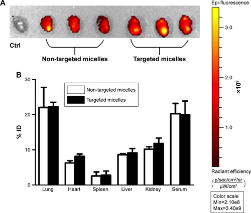
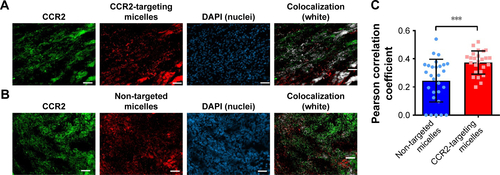
Figure S2 Colocalization studies of DiD-labeled (A) CCR2-targeting micelles and (B) non-targeted micelles with CCR2-labeled inflammatory cells in the infarcted myocardium. The colocalization of CCR2-positive cells and DiD-labeled micelles is shown in white. (C) The PCC was used to quantify the degree of colocalization between CCR2-positive inflammatory cells and the DiD-labeled CCR2-targeting and non-targeted micelles.
Notes: A PCC of 1 indicates perfect correlation, a PCC of 0 means no correlation, and a PCC of −1 means perfect inverse correlation. The CCR2-targeting micelles showed a significantly greater colocalization with CCR2-positive cells than non-targeted micelles (P=0.0004). Three days after inducing myocardial infarction, mice were treated with DiD-labeled CCR2-targeting (n=3) and non-targeted micelles (n=3). 6 hours post administration, the hearts were removed and embedded in Tissue-Tek O.C.T. Frozen heart sections with 10 μm thickness were fixed and permeabilized with ice-cold acetone, and blocked with 5% bovine serum albumin in TBST. The sections were stained with an anti-mouse CCR2 antibody (Thermo Fisher Scientific, PA5-23043) at a 1:50 dilution for 1 hour at room temperature, followed by 45 minutes incubation with a secondary antibody labeled with Alexa Fluor 568 at 1:200 dilution. The nuclei were stained with DAPI. The analysis was performed in ImageJ using the Coloc 2 function. On average, three cryosections per heart were analyzed. For each cryosection, three representative images were taken. Data are presented as mean ± SD. A two-tailed t-test was used to determine statistical significance (P=0.0004). ***P≤0.001.
Abbreviation: PCC, Pearson correlation coefficient.
Acknowledgments
We would like to thank Dr Joseph Spernyak (Roswell Park Cancer Institute) for technical assistance in the use of the IVIS Spectrum in vivo imager and funding by the NIH (S10 OD 016450). We acknowledge support by the NIH through awards HL-126082 (JN&JMC), EB-021454 (JN), EB-023262 (JN), HL-61610 (JMC), the National Center for Advancing Translational Sciences UL1-TR-001412 and the Department of Veterans Affairs 1IO1BX002659 (JMC).
Disclosure
The authors report no conflicts of interest in this work.