

Abstract
Background
Optical coherence tomography (OCT) is an intravascular, high-resolution imaging technique that is used to characterize atherosclerotic plaques. However, the identification of macrophages as important markers of inflammation and plaque vulnerability remains difficult. Here, we investigate whether the uptake of very small iron oxide particles (VSOP) in macrophages, that cluster in phagolysosomes and allow high-quality magnetic resonance imaging (MRI) of atherosclerotic plaques, and uptake of ferumoxytol nanoparticles enhance detection of macrophages by OCT.
Materials and methods
RAW 264.7 macrophage cells were incubated with VSOP (1 and 2 mM Fe) that have been clinically tested and ferumoxytol (8.9 mM Fe) that is approved for iron deficiency treatment and currently investigated as an MRI contrast agent. The light scattering of control macrophages, nanoparticle-labeled macrophages (2,000,000 in 500 µL) and nanoparticle suspensions was measured in synchronous wavelength scan mode using a fluorescence spectrophotometer. For OCT analyses, pellets of 8,000,000 non-labeled, VSOP-labeled and ferumoxytol-labeled RAW 264.7 macrophages were imaged and analyzed on an OPTIS™ OCT imaging system.
Results
Incubation with 1 and 2 mM VSOP resulted in uptake of 7.1±1.5 and 12±1.5 pg Fe per cell, which increased the backscattering of the macrophages in spectrophotometry 2.5- and 3.6-fold, whereas incubation with 8.9 mM Fe ferumoxytol resulted in uptake of 6.6±2 pg Fe per cell, which increased the backscattering 1.5-fold at 700 nm. In contrast, backscattering of non-clustered nanoparticles in suspension was negligible. Accordingly, OCT imaging could visualize significantly increased backscattering and signal attenuation of nanoparticle-labeled macrophages in comparison with controls.
Conclusion
We conclude that VSOP and, to a lesser extent, ferumoxytol increase light scattering and attenuation when taken up by macrophages and can serve as a multimodal imaging probe for MRI and OCT to improve macrophage detection in atherosclerotic plaques by OCT in the future.
Supplementary material
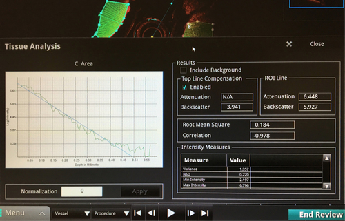
Figure S1 Example for quantification of the OCT images with the ILUMIENTM OPTISTM Offline Review Workstation Tissue Properties Supplement.
Notes: The ROIs were drawn in the region of the macrophage cells in the reaction tubes in a semi-automatic way by selecting ROIs beginning at the bottom of the macrophage cell pellets. The software reduced the ROI at the distant side when reaching a lower signal threshold. The values of Attenuation and Backscatter (ROI Line) were used and averaged from five sections of each pellet of the OCT pullbacks.
Abbreviations: OCT, optical coherence tomography; NSD, normalized standard deviation.
Acknowledgments
We thank the group of Prof M Taupitz for supplying the VSOP.
Disclosure
The authors report no conflicts of interest in this work.