

Abstract
Background
For effective bone regeneration, it is necessary to implant a biocompatible scaffold that is capable of inducing cell growth and continuous osteogenic stimulation at the defected site. Here, we suggest an injectable hydrogel system using enzymatic cross-linkable gelatin (Gel) and functionalized gold nanoparticles (GNPs).
Methods
In this work, tyramine (Ty) was synthesized on the gelatin backbone (Gel-Ty) to enable a phenol crosslinking reaction with horseradish peroxidase (HRP). N-acetyl cysteine (NAC) was attached to the GNPs surface (G-NAC) for promoting osteodifferentiation.
Results
The Gel-Ty hydrogels containing G-NAC (Gel-Ty/G-NAC) had suitable mechanical strength and biocompatibility to embed and support the growth of human adipose derived stem cells (hASCs) during a proliferation test for three days. In addition, G-NAC promoted osteodifferentiation both when it was included in Gel-Ty and when it was used directly in hASCs. The osteogenic effects were demonstrated by the alkaline phosphatase (ALP) activity test.
Conclusion
These findings indicate that the phenol crosslinking reaction is suitable for injectable hydrogels for tissue regeneration and G-NAC stimulate bone regeneration. Based on our results, we suggest that Gel-Ty/G-NAC hydrogels can serve both as a biodegradable graft material for bone defect treatment and as a good template for tissue engineering applications such as drug delivery, cell delivery, and various tissue regeneration uses.
Supplementary materials
Figure S1 Evaluation of ALP activity of hASCs at various concentrations of GNPs below 40 µM. Results are mean ± SD of triplicate experiments: *P<0.05, **P<0.01, and ***P<0.001 represent significant difference compared with control group.
Abbreviations: GNPs, gold nanoparticles; hASCs, human-derived stem cells.
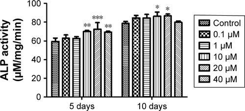
Figure S2 Evaluation of viability (A) and ALP activity (B) of hASCs at various concentrations of G-NAC. Results are mean ± SD of triplicate experiments: *P<0.05, **P<0.01, and ***P<0.001 represent significant differences compared with control group, and #P<0.05 and ##P<0.01 represent significant differences compared with 10 µM group.
Abbreviations: G-NAC, gold nanoparticles-N-acetyl cysteine; hASCs, human-derived stem cells.
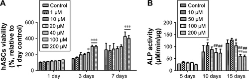
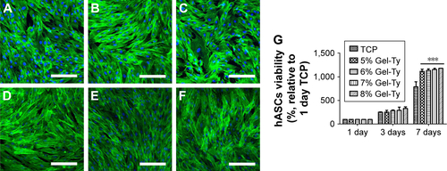
Figure S3 Viability of hASCs on the Gel-Ty hydrogel surface. The CLSM images of 5% (A, D), 7.5% (B, E), and 10% Gel-Ty hydrogel (C, F) after culture during 1 (A–C) and 3 days (D–F). F-actin of hASCs is green and the nucleus of hASCs is blue. Scale bar is 200 µm. Images collected at 200× magnification. The cell viability assay on the Gel-Ty hydrogels for 7 days using EZ-Cytox assay (G). Results are mean ± SD of triplicate experiments: ***P<0.001 represent significant differences compared with tissue culture plate group.
Abbreviations: CLSM, confocal laser scanning microscopy; Gel-Ty, gelatin–tyramine; hASCs, human-derived stem cells; TCP, tissue culture plate.
Acknowledgments
This research was supported by the Bio & Medical Technology Development Program of the National Research Foundation (NRF) and funded by the Korean government (MSIP&MOHW) (No 2017M3A9E4048170).
Disclosure
The authors report no conflicts of interest in this work.