

Abstract
Background
For the past few years, gene-therapy has recently shown considerable clinical benefit in cancer therapy, and the applications of gene therapies in cancer treatments continue to increase perennially. EZH2, an ideal candidate for tumor gene therapy, plays an important role in the tumorigenesis.
Methods
In this study, we developed a novel gene delivery system with a self-assembly method by Methoxy polyethylene glycol-polycaprolactone (MPEG-PCL) and DOTAP(DMC). And EZH2si-DMC was used to research anti-glioma both in vitro and in vivo.
Results
DMC with zeta-potential value of 36.7 mV and size of 35.6 nm showed good performance in the delivery siRNA to glioma cell in vitro with high 98% transfection efficiency. EZH2si-DMC showed good anti-glioma effect in vitro through inducing cell apoptosis and inhibiting cell growth. What’s more, treatment of tumor-bearing mice with DMC-EZH2si complex had significantly inhibited tumor growth at the subcutaneous model in vivo by inhibiting EZH2 protein expression, promoting apoptosis and reducing proliferation.
Conclusion
The EZH2 siRNA and DMC complex may be used to treat the glioma in clinical as a new drug.
Supplementary materials
Figure S1 RT-PCR and MTT test of cell activity.
Notes: When GL261 cells were transfected with DMC, Consi-DMC or EZH2si-DMC for 72 hours, EZH2 expression was tested by RT-PCR (A) and cell activity was tested by MTT test (B). (siEZH2-1:GGATACAGCCTGTGCACAT; siEZH2-2:GCTTTGGACAACAAGCCTT; siEZH2-3:GCAAATTCTCGGTGTCAAA).
Abbreviation: RT-PCR, reverse transcription PCR.
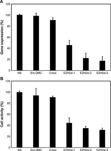
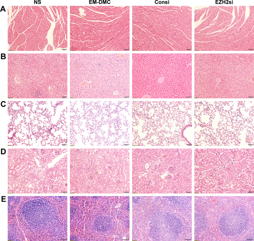
Figure S2 Toxicity assessment in vivo with pathological section.
Notes: Histological examinations of HE-stained (A) heart, (B) liver, (C) lung, (D) kidney, and (E) spleen. No significant pathological changes were detected. Scale bar is 50 µm.
Acknowledgments
This work was supported by the National Natural Science Foundation of China (NSFC81502165 and NSFC81402240).
Disclosure
The authors report no conflicts of interest in this work.