

Abstract
Background
Despite tremendous advancement, cancer still remains one of the leading causes of death worldwide. Inefficiency of current drug delivery regimens is one important factor that limits the therapeutic efficacy of existing drugs, thus contributing to cancer mortality. To address this limitation, synthetic nanotechnology-based delivery systems have been developed; however, they raise concern of inducing adverse immunogenic reactions. Exosomes (Exos) are nonimmunogenic nanosized vesicles that have received significant attention as efficient drug delivery system.
Methods
Drug loading in Exos were achieved by incubating different cell types viz pancreatic cancer cells (PCCs), pancreatic stellate cells (PSCs), and macrophages (MØs) with Doxorubicin (DOX). Differential ultracentrifugation was performed to isolate exosome and their size was determined by dynamic light scattering analysis. The efficacy of drug packaging into Exos was evaluated by HPLC. Flow cytometry was performed to examine the apoptosis. Cell viability was determined using the WST-1 assay.
Results
PCCs shed the most Exos and were the most efficient in drug loading followed by MØs and PSCs as examined by HPLC quantification. However, when compared for antitumor efficacy, MØ-derived Exos loaded with DOX (MØ-Exo-DOX) showed highest activity followed by PSCs and PCCs.
Conclusion
These varying antitumor activities likely resulted from nondrug contents of Exos since we did not observe any significant differences in their uptake by the cancer cells. Altogether, our data suggest that donor cell-specific differences exist in Exos, which could influence their utility as drug carrier for therapeutic purposes.
Supplementary materials
Figure S1 Immunoblot analyses of exosomes shed by PCCs, PSCs, and MØs demonstrate the purity of Exos.
Notes: Equivalent amount of Exos shed by PCCs, PSCs, and MØs was lysed and resolved by SDS-PAGE. Immunoblotting revealed the presence of exosomal marker (CD9) but the absence of microvesicle marker (Arf6).
Abbreviations: Exos, exosomes; MØs, macrophages; PCCs, pancreatic cancer cells; PSCs, pancreatic stellate cells.
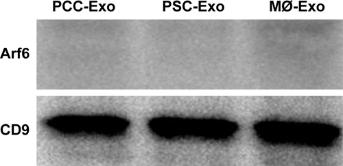
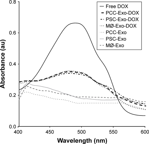
Figure S2 UV–Vis absorption spectra of Exo-DOX show the absorption corresponding to DOX.
Notes: PCCs, PSCs, and MØs treated with 1 µM DOX for 48 h and Exos were extracted by ultra-centrifugation. Obtained Exo pellets were resuspended in PBS and scanned on UV–Vis spectrophotometer to check the absorption.
Abbreviations: DOX, doxorubicin; Exos, exosomes; MØs, macrophages; PCCs, pancreatic cancer cells; PSCs, pancreatic stellate cells; UV, ultraviolet; Vis, visible.
Acknowledgments
We would like to acknowledge the funding support from the National Institute of Health/National Cancer Institute (CA204801 [to SS] and CA224306 and CA175772 [to APS]) as well as the institutional support from University of South Alabama Mitchell Cancer Institute.
Disclosure
APS and SS are cofounders and serve on the executive management team of Tatva Biosciences LLC, which is involved in the development of tools and models for biological research. The authors report no other conflicts of interest in this work.