
 ?Mathematical formulae have been encoded as MathML and are displayed in this HTML version using MathJax in order to improve their display. Uncheck the box to turn MathJax off. This feature requires Javascript. Click on a formula to zoom.
Abstract
Background
It has been difficult to find bioactive compounds that can optimize bone repair therapy and adequate osseointegration for people with osteoporosis. The nano-hydroxyapatite (nHAp)/carbon nanotubes with graphene oxides, termed graphene nanoribbons (GNR) composites have emerged as promising materials/scaffolds for bone regeneration due to their bioactivity and osseointegration properties. Herein, we evaluated the action of nHAp/GNR composites (nHAp/GNR) to promote bone regeneration using an osteoporotic model.
Materials and methods
First, three different nHAp/GNR (1, 2, and 3 wt% of GNR) were produced and characterized. For in vivo analyses, 36 Wistar rats (var. albinus, weighing 250–300 g, Comissão de Ética no Uso de Animais [CEUA] n.002/17) were used. Prior to implantation, osteoporosis was induced by oophorectomy in female rats. After 45 days, a tibial fracture was inflicted using a 3.0-mm Quest trephine drill. Then, the animals were separated into six sample groups at two different time periods of 21 and 45 days. The lesions were filled with 3 mg of one of the above samples using a curette. After 21 or 45 days of implantation, the animals were euthanized for analysis. Histological, biochemical, and radiographic analyses (DIGORA method) were performed. The data were evaluated through ANOVA, Tukey test, and Kolmogorov-Smirnov test with statistical significance at P<0.05.
Results
Both nHAp and GNR exhibited osteoconductive activity. However, the nHAp/GNR exhibited regenerative activity proportional to their concentration, following the order of 3% >2% >1% wt.
Conclusion
Therefore, it can be inferred that all analyzed nanoparticles promoted bone regeneration in osteoporotic rats independent of analyzed time.
Introduction
The increase in the life expectancy of the global population, unfortunately, results in the rise of several adverse physiological conditions, such as osteopenia, which is characterized by the reduction of bone mass.Citation1 When left untreated, this may evolve into osteoporosis, a condition that promotes the deterioration of the microarchitecture of bone tissue, increasing the fragility of its structure and, therefore, susceptibility to fractures. This condition mainly affects women and the elderly,Citation2 and osteoporosis fractures are highly prevalent, thus representing an important public health problem.Citation3 The development of novel materials for bone growth is particularly important, as the World Health Organization has predicted an increase in the number of worldwide osteoporosis cases. However, to date, bioactive materials that can promote bone repair as a therapeutic method for treating osteoporosis are still missing.Citation4 Thus, innovative biomaterials with improved chemical composition and bioactive properties are a pressing need for biomedical applications.Citation5 Biomaterials have different characteristics on a nanometric scale and ideally influence the bone regeneration process by giving a rapid, controlled, and predictable response to biological tissues.Citation6
In this context, ceramic biomaterials based on nano-hydroxyapatite (nHAp) have piqued the interest of several researchers.Citation7 These are used as a bioactive scaffolding during the regeneration of bone tissue, as their physical and chemical properties resemble bone structure, and they exhibit excellent biocompatibility, osseointegration, and osteoconductivity.Citation8 The chemical structure of hydroxyapatite (HAp) is represented by Ca10(PO4)6(OH)2 with a Ca/P molar ratio of 1.67, and it is used in clinical practices as a substitute for damaged hard tissues, periodontal defects, orthopedic surgeries, and other similar applications.Citation4,Citation9 Junior et alCitation10 reported an improvement in the osteoconduction of nHAp in adult rats compared to biological HAp. Carmo et alCitation11 found similar results to those reported previously when using HAp-based nanocomposites and carbonate structures during osteoconduction and bone repair. However, its fragility and low resistance to mechanical stress have limited its use in orthopedic interventions.Citation12,Citation13
Carbonaceous materials, such as carbon nanotubes (CNTs), have also been used in regenerative medicine because they can afford the preservation of biological properties, cell spreading, and adhesion, exhibit excellent cellular biocompatibility and support the growth of osteoblast cells, and stimulate the production of bone matrix, especially those that are hydrophilic. While studying biomaterials based on nanocarbon fiber-reinforced polyether ether ketone-nHAp, Xu et alCitation14 observed their remarkable ability to promote the proliferation and differentiation of MG-63 cells in addition to their ability to stimulate in vivo osseointegration between the implant and bone. One method of improving the biocompatibility of CNTs is to exfoliate them and functionalize them with hydrophilic groups, forming unzipped nanotubes with a structural atomic organization similar to graphene oxide (GO) at their ends, termed graphene nanoribbons (GNR).Citation15 Song et alCitation16 evaluated the influences of GO on biofilm formation, and their growth profile and viability assays indicated that the GO exhibited high antibacterial activity, improving the coating biocompatibility higher than that of pure HAp and Ti substrates.
Scaffolds of nHAp and GNR composites (nHAp/GNR) have emerged as promising biomaterials for bone regeneration, exhibiting appealing properties for osseointegration including the ability to preserve biological properties such as cell growth, spreading, adhesion, and differentiation.Citation17 Herein, our group synthesized and evaluated both the in vitro and in vivo properties of these new nanocomposites for bone engineering. Rodrigues et alCitation15 characterized the chemical, structural, and biological properties of different concentrations of nHAp/GNR and used simulated body fluid to evaluate their bioactivity and human osteoblasts (bone-forming cells). Recently, Medeiros et alCitation18 showed that nHAp/GNR induced in vitro and in vivo osteogenesis process after 15 days. However, an understanding of the influence of nHAp/GNR on bone regeneration using an in vivo model has not yet been reported, and is imperative for determining the potential benefits of these composites for treating osteoporosis.
Herein, we present in vivo studies of nHAp/GNR using an in vivo osteopenic model. First, nHAp/GNR (1, 2, and 3 wt% of GNR) were extensively characterized by Fourier-transform infrared (FTIR) spectroscopy, Raman spectroscopy, and X-ray diffraction. The total carbonate amount was calculated and correlated with biological properties. The in vivo experiments were carried out using 60 osteopenic rats. After each analyzed time (21 or 45 days), the animals were euthanized, and the size of nanocrystals and the bone density were verified through radiographic analysis (DIGORA imaging). The regeneration process was examined through histological, biochemical (alkaline phosphatase [ALP]), and radiographic analyses. We emphasize that the study presented here advances recent literature, enhancing bone regeneration in osteoporosis and offering the potential for the development of new commercial biomaterials based on nHAp/GNR for bone tissue engineering.Citation19
Materials and methods
Production of nHAp/GNR
The GNRs were produced through exfoliation of multiwalled CNTs (MWCNTs). The MWCNTs were purified to reduce the iron (Fe) from the interior of the nanotubes using an acid bath with an H2SO4:HNO3 solution (3:1) under ultrasound irradiation (5 hours, Elmasonic S10H; Elma Electronic Inc., Wetzikon, Switzerland). The functionalization and exfoliation of MWCNTs to obtain GNRs were performed using an oxygen plasma treatment.Citation15 Next, the GNR powder, in the required percentage, was dissolved in an aqueous solution of (NH4)⋅H2PO4 and Ca(NO3)2⋅4H2O using an ultrasonic probe (Sonics, VCX 500W) for 30 minutes. A pH of 10 was attained by adding an ammonium hydroxide solution (NH4OH: 25%). The formed precipitate was then aged over 120 hours, after which it was filtered, washed using distilled water, and dried in an oven for 48 hours at 60°C. After drying, the material was milled (A11 mill from IKA [Staufen im Breisgau, Germany], with an engine speed of 28,000 rpm) to generate the groups of nHAp/GNR at 1, 2, and 3 wt%.Citation20
Characterization of nHAp, GNR, and nHAp/GNR
The molecular structure of the samples was analyzed using a Senterra-model Raman spectrometer from Bruker Corporation (Billerica, MA, USA), with an attached Olympus BX50 microscope and a charge-coupled device as a detector. For excitation, the spectrometer used a laser with a wavelength of 532 nm and an output power of 10 mW. The spectrometer was adjusted to obtain a spectral resolution of 3 cm−1 covering the range of 300–2,000 cm−1. The attenuated total reflectance (ATR)-FTIR spectra were obtained within a spectral range of 1,800–600 cm−1 using a Bruker spectrometer (Vertex 70 v) with the ATR accessory using a germanium crystal. All measurements were collected at room temperature. The crystallinity of the samples was analyzed by X-ray diffraction using an XRD 6000 (Shimadzu Corporation, Kyoto, Japan) powder diffractometer with Cu-Kα radiation (λ=1.5406 Å). Data were collected at 2θ from 20° to 70° with a scanning speed of 1°/min. The X’Pert HighScore (Plus) software was used to identify the phases of the crystalline structure.
The crystallite size of the nanocrystals was determined using the Debye-Scherrer equation (EquationEquation 1(1) ).Citation21
The crystallinity indices were also calculated using Raman spectroscopy, and they were obtained through the profile of the highest intensity band, 961 cm−1, referring to the symmetrical stretching vibration of the group of phosphate. The FWHM values were collected after deconvolution of the 961 cm−1 band using standard and Lorentz-type profiles. The crystallinity index is calculated by using EquationEquation 3
(3) .
The value of 4.9 refers to the average FWHM of the magmatic apatite standard with high crystallinity, and Γ isthe FWHM of the peak 961 cm−1.Citation23
In vivo assays
Surgical procedure
Thirty-six female Wistar rats with initial body weights between 250 and 300 g were used; they were obtained from the colony of the Vivarium of the University Center of Health, Human, and Technological Sciences of Piauí (UNINOVAFAPI). The rats were kept in a room (25°C ± 2°C) with a photoperiod of light and darkness (12/12 hour) in collective cages (four animals/box) with standard food rations (Labina) and free access to water. These samples were submitted to the oophorectomy protocol.Citation24–Citation26 After this period, the rats were divided into 12 groups () and evaluated for a period of 21 (G1–G6) or 45 days (G7–G12).
Table 1 Distribution of groups of animals and evaluation periods after implantation
For the nHAp/GNR and the respective controls, the rats were intraperitoneally anesthetized with ketamine (40 mg/kg) and xylazine (5 mg/kg) mixture. Surgery was performed on the right tibia and began with the trichotomy of the region. The surgical procedures were conducted for all rats, which consisted of the insertion of the composites in the form of a powder at the site of the bone defect.Citation29 This defect was elliptical, with dimensions that allowed it to reach the level of the spinal canal. This procedure was conducted with a trephine type drill and a surgical micromotor (model AEU-707A; Aseptico, Woodinville, WA, USA). The surgical wound was closed by planes, and the suturing of the tissues was performed with silk thread so that the periosteum was positioned on the inserted materials.
X-ray examinations, histological and biochemical analyses
The tibia of rats was removed and fixed under 24 hours. The soft tissue was removed, and bone pieces were examined by radiograph and histological analyses.Citation19 For biochemical analysis, ALP kit was used (Labtest Diagnostica SA, Lagoa Santa, Brazil). The absorbance was measured at 590 nm. The results were presented as normality test graphs.
Statistical analysis
The data were statistically analyzed using the Minitab 16 software. The data presented a normal distribution pattern (Kolmogorov-Smirnov test). After that, the groups were compared using one-way ANOVA applying a post-hoc Tukey’s test (level of significance at a 95% CI, P<0.05).
Ethical principles
This study is part of the project entitled: Nano biomaterials based on CNTs, graphene, nanoceramics, and bioresorbable polymers for tissue engineering, approved by the ethical committee of CEUA/UNINOVAFAPI number 002/17. The ethical norms in Animal Research law n.11.794 of 2008 were observed.
Results and discussion
illustrates the characterization of the nHAp- and GNR-based composites, as well as those of their individual components. The GNRs were verified by the transmission electron microscopy (white arrow ), along with the presence of nHAp crystals in the composites (red circle ). shows the results of X-ray analyzes of pure nHAp and at different concentrations of GNR. The main reflection peak characteristics of hexagonal HAp in 2θ were present at ~25.9°, referring to the plane (0 0 2) (); at ~31.7°, ~32.1°, and ~32.9°, referring to the planes (2 1 1), (1 1 2), and (3 0 0), respectively; and at ~34.1°, referring to the plane (2 0 2), which are in accordance with the crystallographic file for nHAp (International Centre for Diffraction Data [JCPDS]: 00-046-0905). In addition, the GNR sample exhibited peaks in 2θ at ~26.9°, ~37.7°, ~42.9°, ~43.7°, ~44.8°, and ~45.9°, which are assigned to carbon in the crystallographic data file (JCPDS: 00-026-1077). Peaks are also observed in 2θ at ~37.8° and ~49.1°, which are characteristic of Fe (used as a growth catalyst) in the GNR sample, according to the crystallographic data sheet (JCPDS: 01-085-0871). From analyzing the diffractograms and CIX-ray (), a decrease in crystallinity with increasing GNR in the nanocomposites was observed, which was further confirmed by Raman spectroscopy () and corroborated with the increase in carbonate observed by ATR-FTIR ().
Figure 1 (A) TEM of the GNR and (B) nHAp/GNR. (C) X-ray diffractogram of the powders. (D) Size of the crystallite obtained according to the Scherrer equation, using the (0 0 2) diffraction plane of HAp (JCPDS 00-046-0905). (E) Average crystallinity index. (F) Spectra of ART-FTIR. (G) Spectra of Raman for: (a) GNR, (b) nHAp, (c) nHAp/GNR1%, (d) nHAp/GNR2%, and (e) nHAp/GNR3%.
Abbreviations: ART-FTIR, attenuated total reflection - Fourier transform infrared; FWHM, full peak width at half height of the maximum; HAp, hydroxyapatite; nHAp, nano-hydroxyapatite; GNR, graphene nanoribbons; nHAp/GNR, nHAp and GNR composites; TEM, transmission electron micrographs.
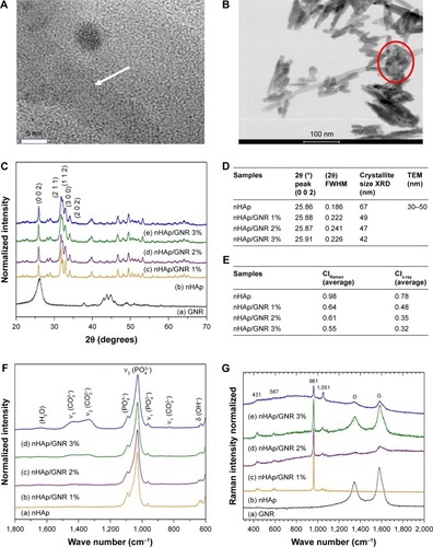
In the Raman spectra presented in (a), D (1,346 cm−1) and G (1,580 cm−1) characteristic bands of amorphous carbon are observed due to the disorder and imperfections of carbon bonds promoted by the exfoliation of MWCNTs. The D and G bands are also observed in the spectra of (c), (d), and (e) due to the differences in the concentrations of GNR present in the samples. All nHAp and nHAp/GNR spectra at different concentrations of GNR have shown a stretching mode of at 961 cm−1, which is characteristic of crystalline HAp. Three bands with lower intensities were also observed at 431, 587, and 1,051, which are attributed to the vibrations of other apatite groups such as octacalcium phosphate, dehydrated calcium phosphate, and the apatite’s phosphate group, respectively, which is only observed in well-crystallized HAp.Citation30 A decrease in the crystallinity of the HAp was observed when the concentration of GNR in the composites increased, as indicated by the decrease in the calculated CIRaman value of the Raman spectra obtained in , previously observed by XRD. The ATR-FTIR spectra () of the nHAp/GNR show a band at 1,642 cm−1, related to the characteristic vibration deformation of water adsorbed during apatite formation. The carbonate group
has appeared in nHAp/GNR and it is assigned to the doublet of bands at 1,332 and 1,447 cm−1, referring to the stretching modes, and a singlet at 826 cm−1, related to the deformation mode. The bands at 1,026–1,090 cm−1 and 962 cm−1 are attributed to the triple unfolded antisymmetric P–O (ν3) and nondegenerate P–O symmetric (ν1) stretching modes, respectively, which all belong to the
group. Also, the band observed at 635 cm−1 is attributed to deformation vibration of the OH− groups from HAp.Citation7,Citation28,Citation29 The ATR-FTIR spectra of the pure HAp sample and the nHAp/GNR show an increase in carbonation with the increase in the concentration of GNR in the nanocomposites () due to the increase in the band intensities of the carbonate group.
In vivo study
The animals used in this study were of the same age (90 days) and gained weight during the 45 days after surgery. No animals were lost throughout the study. The weight gain verified in our study is consistent with the data described in the literature,Citation26,Citation31–Citation36 that oophorectomy produces an increase in weight due to a loss of estrogen, as estrogen increases energy consumption and thereby decreases body weight. Therefore, the energy consumption in animals with estrogenic suppression will be lower, and they will gain weight.Citation26 The spherical bone lesion model was used as it provides identical lesions based on position and size ().Citation36
Figure 2 Radiography of tibias: (A) control and (B and C) experimental groups with GNR after 21 and 45 days, respectively, using the DIGORA system. The circle indicates the location of the defect. Statistical data on bone density are presented in (D) and (E) recorded as mean ± SD (n=3). Different letters show significant differences for P<0.05.
Abbreviation: GNR, graphene nanoribbons.
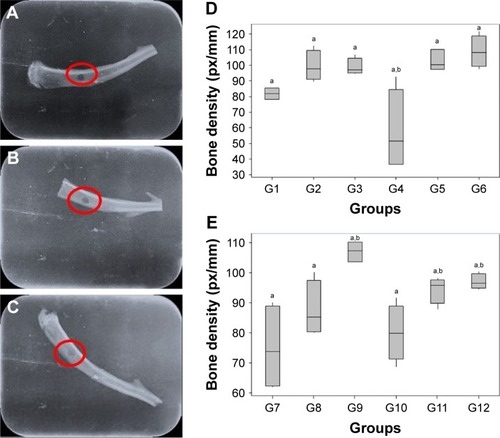
shows radiographic images of the tibias of the rats that were oophorectomized and implanted with different analyzed groups during the two periods of investigation, captured using the DIGORA system.Citation29
No deformation was detected in the bone structures for all studied groups that would compromise the selected methodology. The evolution of bone density in the tibia of rats with osteopenia could also be observed from the images showing the filling of the hole inflicted upon these animals (circle, ). The tibial bone density for the abovementioned periods are quantified (), which show the highest bone density in group G6 () for the first analyzed period (21 days); however, the overall bone density was highest in group G9 for the period of 45 days ().
The histological results of the tibial surgical defect sections of the control and implanted groups with nHAp, GNR, and nHAp/GNR at 3% after 21 days are presented in . The presence of both GNR and nHAp/GNR was verified along the tissues for both periods, and it is distributed in an agglomerated form. Bone matrix formation was observed in all analyzed groups, indicating medullary endochondral ossification. In addition, several regions with a vascular cortical tissue were observed in the experimental groups after 21 days from implantation in relation to the control group, indicative of regeneration in both cases.
Figure 3 Optical microscopy photographs of the rat tibias from the control group and after 21 days from the implantation: G1 (A, A1, and A2), G2 (B, B1, and B2), G3 (C, C1, and C2), and G6 (D, D1, and D2). H&E: 10, 20, and 40×, respectively. Scale bar of 200 µm. Periosteum (circle), trabecular bone (➔), osteocytes (→), bone marrow (▲), and nHAp/GNR (square).
Abbreviations: CB, compact bone; H&E, hematoxylin and eosin; nHAp/GNR, nHAp and GNR composites.
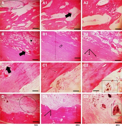
The animals analyzed after 45 days from the implantation exhibited a difference in the cortical thickness and vascularization compared to that of the control group. For groups containing GNR and nHAp/GNR at 2 and 3 wt%, respectively, the structures were compacted, trabecular, and less vascularized, indicating the action of remodeling by an osteoclast.Citation33 However, the group containing 1% GNR exhibited a highly vascularized structure, similar to those related to the control group, which could be due to a delay in the repair period of the bone lesions. This is consistent with the ATR-FTIR and Raman data, which imply lower carbonation ().
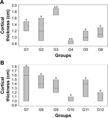
Differences in bone thickness between the studied groups were detected from the quantitative analyses of bone healing through the measurement of the cortical thickness of each bone (). For the 21-day period (), the cortical thickness between the control (G1) and experimental groups varied, except for G4 (nHAp/GNR 1%), which exhibited the lowest growth. The group implanted with just GNR (G3) exhibited a more pronounced regenerative process than the others, which verified the promotion of bone regeneration with increasing GNR concentration (G6> G5> G4) for the period of 21 days. For the period of 45 days, there was a reduction in the cortical thickness for all experimental groups compared to the control group (). The lowest cortical thickness was observed in the group containing 1% GNR (G10) compared to the other groups.
Figure 4 Quantification of the cortical thickness of the tibia bones in rats implanted with GNR, nHAp, and nHAp/GNR after (A) 21 and (B) 45 days of implantation recorded as mean ± SD (n=3). Different letters show significant differences for P<0.05.
Abbreviations: GNR, graphene nanoribbons; nHAp, nano-hydroxyapatite; nHAp/GNR, nHAp and GNR composites.
The results of enzymatic dosing showed low ALP expression in control group (G1 and G7) for both the periods (). There was an increase in enzyme concentration for all groups after 21 days of implantation compared to the control group (). For the analyzed groups after 45 days, the ALP only increased in G9, G10, and G12 groups compared to the control group (); the other experimental groups (G8 and G11) exhibited a reduction in their enzymatic levels for this biochemical marker. Upon comparison between the two investigation time periods, the 45-day time period presented overall lower enzymatic values than the 21-day period for almost all the experimental groups. The experimental group G10 (), however, registered higher enzymatic levels, which was probably due to a delay in the healing process, also suggested by the lower cortical thickness () of this group compared to others.Citation34
Figure 5 Quantitative analysis of the ALP of rats that were oophorectomized and implanted with GNR, nHAp, and nHAp/GNR after (A) 21 and (B) 45 days of implantation presented as mean ± SD (n=3). Different letters show significant differences for P<0.05.
Abbreviations: ALP, alkaline phosphatase; GNR, graphene nanoribbons; nHAp, nano-hydroxyapatite; nHAp/GNR, nHAp and GNR composites.
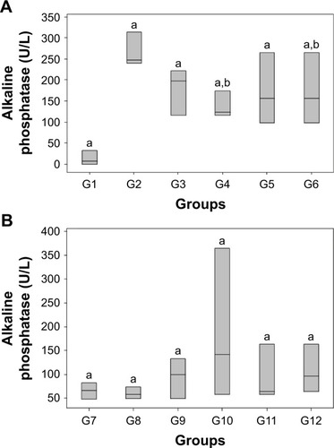
ALP may increase the local concentration of inorganic phosphorus or activate collagen fibers, causing calcium salts to be deposited in these tissues.Citation35 The analysis of cortical thickness is consistent with the data obtained from the biochemical analyses, which verify that mineralization was more evident in the groups that contained pure nHAp and GNR compared to the composites and control groups (more at 21 days). This suggests that the increase in the levels of ALP in G2 (only nHAp) is because it increases the deposition of inorganic phosphorus ions on bone tissues to a greater extent than GNR due to its chemical composition ().Citation36
Meanwhile, for the nHAp/GNR, the lowest ALP level was exhibited for the 1% group, which is consistent with the observed lower cortical thickness and bone density for that group (). This behavior can be attributed to a delay in the repair period of bone lesions.Citation26 The higher bone density (), ALP (), and cortical thickness () values suggest a direct relationship between the efficiency of mineralization and the concentration of GNR for the 21-day period,Citation28,Citation37 which is also consistent with the concentration data presented by the ATR-FTIR and Raman spectra ().
The reduction in the ALP levels and bone density of the composites after 45 days of implantation is consistent with the histological data, suggesting the cellular remodeling of osteoclasts.Citation38 It is also believed that the decrease in the crystallinity of HAp with the increase of concentration of GNR in the nHAp/GNR may indicate a higher efficiency of the nHAp/GNR 3% compared to the other analyzed groups and controls.Citation11
Conclusion
Herein we showed that a mineralization occurred for all analyzed groups independently of GNR composition and respective controls, suggesting that nHAp and GNR have chemical properties to promote bone growth in osteoporotic animals. This was particularly evident for the groups containing pure nHAp and GNR, and their nHAp/GNR 3% during the 21-day period. Therefore, this is a promising alternative for the regeneration of bone tissue, and further studies are required. New studies are being conducted to compare GNRs with other biomaterials.
Acknowledgments
AOL and FRM thank the National Council for Scientific and Technological Development (CNPq grants numbers AOL#303752/2017-3 and FRM#304133/2017-5), Coordination for the Improvement of Higher Education Personnel (CAPES, grant numbers AOL#88881.120138/2016-01 and FRM#88881.120221/2016-01), and Universidade Brasil for the scholarships.
Disclosure
The authors report no conflicts of interest in this work.
References
- LiuLWebsterTJIn situ sensor advancements for osteoporosis prevention, diagnosis, and treatmentCurr Osteoporos Rep201614638639527815807
- RadominskiSCBernardoWPaula deAPDiretrizes brasileiras para o diagnóstico e tratamento da osteoporose em mulheres na pós-menopausa. [Brazilian guidelines for the diagnosis and treatment of osteoporosis in postmenopausal women]Revista Brasileira de Reumatologia201757Suppl 3452466
- LouresMARZerbiniCAFDanowskiJSDiretrizes da sociedade brasileira de reumatologia para diagnóstico e tratamento da osteoporose em homens. [Brazilian guidelines for the diagnosis and treatment of osteoporosis in men]Revista Brasileira de Reumatologia201757S249751428800970
- dos Santos AlmeidaRdosAnjos RibeiroIÍda SilvaMHPda RochaDNMiguelFBRosaFPEvaluation of the initial phase of bone repair after implantation of biomaterialsRev Ciênc Méd Biol201413n.3331336
- PiresABierhalaAMoraesAMBiomaterials: type, applications and marketQuim Nova2015387957971
- ShuklaADasguptaNRanjanSSinghSChidambramRNanotechnology towards prevention of anaemia and osteoporosis: from concept to marketBiotechnology & Biotechnological Equipment2017315863879
- ZaninHSaitoEMarcianoFRFast preparation of nanohydroxyap-atite/superhydrophilic reduced graphene oxide composites for bioactive applicationsJ Mater Chem B20131384947
- LoboAOCoratMAFRamosSCFast preparation of hydroxyapatite/superhydrophilic vertically aligned multiwalled carbon nanotube composites for bioactive applicationLangmuir20102623183081831420961085
- RajkumarMSundaramNMRajendranVPreparation of size controlled, stoichiometric and bioresorbable hydroxyapatite nanorod by varying initial pHCa/P ratio and sintering temperature201161169179
- JuniorSAAllegriniMRFYoshimotoMSallesM B de AReparação óssea utilizando hidroxiapatia natural e nanometrica “in vivo”. [Bone repair using natural and nanometric hydroxyapatite “in vivo]J Biodentistry Biomater20144124
- CarmoABXDSartorettoSCAlvesATNNAlveolar bone repair with strontium-containing nanostructured carbonated hydroxyapatiteJ Appl Oral Sci201826e2017008429364342
- SuruagyAAAlvesATSartorettoSCCalasans-MaiaJAGranjeiroJMCalasans-MaiaMDPhysico-chemical and histomorphometric evaluation of zinc-containing hydroxyapatite in rabbits calvariaBraz Dent J201627671772627982185
- XuALiuXGaoXDengFDengYWeiSEnhancement of osteogenesis on micro/nano-topographical carbon fiber-reinforced polyetheretherketone–nanohydroxyapatite biocompositeMater Sci Eng C201548592598
- LiMLiuQJiaZXuXGraphene oxide/hydroxyapatite composite coatings fabricated by electrophoretic nanotechnology for biological applicationsCarbon201467185197
- RodriguesBVLeiteNCCavalcantiBGraphene oxide/multi-walled carbon nanotubes as nanofeatured scaffolds for the assisted deposition of nanohydroxyapatite: characterization and biological evaluationInt J Nanomedicine2016112569258527358560
- SongCYangC-MSunX-FInfluences of graphene oxide on biofilm formation of gram-negative and Gram-positive bacteriaEnviron Sci Pollut Res201825328532860
- MuruganNSundaramurthyAChenS-MSundramoorthyAKGraphene oxide/oxidized carbon nanofiber/mineralized hydroxyapatite based hybrid composite for biomedical applicationsMater Res Express2017412124005
- S MedeirosJOliveiraAMde CarvalhoJONanohydroxyapatite/graphene nanoribbons nanocomposites induce in vitro osteogenesis and promote in vivo bone neoformationACS Biomater Sci Eng20184515801590
- CarvalhoJCOliveiraFCFreitasSAPCarbon nanomaterials for treating osteoporotic vertebral fractures introduction: a perspective of carbonCurr Osteoporos Rep201816562663430203250
- LoboAOZaninHSiqueiraIEffect of ultrasound irradiation on the production of HAp/MWCNT nanocompositesArq Bras Endocrinol Metabol201433243054312
- RaynaudSChampionEBernache-AssollantDThomasPCalcium phosphate apatites with variable Ca/P atomic ratio I. Synthesis, characterisation and thermal stability of powdersBiomaterials20022341065107211791909
- BarbosaMCMessmerNRBrazilTRMarcianoFRLoboAOThe effect of ultrasonic irradiation on the crystallinity of nano-hydroxyapatite produced via the wet chemical methodMater Sci Eng C Mater Biol Appl20133352620262523623076
- PucéatEReynardBLécuyerCCan crystallinity be used to determine the degree of chemical alteration of biogenic apatites?Chemical Geology20042051–28397
- Kaczmarczyk-SedlakIKlasik-CiszewskaSWojnarWGlabridin and glycyrrhizic acid show no beneficial effect on the chemical composition and mechanical properties of bones in ovariectomized rats, when administered in moderate dosePharmacol Rep20166851036104127434879
- HaddadPTSalazarMHernandesLHistomorfometria da matriz orgânica do fêmur de ratas ovariectomizadas tratadas com alendronato de sódio. [Histomorphometry of the organic matrix of the femur in ovariectomized rats treated with sodium alendronate]Revista Brasileira de Ortopedia201550110010426229885
- VasconcellosLSLeiteJMSabinoKRPetroianuAInfluência da ooforectomia na variação ponderal em ratas jovens e adultas. [Influence of oophorectomy on the weight variance in young and adult female rats]Arq Bras Endocrinol Metab2004482299304
- MesquitaPFelinoARaposoHAfonsoAAvaliação in vitro do comportamento de osteoblastos sobre implantes com diferentes tratamentos de superfície. [In vitro osteoblastic cells behavior evaluation cultured on implant surfaces with different treatments]ElSEVIER201556295102
- LoboAOSilvaGRMarcianoFRPacheco-SoaresCOsteoblastos humanos cultivados sobre arcabouços à base de nanotubos de carbono superhidrofílicos e nanohidroxiapatita. [Human osteoblasts cultivated on biomineralizated nanohydroxyapatite/superydrophilic carbon nano-tube scaffolds]Rev Bras Apl Vácuo2014331–212
- CunhaFVMMoura-FilhoOFMouraFSMMEffects os exercise and testosterone administration on tibia fracture healing in rats. [Effects os exercise and testosterone administration on tibia fracture healing in rats]PhysTher in Mov2012254777784
- LegerosRZCalcium phosphates in oral biology and medicineMonogr Oral Sci19911512011870604
- AtmacaHAydınAMusaoğluRExperimental model of osteoporosis: comparison between ovariectomy and botulinum toxin AActa Ortop Bras [Internet]2013216340343
- MoraisGQBurgosMGP de AImpacto dos nutrientes na saúde óssea: novas tendências. [Nutrients impact on bone health: new trends]Rev Bras Ortop2007427189194
- SartoriARMoreiraJASantosAMMCintraDECBone repair process in normal and osteopeni female rats tibiae: a comparative studyBrazOrthop Acta20081613740
- KhajuriaDKRazdanaRRoy MahapatrabDAdditive effects of zoledronic acid and propranolol on bone density and biochemical markers of bone turnover in osteopenicovariectomized ratsElSEVIER2015552103112
- CabralHWSAndolphiBFGFerreiraBVCThe use of biomarkers in clinical osteoporosisRev Assoc Med Bras201662436837627437684
- GranatoAECRodriguesBVMRodrigues-JuniorDMMarcianoFRLoboAOPorcionattoMAMagnetic super-hydrophilic carbon nano-tubes/graphene oxide composite as nanocarriers of mesenchymal stem cells: insights into the time and dose dependencesMater Sci Eng C201667694701
- MendesHMFAlvesGESMachadoIRLFaleirosRRNanotubos de carbono: potencial de uso em medicina veterinária. [Carbon nano-tubes: Potential use in veterinary medicine]Ciência Rural2014441018231829
- WangTYangXQiXJiangCOsteoinduction and proliferation of bone-marrow stromal cells in three-dimensional poly (ε-caprolactone)/hydroxyapatite/collagen scaffoldsJ Transl Med201513111125591711