

Abstract
Background:
Exosomes are ubiquitous naturally secreted stable nanovesicles that can be engineered to target and deliver novel therapeutics to treat a host of human diseases.
Methods:
We engineered the surfaces of cell-derived nanovesicles to act as decoys in the treatment of inflammation by antagonizing the major proinflammatory cytokine, tumor necrosis factor alpha (TNFα).
Results:
Decoy exosomes were generated by displaying the TNFα binding domain of human TNF receptor-1 (hTNFR1) on the outer surface of exosomes using stably transfected HEK293 cells. We developed an efficient method to purify the engineered exosomes from conditioned medium based on sequential centrifugation, ultrafiltration, and precipitation. We characterized decoy exosomes using immune-quantification, nanoparticle tracking analysis, and confocal microscopy to confirm that they retain the correct orientation, size, and shape of naturally produced exosomes. We demonstrated the engineered decoy exosomes specifically antagonize activities of TNFα using an inflammatory reporter cell line.
Conclusions:
Decoy exosomes produced in human cells serve as a novel biologic reagent for antagonizing inflammatory signaling mediated by TNFα.
Acknowledgment
We thank Dr. Yan Jiang for critically reading and editing the manuscript. This work was supported by internal funds from the School of Engineering, Santa Clara University. GM acknowledges support from the Tsinghua-Berkeley Shenzhen Institute. The funding institute plays no role in the design of the study and collection, analysis, and interpretation of data.
Data availability
All data generated or analyzed in this study are included in this published article and its Supplementary information files.
Disclosure
The authors report no conflicts of interest in this work.
Supplementary materials
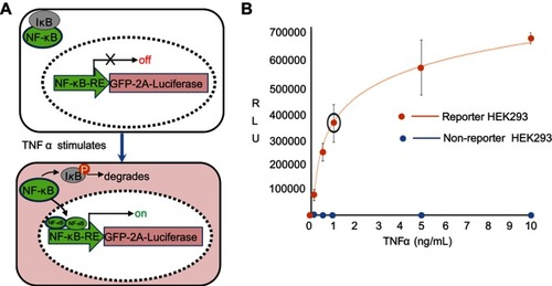
Figure S1 Reporter cell line and its dose-response to TNFα. (A) Molecular mechanism of the reporter line. A genetically encoded reporter circuit (NF-κB-RE-driven GFP and fire luciferase gene with a self-splicing 2A peptide) was incorporated into the genome of HEK293 cells. In the absence of TNFα, the transcription factor NF-κB remains in cytosol and associates with its inhibitor protein IκB. Therefore, the reporter genes has little expression due to lack of NF-κB binding to its response element (RE). In the presence of TNFα, TNFα binding to its receptor results in the phosphorylation of IκB, which leads to its degradation and a release of NF-κB. The freed NF-κB then enters into nucleus and binds the RE in the promoter of reporter, resulting in an increase of luciferase activities. (B) A dose-response of TNF α. HEK293 cells were stimulated with incremental amount of TNFα for 24 hours. Reporter cells were then collected for the luciferase activities. A dose-dependent response to TNFα was evident (red line) as compared the non-reporter parental HEK293 (blue dots).
Supplementary sequences
N-CD63-hTNFR1-EC-C-CD63-GFP coding sequences
N-CD63-hTNFR1-EC-C-CD63-GFP chimeric protein sequences
1. N-CD63-hTNFR1-EC-C-CD63-GFP coding sequences
ATGGCGGTGGAAGGAGGAATGAAATGTGTGAAGTTCTTGCTCTACGTCCTCCTGCTGGCCTTTTGCGCCTGTGCAGTGGGACTGATTGCCGTGGGTGTCGGGGCACAGCTTGTCCTGAGTCAGACCATAATCCAGGGGGCTACCCCTGGCTCTCTGTTGCCAGTGGTCATCATCGCAGTGGGTGTCTTCCTCTTCCTGGTGGCTTTTGTGGGCTGCTGCGGGGCCTGCAAGGAGAACTATTGTCTTATGATCACGTTTGCCATCTTTCTGTCTCTTATCATGTTGGTGGAGGTGGCCGCAGCCATTGCTGGCTATGTGTTTAGAGATAAGGTGATGTCAGAGTTTAATAACAACTTCCGGCAGCAGATGGAGAATTACCCGAAAAACAACCACACTGCTTTCGAATCTGGCATGGGCCTCTCCACCGTGCCTGACCTGCTGCTGCCACTGGTGCTCCTGGAGCTGTTGGTGGGAATATACCCCTCAGGGGTTATTGGACTGGTCCCTCACCTAGGGGACAGGGAGAAGAGAGATAGTGTGTGTCCCCAAGGAAAATATATCCACCCTCAAAATAATTCGATTTGCTGTACCAAGTGCCACAAAGGAACCTACTTGTACAATGACTGTCCAGGCCCGGGGCAGGATACGGACTGCAGGGAGTGTGAGAGCGGCTCCTTCACCGCTTCAGAAAACCACCTCAGACACTGCCTCAGCTGCTCCAAATGCCGAAAGGAAATGGGTCAGGTGGAGATCTCTTCTTGCACAGTGGACCGGGACACCGTGTGTGGCTGCAGGAAGAACCAGTACCGGCATTATTGGAGTGAAAACCTTTTCCAGTGCTTCAATTGCAGCCTCTGCCTCAATGGGACCGTGCACCTCTCCTGCCAGGAGAAACAGAACACCGTGTGCACCTGCCATGCAGGTTTCTTTCTAAGAGAAAACGAGTGTGTCTCCTGTAGTAACTGTAAGAAAAGCCTGGAGTGCACGAAGTTGTGCCTACCCCAGATTGAGAATGTTAAGGGCACTGAGGACTCAGGCACCACAGGGCTCGATTTAAATTCGATCCTGGACAGGATGCAGGCAGATTTTAAGTGCTGTGGGGCTGCTAACTACACAGATTGGGAGAAAATCCCTTCCATGTCGAAGAACCGAGTCCCCGACTCCTGCTGCATTAATGTTACTGTGGGCTGTGGGATTAATTTCAACGAGAAGGCGATCCATAAGGAGGGCTGTGTGGAGAAGATTGGGGGCTGGCTGAGGAAAAATGTGCTGGTGGTAGCTGCAGCAGCCCTTGGAATTGCTTTTGTCGAGGTTTTGGGAATTGTCTTTGCCTGCTGCCTCGTGAAGAGTATCAGAAGTGGCTACGAGGTGATGatggagagcgacgagagcggcctgcccgccatggagatcgagtgccgcatcaccggcaccctgaacggcgtggagttcgagctggtgggcggcggagagggcacccccaagcagggccgcatgaccaacaagatgaagagcaccaaaggcgccctgaccttcagcccctacctgctgagccacgtgatgggctacggcttctaccacttcggcacctaccccagcggctacgagaaccccttcctgcacgccatcaacaacggcggctacaccaacacccgcatcgagaagtacgaggacggcggcgtgctgcacgtgagcttcagctaccgctacgaggccggccgcgtgatcggcgacttcaaggtggtgggcaccggcttccccgaggacagcgtgatcttcaccgacaagatcatccgcagcaacgccaccgtggagcacctgcaccccatgggcgataacgtgctggtgggcagcttcgcccgcaccttcagcctgcgcgacggcggctactacagcttcgtggtggacagccacatgcacttcaagagcgccatccaccccagcatcctgcagaacgggggccccatgttcgccttccgccgcgtggaggagctgcacagcaacaccgagctgggcatcgtggagtaccagcacgccttcaagacccccatcgccttcgccagatcccgcgctcagtcgtccaattctgccgtggacggcaccgccggacccggctccaccggatctcgcCATCATCATCATCATCATTAA
Abbreviations: N-CD63, N-terminus of CD63 coding sequences; C-CD63, C-terminus of CD63 coding sequences; hTNFR1-EC, human TNFα receptor 1 extracellular domain coding sequences; GFP, green fluorescent protein coding sequences
2. N-CD63-hTNFR1-ED-C-CD63-GFP chimeric protein sequences
MAVEGGMKCVKFLLYVLLLAFCACAVGLIAVGVGAQLVLSQTIIQGATPGSLLPVVIIAVGVFLFLVAFVGCCGACKENYCLMITFAIFLSLIMLVEVAAAIAGYVFRDKVMSEFNNNFRQQMENYPKNNHTAFESGMGLSTVPDLLLPLVLLELLVGIYPSGVIGLVPHLGDREKRDSVCPQGKYIHPQNNSICCTKCHKGTYLYNDCPGPGQDTDCRECESGSFTASENHLRHCLSCSKCRKEMGQVEISSCTVDRDTVCGCRKNQYRHYWSENLFQCFNCSLCLNGTVHLSCQEKQNTVCTCHAGFFLRENECVSCSNCKKSLECTKLCLPQIENVKGTEDSGTTGLDLNSILDRMQADFKCCGAANYTDWEKIPSMSKNRVPDSCCINVTVGCGINFNEKAIHKEGCVEKIGGWLRKNVLVVAAAALGIAFVEVLGIVFACCLVKSIRSGYEVMMESDESGLPAMEIECRITGTLNGVEFELVGGGEGTPKQGRMTNKMKSTKGALTFSPYLLSHVMGYGFYHFGTYPSGYENPFLHAINNGGYTNTRIEKYEDGGVLHVSFSYRYEAGRVIGDFKVVGTGFPEDSVIFTDKIIRSNATVEHLHPMGDNVLVGSFARTFSLRDGGYYSFVVDSHMHFKSAIHPSILQNGGPMFAFRRVEELHSNTELGIVEYQHAFKTPIAFARSRAQSSNSAVDGTAGPGSTGSRHHHHHH
Abbreviations: N-CD63, N-terminus of CD63; C-CD63, C-terminus of CD63; hTNFR1-EC, human TNFα receptor 1-extracellular domain; GFP, green fluorescent protein.