

Abstract
Purpose:
Combining siRNA and other chemotherapeutic agents into one nanocarrier can overcome the multidrug resistance (MDR) phenomenon by synergistically MDR relative genes silencing and elevated chemotherapeutic activity. Most of these systems are typically fabricated through complicated procedures, which involves materials preparation, drug loading and modifications. Herein, the purpose of this study is to develop a new and fast co-delivery system of siRNA and doxorubicin for potentially synergistic cancer treatment.
Methods:
The co-delivery system is constructed conveniently by a stable complex consisting of doxorubicin bound to siRNA via intercalation firstly, followed by interacting with (3-Aminopropyl)triethoxysilane (APTES) electrostatically and Tetraethyl orthosilicate (TEOS) co-condensed, and the characterizations of the resultant nanocarrier are also investigated. Furthermore, this study evaluates the synergistic anti-cancer efficacy in MCF-7/MDR cells after treatment of siRNA and doxorubicin ‘two in one’ nanocarriers.
Results:
We establish a new and fast method to craft a co-delivery system of siRNA and doxorubicin with controllable and nearly uniform size, and the entire fabrication process only costs in about 10 minutes. The resultant co-delivery system presents high loading capacities of siRNA and doxorubicin, and the encapsulated doxorubicin plays a pH-responsive control release. Further, biological functionality tests of the synthesized co-delivery nanocarriers show high inhibition of P-gp protein encoded by MDR-1 gene in MCF-7/MDR cells (a variant of human breast cancer cell line with drug resistance) after transfection of these nanocarriers carrying MDR-1 siRNA and doxorubicin simultaneously, which sensitize the MCF-7/MDR cells to doxorubicin, overall leading to improved cell suppression.
Conclusion:
Collectively, this co-delivery system not only serves as potent therapeutics for synergistic cancer therapy, it also may facilitate the bench-to-bedside translation of combinatorial delivery system as a robust drug nanocarrier by allowing for fabricating a simply and fast nanocarrier for co-delivery of siRNA and doxorubicin with predictable high production rate.
Results and discussion
Formation of the siRNA-Dox complex and its cytotoxicity
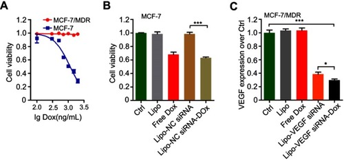
Physical interactions between doxorubicin (thereafter referred as Dox) and nucleic acids are well known, with Dox preferentially binding to the 5ʹ-GC-3ʹ or 3ʹ-GC-5ʹ sequences, owing to the presence of flat aromatic rings. Taking advantage of such properties, researchers sought to develop certain formulations by complexing plasmid/apatmer with Dox, and most of these complexes, when applied by delivery systems, had shown a marked inhibition of tumor growth.Citation14–Citation17 Inspired by these exciting studies, we hypothesized that Dox could also intercalate into double-stranded regions of siRNA to form a physical complex. In the current study, we fixed the siRNA concentration, added Dox at increasing concentrations but at a fixed volume, and monitored the fluorescence spectrum of Dox. Bare siRNA exhibited no fluorescence, but when the concentration of Dox increased from 1: 12.5 to 1: 25 (siRNA to Dox, M/M), the fluorescence signals remained nearly unchanged, suggesting that Dox intercalated with the double-stranded siRNA and hence quenched fluorescence (). Fluorescence signal started to increase again when the Dox concentration reached 1:50 or higher. The spectrum of free Dox was also included for comparison (). We chose the 1: 12.5 molar equivalence of siRNA to Dox for further experiments.
Next, whether the prepared siRNA-Dox complex formation could influence each component’s function remained to be determined. Here, we chose MCF-7, a common breast cancer cell line and its derived drug-resistant MCF-7/MDR cells for the following studies. First, we determined the cytotoxicity of Dox in MCF-7 and MCF-7/MDR cells. As a result, MCF-7/MDR cells were much more resistant to Dox than MCF-7 cells with IC50 at around 1000 ng/mL (A). Then, we conducted cytotoxicity assay in MCF-7 cells, showing that LipofectamineTM2000-NC siRNA-Dox (applied NC siRNA concentration at 125 nM with Dox concentration at 1000 ng/mL) had similar cytotoxicity as free Dox at equivalent concentration, and the control groups (control, Lipofecatamine and LipofectamineTM2000-NC siRNA) presented negligible toxicity (B). Meanwhile, in vitro RNAi efficacy experiment revealed that LipofectamineTM2000-VEGF siRNA-Dox (applied VEGF siRNA at 125 nM with Dox concentration at 1000 ng/mL) was as effective in silencing the target gene as LipofectamineTM2000-VEGF siRNA at equivalent siRNA dosage in MCF-7/MDR cells (instead of MCF-7 cells, in order to eliminate the toxic influence involved with the use of Dox). In the three other groups (control, Lipofectamine and Dox), no gene knockdown was observed (C). Collectively, these results suggested that Dox could intercalate into siRNA groove readily via noncovalent conjugation to form the physical complex and that the formation of such complex did not affect each component’s own function in cells.
Figure S1 Biological function tests of siRNA-Dox complex. (A) Cytotoxicity of MCF-7 or MCF-7/MDR cell lines in treatment of various Dox concentrations (rang from 100 to 2000 ng/mL). (B) Cytotoxicity of MCF-7 cells treated with various formulations. Lipo, Lipofectaminetm2000; Lipo-NC siRNA, NC siRNA loaded Lipofectaminetm2000; Lipo-NC siRNA-Dox, NC siRNA-Dox complex loaded LipofectamineTM2000. (C) VEGF expressions in MCF-7/MDR cells regulated by various formulations. Lipo, LipofectamineTM2000; Lipo-VEGF siRNA, VEGF siRNA loaded lipofectamineTM2000; Lipo-VEGF siRNA-Dox, VEGF siRNA-Dox complex loaded LipofectamineTM2000. Data are represented as the means ± S.D. (*p<0.01; ***p<0.0001; n=3).
Abbreviations: siRNA, small interfering RNA; Dox, doxorubicin; NC siRNA, negative control small interfering RNA; VEGF, vascular endothelial growth factor; VEGF siRNA, vascular endothelial growth factor small interfering RNA.
Preparation and characterization of siRNA-Dox-SiO2 and determination of dox release from the complex
The synthesized complex of siRNA-Dox provided a convenient tool for targeted and combination cancer therapy. Inspired by previous studies in which anionic surfactants such as DNA or RNA could be used as templates to synthesize anionic-surfactant-templated mesoporous silicas mediated by CSDA,Citation19–Citation21 we speculated that siRNA or siRNA-Dox complex could also be appropriate to serve as novel anionic surfactants for the synthesis of siRNA/siRNA-Dox-based silica nanoparticles, by the above-mentioned self-assembly method. To this end, we adopted a sequential assembly approach (): first the siRNA-Dox complex was produced via non-covalent physical interactions, electrostatic interactions between the complex and the positively charged amino site of APTES, and co-condensation with TEOS assembled subsequently to form the silica framework at an appropriate molar proportion (siRNA: Dox: APTES: TEOS=1: 12.5: 3: 3.75). Interestingly, the amount of encapsulated siRNA and Dox in this nanocarrier was determined by the reaction time. As shown in , loaded quantities of siRNA and Dox in the nanocarrier increased steadily over time: specifically at the 8-min timepoint, loaded siRNA and Dox reached about 70% and about 50% of the total capacity, respectively; over 15–30 mins, the loaded siRNA and Dox almost maintained at a steady level (~85% or higher). In parallel, the nanoparticle size increased as the siRNA and Dox-loaded volumes increased over reaction time (). Taken together, the loading capacities of siRNA and Dox and nanocarrier size could be readily controlled by reaction time. For the current study, we chose 8 mins as the reaction termination time point, producing nanoparticles that consistently contained siRNA (~75% of total) and Dox (~50% of total), with a uniform size of about 250 nm. We characterized the physical properties of these nanoparticles. TEM image of siRNA-Dox-SiO2 showed the nanoparticle size to be about 220 nm (). The presence of Dox in this nanocarrier was evidenced by the red precipitation compared with that before centrifugation (). DLS analysis revealed the nanocarrier’s diameter at 240±1.5 nm, along with zeta potential of the nanoparticle that was approximately −38 mV (), which was generally regarded suitable for cellular endocytosis via the strong electrostatic interactions between silanol groups of silica nanoparticles and trimethyl-ammonium head groups of the cellular membrane.Citation22 In comparison with several published siRNA/Dox co-delivery systems,Citation4,Citation13,Citation23 our method has the ability to synthesize co-delivery vectors conveniently in about 10 mins without prior preparation of inorganic materials and eliminate drug encapsulation, leading overall to improved drug loading and decreased preparation time.
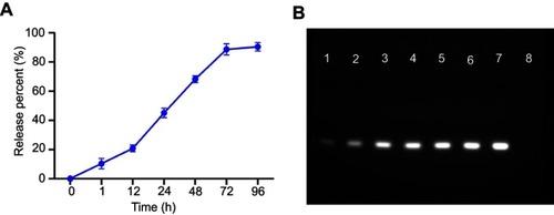
In vitro Dox release study was conducted at 37℃ in sterile PBS buffer at pH 4.5 and 7.4, simulating the acidic lysosomes/endosomes and the normal physiological environment, respectively. Results confirmed that Dox was slowly and steadily released (~8%) at pH 7.4, extending up to 96 hrs, whereas at pH 4.5, Dox was released more rapidly (>30%) within 96 hrs, suggesting Dox release from the nanoparticle was both time- and pH-dependent (-a and b). pH sensitivity of siRNA-Dox-SiO2 was an advantage for drug delivery. During circulation or in the extracellular space with a pH of approximately 7.4, Dox was only released from the nanocarrier at a basal level. Contrarily, when siRNA-Dox-SiO2 was internalized by tumor cells and entered endosomal/lysosomal compartments, Dox was quickly released from the particles to facilitate the killing of cancer cells. To test the stability of siRNA in siRNA-Dox-SiO2 nanocarrier, siRNA-release behavior in PBS was monitored. Results reflected that the amount of siRNA was slowly released from the nanocarrier and eventually reached the plateau at 72 hrs with about 85% of the total loaded siRNA (A), as corroborated by the agarose gel electrophoresis result (B) that revealed the highly effective retardation and protection of siRNA by the nanocarrier.
Figure S2 (A) siRNA release profiles of siRNA-Dox-SiO2 nanocarrier in PBS at different time points (1, 12, 24, 48, 72 and 96 hrs). (B) Agarose gel electrophoretic analysis to evaluate the siRNA release at different time points. Lane 1: 1 hr; Lane 2: 12 hrs; Lane 3: 24 hrs; Lane 4: 48 hrs; 5: 72 hrs; Lane 6: 96 hrs; 7: free siRNA (equivalent to the total loaded siRNA in siRNA-Dox-SiO2 nanocarriers); 8: DNA ladder (50 bp).
Abbreviations: siRNA, small interfering RNA; Dox, doxorubicin; SiO2, silica; PBS, phosphate-buffered saline; DNA, deoxyribonucleic acid.
Cytotoxicity and gene silencing efficacy of siRNA-Dox-SiO2
Although silica-based materials are generally regarded biocompatible and suitable for in vivo use,Citation24,Citation25 the cytotoxicity of siRNA-SiO2 delivery system (similarly prepared as siRNA-Dox-SiO2 but without Dox) remains to be tested prior to further studies. The cell viability of MCF-7 and MCF-7/MDR incubated with different concentrations of NC siRNA-SiO2 (applied siRNA concentration, from 0 to 600 nM) for 24 hrs were measured by the CCK-8 assay (). All concentrations exhibited negligible cytotoxicity even at 600 nM for both MCF-7 and MCF-7/MDR cell lines compared to that of nearly 100% cell death in treatment of 2% Triton X-100 that has widely been used as a positive control group,Citation26,Citation27 indicating that siRNA-SiO2-based nanocarrier presented great biocompatibility with cells, and possibly could be suitable for in vitro/in vivo biological applications.
Figure 2 The biological function tests of siRNA-Dox-SiO2 nanoparticles. (A) Cytotoxicity of NC siRNA-SiO2 in MCF-7 and MCF-7/MDR cell lines with various nanocarrier concentrations (applied siRNA concentrations: 0, 38, 75, 150, 300, 450, 600 nM). (B) Cytotoxicity of MCF-7 cells treated with various formulations. (C) VEGF expressions in MCF-7/MDR cells regulated by various formulations. Lipo, LipofectamineTM2000; Lipo-NC siRNA, NC siRNA loaded LipofectamineTM2000; Lipo-VEGF siRNA, VEGF siRNA loaded LipofectamineTM2000. Data are represented as the means ± S.D. (**p<0.001; n=3; ***p<0.0001; n=3). (D) Evaluation of endosomal escapability of FAM-NC siRNA-Dox-SiO2. Fluorescence images of MCF-7/MDR cells incubated with FAM-NC siRNA-Dox-SiO2 vectors for 4 hrs and 24 hrs using confocal laser scanning microscopy (CLSM).
Abbreviations: NC siRNA, negative small interfering RNA; Dox, doxorubicin; SiO2, silica.
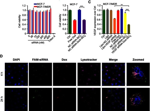
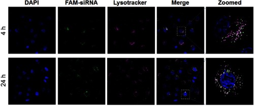
Following biocompatible analysis of siRNA-SiO2 nanoparticles, a similar study on siRNA-Dox complex that whether siRNA and Dox in the siRNA-Dox-SiO2 nanoparticle could interfere with their own functions should be confirmed. We again measured the cytotoxicity of NC-siRNA-Dox in MCF-7 cells and chose the VEGF gene and tested for target gene silencing efficacy on MCF-7/MDR cells (to eliminate the toxic influence from Dox). In MCF-7 cells, NC siRNA-Dox-SiO2 (applied 162.5 nM NC siRNA with Dox concentration at 1000 ng/mL) showed very similar cytotoxicity levels compared with an equivalent amount of free Dox, while the control groups presented undetectable toxicity (). In parallel, VEGF siRNA-Dox-SiO2 (applied 162.5 nM VEGF siRNA with Dox concentration at 1000 ng/mL) caused significantly better VEGF silencing than LipofectamineTM2000-VEGF siRNA did at an equivalent siRNA dosage (). Five other groups (Control, LipofectamineTM2000, LipofectamineTM2000-NC siRNA, Dox, NC siRNA-SiO2) showed no VEGF knockdown in MCF-7/MDR cells, as shown in . Interestingly, VEGF siRNA-Dox-SiO2 showed better VEGF knockdown, plummeting to about 30% of the control, than VEGF siRNA-SiO2 did in which VEGF level only dropped to about 85% of the control group (). This improved gene silencing capacity of VEGF siRNA-Dox-SiO2 in comparison to VEGF siRNA-SiO2 plausibly resulted from Dox intercalation that not only stabilized siRNA, but also mediated timely escape of siRNA from endosomes/lysosomes.Citation26,Citation28,Citation29 Initial evidence of this enhanced siRNA escape was provided by confocal microscopy visualizing the cellular localization FAM-labeled siRNA from the two types of nanoparticles siRNA-Dox-SiO2 () and siRNA-SiO2 (), in which the most FAM-labeled siRNA carried by siRNA-SiO2 nanoparticles were trapped in endosomes/lysosomes after transfection (), whereas the siRNA encapsulated by siRNA-Dox-SiO2 nanoparticles were nearly completely separated from endosomes/lysosomes (). Taken together, these experiments demonstrated, in principle, siRNA-Dox-SiO2 as a novel co-delivery system to simultaneously deliver Dox and siRNA into cancer cells. Such a system could produce effective target gene silencing and induce marked cytotoxicity, suggesting its potential use for combination cancer therapy, especially when the target gene was related to MDR.
Figure S3 Evaluation of endosomal escapability of FAM-NC siRNA-SiO2. Fluorescence images of MCF-7/MDR cells incubated with FAM-NC siRNA-SiO2 vectors for 4 hrs and 24 hrs using confocal laser scanning microscopy (CLSM).
Abbreviations: NC siRNA, negative small interfering RNA; Dox, doxorubicin; SiO2, silica.
Cytotoxicity induced by MDR-1 siRNA-Dox-SiO2 in MCF-7/MDR cells
To demonstrate that our co-delivery could target MDR, we chose the target gene of MDR-1. MDR to chemotherapeutic agents, such as Dox, anthracyclines and taxanes, was commonly associated with cancer cell overexpression of drug transporter proteins such as P-gp, which is encoded by the ABCB2 gene.Citation30,Citation31 MCF-7 expressed negligible P-gp while MCF-7/MDR expressed it at a higher level, as confirmed by flow cytometry assay (A). By contrast, higher P-gp expression in MCF-7/MDR cells was highly associated with reduced Dox retention in cytoplasm compared with that tested in MCF-7 cells (B). We then tested four MDR-1 siRNAs to target human MDR-1 gene. Of the four siRNAs, MDR-1 siRNA-3 (L-S3) potently suppressed the expression of P-gp proteins in MCF-7/MDR cells as compared with the other siRNAs, after transfection with LipofectamineTM2000 (-a). Based on these results, we selected MDR-1 siRNA-3 for the following experiments (thereafter, MDR-1 siRNA-3 denoted as MDR-1 siRNA). Next, we combined MDR-1 siRNA with Dox to be assembled subsequently to form MDR-1 siRNA-Dox-SiO2 after addition of APTES and TEOS. MDR-1 siRNA-Dox-SiO2 exhibited highly effective inhibition of P-gp expression compared to MDR-1 siRNA-SiO2 (-b). This high gene-silencing efficacy of MDR-1 siRNA-Dox-SiO2 was considered as a result of Dox intercalation, also corroborated by the findings in .
Figure 3 The cytotoxicity of MDR-1 siRNA-Dox-SiO2 nanoparticles against MCF-7/MDR cells. (A) (a) P-gp expression determined through Western blot in treatment of various MDR-1 siRNAs transfected by LipofectamianeTM2000 in MCF-7/MDR cells. WT, without treatment; L-NC, LipofectamianeTM2000-NC siRNA; L-S1, LipofectamianeTM2000-MDR-1 siRNA-1; L-S2, LipofectamianeTM 2000-MDR-1 siRNA-2; L-S3, LipofectamianeTM2000-MDR-1 siRNA-3; L-S4, LipofectamianeTM2000-MDR-1 siRNA-4. (b) P-gp expression measured by Western blot in treatment of various silica-based siRNA formulations in MCF-7/MDR cells. WT, without treatment; 1, NC siRNA-Dox-SiO2; 2, MDR-1 siRNA-Dox-SiO2; 3, MDR-1 siRNA-SiO2. (B) Cytotoxicity of MDR-1 siRNA-Dox-SiO2 (MDR siRNA refer to MDR siRNA-3) containing different Dox concentrations determined by CCK-8 assay in MCF-7/MDR cells. MDR-1 siRNA-Dox-SiO2-1, Dox concentration at 500 ng/mL; MDR-1 siRNA-Dox-SiO2-2, Dox concentration at 1000 ng/mL; MDR-1 siRNA-Dox-SiO2-3, Dox concentration at 2000 ng/mL. Free Dox group with concentration of 2000 ng/mL. All of groups contained the same siRNA value. *p<0.01; **p<0.001;***p<0.0001, n=5. (C) The cellular Dox retention observed by CLSM (confocal laser scanning microscopy) 60 hrs after treating with Dox (a), NC siRNA-Dox-SiO2 (b) and MDR-1 siRNA-Dox-SiO2 (c). (d) The cellular Dox retention determined by FCM (flow cytometry) 60 hrs after treating with Dox (green line), NC siRNA-Dox-SiO2 (blue line) and MDR-1 siRNA-Dox-SiO2 (red line); the black line denoted as the control group (without any nanocarrier and Dox). Bar=50 μm.
Abbreviations: NC siRNA, negative small interfering RNA; Dox, doxorubicin; SiO2, silica.
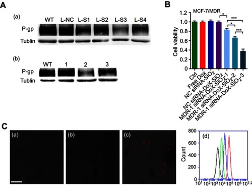
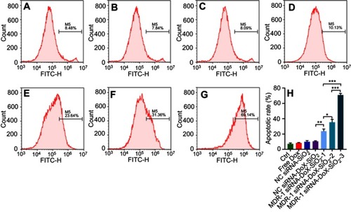
Similarly, synergism of MDR-1 siRNA and Dox via the siRNA-Dox-SiO2 nanocarrier was also evident in cytotoxicity assays, where the combination treatment caused a significant inhibition of cell viability compared to each treatment alone or nanocarrier containing NC siRNA in MCF-7/MDR cells, as shown in . Meanwhile, we prepared various MDR-1 siRNA-Dox-SiO2 formulations that included fixed siRNA concentration (162.5 nM) with Dox concentrations at 500, 1000 and 2000 ng/mL, respectively. Cytotoxicity assay in MCF-7/MDR cells 60 hrs after incubation with these formulations showed that highest cell inhibition was observed at the Dox concentration of 2000 ng/mL (***p<0.0001), followed by 1000 and 500 ng/mL, at a concentration-dependent manner (). In contrast, free doxorubicin at 2000 ng/mL posed negligible toxicity to MCF-7/MDR cells ( and A). In parallel, quantitation of the apoptotic rate of different formulations against MCF-7/MDR cells were measured by flow cytometry using Annexin V staining method (). Consequently, co-occurrence of Dox and MDR-1 siRNA via nanocarrier in MCF-7/MDR cells exhibited the highest cell apoptosis at the Dox concentration of 2000 ng/mL, followed by 1000 and 500 ng/mL. These results illustrated that knockdown of MDR-1 re-sensitized the multidrug-resistant MCF-7/MDR cells. To better determine the intracellular Dox concentration, flow cytometry and confocal microscopy were performed after treatment with MDR-1 siRNA-Dox-SiO2, NC siRNA-Dox-SiO2 and free doxorubicin. Abundant red signals distributed in the cytoplasm and the nucleus were detected in the MDR-1 siRNA-Dox-SiO2 group, as compared with weaker red spots in NC siRNA-Dox-SiO2 and free doxorubicin groups (-a–c). In parallel, total red fluorescence among various groups was determined by flow cytometry, which indicated that the intracellular Dox content after treatment with MDR-1 siRNA-Dox-SiO2 in MCF-7/MDR cells was approximate 9 times higher than free Dox group (-d). In conclusion, we showed the synergistic effect of MDR-1 siRNA/Dox combination delivered by self-assembly inorganic silica nanocarrier in overcoming drug resistance in MCF-7/MDR cells. This nanocarrier could suppress the P-gp levels and promote the Dox retention in MCF-7/MDR cells, overall leading to enhanced cell inhibition. Nevertheless, these results demonstrated in principle the usefulness of the nanocarrier to inhibit cancer cell growth in vitro alone. Future in vivo investigations are warranted.
Figure S4 (A) P-gp expression determined through flow cytometry in MCF-7 and MCF-7/MDR cell lines (a). The corresponding statistics analysis (b). (B) Intercellular Dox retention measured by flow cytometry in MCF-7 and MCF-7/MDR cell lines (a). The corresponding statistics analysis (b). (*p<0.05; n=3).
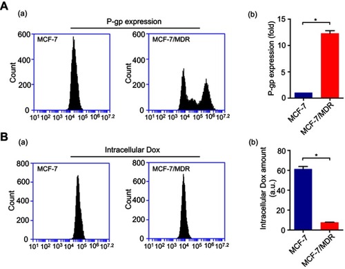
Figure S5 Evaluation of apoptosis of MDR-1 siRNA-Dox-SiO2 containing different Dox concentrations determined by flow cytometry using annexin V method in MCF-7/MDR cells. (A) control, without any treatment; (B) Free dox, dox concentration of 2000 ng/mL; (C) NS siRNA-SiO2; (D) NS siRNA-Dox-SiO2, Dox concentration of 2000 ng/mL; (E) MDR-1 siRNA-Dox-SiO2-1, Dox concentration at 500 ng/mL; (F) MDR-1 siRNA-Dox-SiO2-2, dox concentration at 1000 ng/mL; (G) MDR-1 siRNA-Dox-SiO2-3, Dox concentration at 2000 ng/mL; (H) Quantitation of apoptotic rate of MCF-7/MDR cells after treatment with different groups. All of groups contained the same siRNA value. M5% represents apoptotic cell population%. *p<0.01; **p<0.001; ***p<0.0001, n=3.
Abbreviations: NC siRNA, negative small interfering RNA; Dox, doxorubicin; SiO2, silica.
Conclusion
In this study, we developed a novel and readily prepared co-delivery system of siRNA and doxorubicin via self-assembly of inorganic silica nanoparticles. Physical interactions between doxorubicin and siRNAs facilitated a fast and reversible formation of siRNA-Dox complex. This complex then could be used as templates for synthesizing silica-based siRNA and Dox co-delivery system by introducing APTES (a kind of co-structure-directing agent, CSDA) and TEOS (a type of inorganic silicon) successively. siRNA and doxorubicin were encapsulated into this nanocarrier with high loading capacities and their own biological functions were not compomised. Consequently, transfection of MDR-1 siRNA-Dox-SiO2 into MCF-7/MDR cells could improve their sensitivity to Dox, overall leading to enhanced chemotherapeutic cytotoxicity.
Acknowledgments
This study is supported by grants from National Natural Science Foundation of China [No. 81701828], Natural Science Foundation of Zhejiang Province [No. LY16H180008], Wenzhou Science and Technology Bureau Foundation [No. Y20150082].
Disclosure
The authors report no conflicts of interest in this work.
Supplementary materials
Supplementary Methods
siRNA releasing from the nanocarrier in vitro
To investigate siRNA release, siRNA-Dox-SiO2 was incubated in PBS buffer for 96 hrs at 37 ℃. At different time points, samples were subjected to a centrifugation speed of 13,200 rpm.25 μL of the supernatant was taken out from the total volume (500 μL) to test the amount of released siRNA by Nanodrop 1000. 25 μL fresh buffer was then replenished to maintain a fixed volume (500 μL).
Apoptosis induced by MDR-1 siRNA-Dox-SiO2 in vitro
The MCF-7/MDR cells were seeded in 6-well plates to about 80% confluence, and each well was treated with MDR-1 siRNA-Dox-SiO2 (containing 162.5 nM MDR-1 siRNA) that encapsulated Dox at various concentrations (500, 1000, 2000 ng/mL, respectively), Dox (2000 ng/mL) group, NC siRNA-SiO2 (containing 162.5 nM NC siRNA) and NC siRNA-Dox-SiO2 (containing 162.5 nM NC siRNA with Dox concentration at 2000 ng/mL). After incubation for 24 hrs, the wells were replaced with fresh culture medium and incubated for another 36 hrs. After that, the adhesive cells were digested to suspension cells and then treated with FITC annexin V reagent following the manufacturer’s instructions. Finally, the cells were analyzed by flow cytometry. No treatment worked as control group. All experiments were performed in hexaplicate.