Abstract
Background and purpose:
Surgery is regarded as the gold standard for patients with advanced ovarian cancer. However, complete surgical removal of tumors remains extremely challenging; fewer than 40% of patients are cured. Here, we developed a new modality of theranostics for ovarian cancer based on a near-infrared light-triggered nanoparticle.
Methods:
Nanoparticles loading IR780 iodide on base of folate modified liposomes were prepared and used for theranostics of ovarian cancer. Tumor targeting of FA-IR780-NP was evaluated in vitro and in an ovarian xenograft tumor model. A fluorescence stereomicroscope was applied to evaluate the tumor recognition of FA-IR780-NP during surgery. FA-IR780-NP mediated photothermal therapy effect was compared with other treatments in vivo.
Results:
FA-IR780-NP was demonstrated to specifically accumulate in tumors. IR780 iodide selectively accumulated in tumors; the enhanced permeability and retention effect of the nanoparticles and the active targeting of folate contributed to the excellent tumor targeting of FA-IR780-NP. With the aid of tumor targeting, FA-IR780-NP could be used as an indicator for the real-time delineation of tumor margins during surgery. Furthermore, photothermal therapy mediated by FA-IR780-NP effectively eradicated ovarian cancer tumors compared with other groups.
Conclusion:
In this study, we present a potential, effective approach for ovarian cancer treatment through near-infrared fluorescence image-guided resection and photothermal therapy to eliminate malignant tissue.
Acknowledgments
This work was financially supported by the National Natural Science Foundation of China (Grant Nos. 31630026, 81630047, 81771847, 81771845, and 81601513).
Disclosure
The authors report no conflicts of interest in this work.
Supplementary material
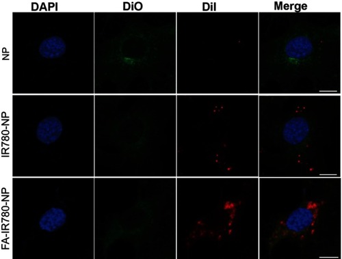
Figure S1 Fluorescence images of a single SKOV3 cell incubated with NPs (NP, IR780-NP, FA-IR780-NP). Blue (DAPI) represents cell nuclei; green (Dio) reveals the cytomembrane; whereas the red (DiI) dots imply DiI-labeled NPs. The scale bars represent 5 μm.