

Abstract
Aim
To determine whether the use of a mixed polymeric micelle delivery system based on vitamin E succinate (VES)-grafted-chitosan oligosaccharide (CSO)/VES-grafted-chitosan (CS) mixed micelles (VES-g-CSO/VES-g-CS MM) enhances the delivery of C-DMSA, a theranostic fluorescent probe, for Hg2+ detection and detoxification in vitro and in vivo.
Methods
Mixed micelles self-assembled from two polymers, VES-g-CSO and VES-g-CS, were used to load C-DMSA and afforded C-DMSA@VES-g-CSO/VES-g-CS MM for cell and in vivo applications. Fluorescence microscopy was used to assess C-DMSA cellular uptake and Hg2+ detection in L929 cells. C-DMSA@VES-g-CSO/VES-g-CS MM was then administered intravenously. Hg2+ detection was assessed by fluorescence microscopy in terms of bio-distribution while detoxification efficacy in Hg2+-poisoned rat models was evaluated in terms of mercury contents in blood and in liver.
Results
The C-DMSA loaded mixed micelles, C-DMSA@VES-g-CSO/VES-g-CS MM, significantly enhanced cellular uptake and detoxification efficacy of C-DMSA in Hg2+ pretreated human L929 cells. Evidence from the reduction of liver coefficient, mercury contents in liver and blood, alanine transaminase and aspartate transaminase activities in Hg2+ poisoned SD rats treated with the mixed micelles strongly supported that the micelles were effective for Hg2+ detoxification in vivo. Furthermore, ex vivo fluorescence imaging experiments also supported enhanced Hg2+ detection in rat liver.
Conclusion
The mixed polymeric micelle delivery system could significantly enhance cell uptake and efficacy of a theranostic probe for Hg2+ detection and detoxification treatment in vitro and in vivo. Moreover, this nanoparticle drug delivery system could achieve targeted detection and detoxification in liver.
Supplementary materials
Determination of critical micelle concentrations
Critical micelle concentrations (CMC) of the VES-g-CSO, VES-g-CS, VES-g-CSO/VES-g-CS (w/w=8:2) were determined on a Lumina fluorescence spectrometer (Thermo Fisher Scientific, Waltham, MA, USA). Solutions containing of 4.94×10⁻⁷ mol/L pyrene and increasing concentrations (1.0×10⁻⁴ −1.0 mg/mL) of the polymer to be tested were prepared. Each solution sample was sonicated for 5 mins at 100 W with the pulse turned off for 1 s at intervals of 1 s, incubated at room temperature for 12 hrs in light-resistance containers before its fluorescence emission spectrum was recorded at excitation wavelength 335 nm. CMC values were determined from plots of the intensity ratio of I1 (373 nm)/I3 (385 nm) against logarithm of polymer concentrations ().
Cytotoxicity study
In vitro cytotoxicity of C-DMSA@VES-g-CSO/VES-g-CS MM was studied on L929 cells by the MTT assay. Cells were seeded at a density of 1×10⁴ cells/well in a 96-well plate. After 24-hr incubation, the growth medium was replaced with 200 μL of medium containing different concentrations of free C-DMSA (1–100 μg/mL), VES-g-CSO/VES-g-CS MM (0.01–1 mg/mL) or C-DMSA@VES-g-CSO/VES-g-CS MM (containing 1–100 μg/mL C-DMSA). After incubation for additional 24 hrs in the dark, the drug-containing medium was replaced with PBS and the samples were incubated in a humidified incubator at 37°C with 5% CO2. PBS was then replaced with fresh medium and MTT solution (500 μg/mL, 200 μL/well) was added, after that the cells were cultured again for 4 hrs. The supernatant was removed, dimethyl-sulfoxide (200 μL/well) was added and the samples were shaken for 10 mins. The absorbance of each well was measured at 570 nm by a microplate reader (Tecan Safire2, Männedorf, Switzerland) ().
Pharmacokinetic profiles
Six male SD rats weighing 170–190 g were randomly divided into two groups for pharmacokinetics studies. Before administration, the rats were fasted for 12 hrs with access to drinking water. Free C-DMSA and C-DMSA@VES-g-CSO/VES-g-CS MM were intravenously injected at an equivalent dose of 10 mg/kg. Blood (0.2 mL) was taken from the orbital venous plexus prior to administration of test substances (0 hr) and after 0.1, 0.25, 0.5, 1, 2, 4, 7, 10 and 24 hrs (n=3 for each time point). The concentration of C-DMSA in the blood samples was determined by a fluorescence spectrophotometer (λEx 403 nm and λEm 480 nm). The related pharmacokinetic parameters were calculated using Kinetic 5.0 software.
The Phase I half-life (t½α) for C-DMSA and C-DMSA@VES-g-CSO/VES-g-CS MM was calculated at 0.12±0.01 and 0.74±0.12 hrs, respectively, Phase II values (t½β) were 1.91±0.20 and 15.23±1.20 hrs, respectively. Furthermore, the AUC0–24 hrs for the drug-loaded micelles was 4.57 folds increased compared to free C-DMSA (). The results demonstrated that the entrapment of C-DMSA in nano drug delivery systems can prolong its circulation.
Figure S1 1H-NMR CS (A) and VES (B).
Abbreviations: VES, vitamin E succinate; CS, chitosan.
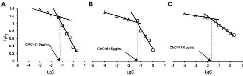
Figure S2 Determination of CMC values of VES-g-CSO/VES-g-CS (w/w=4:1) (A), VES-g-CSO (B) and VES-g-CS (C) solutions.
Abbreviations: CMC, critical micelle concentration; VES-g-CSO, vitamin E succinate-grafted-chitosan oligosaccharide; VES-g-CS, vitamin E succinate-grafted-chitosan; VES-g-CSO/VES-g-CS, vitamin E succinate-grafted-chitosan oligosaccharide/vitamin E succinate-grafted-chitosan.
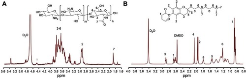
Figure S3 Cell viability of L929 cells treated with the blank mixed micelles (VES-g-CSO/VES-g-CS MM) or C-DMSA@VES-g-CSO/VES-g-CS MM (A). Cell viability of L929 cells treated with C-DMSA or C-DMSA@VES-g-CSO/VES-g-CS MM (B).
Note: Data were presented as mean±SD (n=3).
Abbreviations: C-DMSA, 3-formyl-7-diethylamino coumarin masked meso-dimercaptosuccinic acid; VES-g-CSO/VES-g-CS MM, vitamin E succinate-grafted-chitosan oligosaccharide/vitamin E succinate-grafted-chitosan mixed micelles; C-DMSA@VES-g-CSO/VES-g-CS MM, C-DMSA loaded vitamin E succinate-grafted-chitosan oligosaccharide/vitamin E succinate-grafted-chitosan mixed micelles.
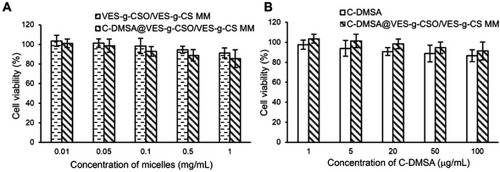
Figure S4 Mean plasma concentration-time curves of C-DMSA after intravenous administration of C-DMSA and C-DMSA@VES-g-CSO/VES-g-CS MM in rats.
Notes: All the rats were received the single dosage at an equivalent dose of 10 mg/kg C-DMSA. Data were presented as mean±SD (n=3).
Abbreviations: C-DMSA, 3-formyl-7-diethylamino coumarin masked meso-dimercaptosuccinic acid; C-DMSA@VES-g-CSO/VES-g-CS MM, C-DMSA loaded vitamin E succinate-grafted-chitosan oligosaccharide/vitamin E succinate-grafted-chitosan mixed micelles.
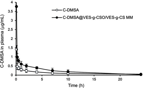
Table 1 Physicochemical properties of C-DMSA loaded micelles
Table 2 Liver coefficients and contents of total mercury in rat liver and blood
Table S1 Elemental analysis of VES-g-CSO and VES-g-CS
Table S2 Pharmacokinetic parameters of C-DMSA after a single dosage intravenous administration to rats
Acknowledgment
This work was supported by the National Natural Science Foundation of China (grant no. 21577037, K. L.) , Shanghai Municipal Natural Science Foundation (contract no. 17ZR1406600), Science and Technology Commission of Shanghai Municipality (contract no. 10DZ2220500) and the Shanghai Committee of Science and Technology (grant no. 11DZ2260600).
Disclosure
The authors report no conflicts of interest in this work.