

Abstract
Background
Diabetic retinopathy (DR) is a complication of diabetes that affects the eyes and vision. It is a leading cause of visual impairment and blindness in working-age people. Vascular endothelial growth factor-A (VEGF-A) is a primary initiator and potential mediator of DR. Matrix metalloproteinase-9 (MMP-9) plays a progressive role in the onset and severity of DR. Interleukin-12 (IL-12) is a cytokine of the chemokine family that could reduce the levels of MMP-9 and VEGF-A and suppress tumor angiogenesis. We hypothesize that IL-12 may also have superior therapeutic efficacy against DR. However, protein drugs are prone to degradation by various proteases after drug injection. Therefore, they have short half-lives and low blood concentrations. The objective of this study was to develop IL-12-loaded nanoparticles for long-term and sustained DR treatment.
Methods
IL-12-loaded poly(lactic-co-glycolic acid) (PLGA) nanoparticles (IL-12-PNP) were developed by double emulsion. The characteristics, anti-DR activity, and mechanisms of IL-12-PNP were examined in vitro and in vivo.
Results
The nanoparticles had suitable particle size (~132.8 nm), drug encapsulation efficiency (~34.7%), and sustained drug release profile. Compared with IL-12 and blank nanoparticles, IL-12-PNP showed better inhibitory efficacy against VEGF-A and MMP-9 expression in rat endothelial cells and DR mouse retina. Intraocular IL-12-PNP administration significantly reduced retinal damage in DR mice as they presented with increased thickness and decreased neovascularization after treatment.
Conclusion
These data indicate that IL-12-PNP is an effective drug delivery platform for DR therapy. It restores the thickness and reduces neovascularization of the retinas of DR mice.
Supplementary material
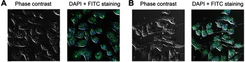
Figure S1 The expression of vWf and CD31 on endothelial cells was confirmed by the immunofluorescence assay. The cells were treated with the first antibodies (anti-rat vWf and anti-rat CD31 antibodies), and treated with FTIC-labeled anti-rabbit secondary antibodies. 4’,6-diamidino-2-phenylindole (DAPI) was used to stain the nuclei. (A) vWf staining. (B) CD31 staining. Bars represent 10 μm
Acknowledgment
This work was supported by the National Natural Science Foundation of China (Project No. 81802714).
Disclosure
The authors report no conflicts of interest in this work.