
Abstract
The clinical development of cell therapies is revealing that extracellular vesicles (EVs) may become very instrumental as subcellular therapeutic adjuncts in human medicine. EVs are released by various types of cells, grown in culture, such as mesenchymal stromal cells, or obtained from patients or allogeneic donors. Some EV populations (especially species of exosomes and shed microvesicles) exhibit inherent roles in cell-cell communication, thanks to their ca. 30~1000-nm nanosize and the physiological expression of cell-specific markers on their lipid bilayer membranes. Biomedical engineers are now attempting to exploit this cellular crosstalk capacity to use EVs as smart drug delivery systems that display substantial benefits in targeting, safety, and pharmacokinetics compared to synthetic nanocarriers. In parallel, the development of a set of nano-instrumentation, biochemical tools, and preclinical assays needed for optimal characterization of both naïve and drug-loaded EVs is ongoing. Although many hurdles remain, owing to the complexity of EV populations, translation of this “subcellular therapy” platform into reality is at hand and may soon change the landscape of the therapeutic arsenal in place to treat human degenerative and metabolic pathologies as well as diseases like cancer. This article provides objective opinions, balanced between unrealistic hopes of the capacity of EVs to resolve multiple clinical issues and concrete hurdles that have to be overcome to ensure that EVs are not lost in the translation phase, so that EVs can fulfill their promise by becoming a reliable therapeutic modality.
Plain Language Summary
The clinical therapeutic development of cell-based therapy is revealing that extracellular vesicles (EVs) released by various types of cells may become very instrumental in human medicine for various indications. There are extensive studies attempting to use EVs (in particular, species of exosomes and shed microvesicles) as smart drug delivery systems. Although there are several challenges, the wide clinical applicability of EVs is at hand and may soon change the landscape of the therapeutic arsenal in place to treat human diseases like cancer and neurological disorders. This article provides objective opinions, balanced between unrealistic hopes of the capacity of EVs to resolve multiple clinical issues and concrete hurdles that have to be overcome to ensure that EVs are clinically available as a reliable therapeutic modality. The future of EVs as therapeutic medicine holds promise but may comprise complex delivery systems, which need extensive development and optimization approaches and collaboration among academicians, biomedical engineers, clinicians, industry, scientists, and regulatory authorities.
Introduction
Extracellular vesicles (EVs) are subcellular nanostructures, surrounded by a lipid bilayer, likely present in essentially all body fluids, and increasingly recognized as important intercellular vectors of biochemical information.Citation1 As such, EVs were gradually unveiled as playing important, so far largely ignored, physiological roles in balancing health and the initiation and evolution of pathologic conditions.Citation2 As their functional importance is being recognized, substantial and rather justified expectations are growing among the scientific and medical communities, as well as the biotechnology and cell therapy industries, that EVs can be turned into therapeutic tools of interest in human medicine.Citation3 EVs, either in their native state or loaded with therapeutic agents to serve as drug delivery vehicles, are increasingly regarded as being worthy of interest for treating various pathological conditions, such as immune disorders, cancer, inflammatory disorders, and degenerative diseases.Citation3,Citation4 EVs can be used as cargo for a variety of functional endogenous and exogenous therapeutic compounds including small molecules, anti-cancer agents, anti-inflammatory compounds, miRNA, mRNA, proteins, and growth factors, etc. Further information on technical achievements and pre-clinical studies performed with EVs as drug delivery system (DDS) of various chemical and biological entities can be found in recent reviews.Citation5–Citation7
Owing to their potential therapeutic benefits, there is also now a growing focus in developing scalable production and isolation technologies of EVs from various cell types, with the intent to evaluate their functional activity in in vitro and animal models, as well as now through clinical trials (ClinicalTrials.gov: NCT02565264).Citation3,Citation8 Technologies are thus currently being developed to optimize (a) the generation of EVs from various cell types, (b) drug-loading methods and quantification approaches, (c) the design of cost-effective and scalable isolation processes, and (d) methods to ensure the degree of quality, safety, and consistency mandatory for clinical applications.
Previous studies of EVs mostly focused on using cells grown in culture. Not surprisingly, specific pragmatic interest for industrial-scale applications exists in the evaluation of EVs generated by various types of mesenchymal stromal cells (MSCs), since clinical-grade ex vivo culture conditions for their expansion are well-established.Citation9,Citation10 One can also expect growing interest in EVs from various differentiated cells, such as T cells, dendritic cells, chondrocytes, neural cells, cancer cells, and macrophages among others, which have currently been expanded in vitro to be used for clinical treatments. Use of EVs generated using procedures of cell expansion that meet clinical requirements can alleviate some of their development challenges and accelerate translational applications. For the same reasons, there is growing interest by blood-transfusion communities in evaluating possible clinical values, especially in the field of regenerative medicine, of platelet-derived EVs that are rich in growth factors and micro (mi)RNAs.Citation11,Citation12 Therapeutic blood cells indeed also constitute an almost ideal “source material” for the production and isolation of clinical-grade EVs, since the collection infrastructure is well established and licensed by competent authorities,Citation13 and producing EVs from red blood cell (RBC) concentrates and platelet concentrates requires no ex vivo expansion.Citation3
Multiple types of EV preparations can be translated into clinical products, and depending upon their cellular origin, membrane decoration, and content, EVs from different cells likely exhibit distinct functional and targeting capacities and as DDSs. As hope in the capacity of EVs to be developed as new and moldable therapeutic platforms is growing among the biotechnology and cell-therapy industries, concrete hurdles need to be overcome for consistent and homogeneous EV production for their safe and reliable clinical use. This expert opinion article intends to provide realistic and objective views, balanced between unrealistic hopes in the capacity of EVs to resolve multiple clinical issues and concrete hurdles that have to be overcome in a pragmatic manner to ensure that EVs are not lost in the translation phase, and can deliver on to their promise by becoming proven and reliable therapeutic modalities.
Defining EVs
It is commonly thought that EVs comprise three subcellular populations: exosomes, apoptotic bodies, and microvesicles (MVs).Citation1 Exosomes are typically characterized by (a) their relatively small dimension of ca. 30~150 nm and (b) their intracellular origin as multivesicular bodies, preexisting within the cellular endosomal system and being expelled from cells. Apoptotic bodies with a size ranging from 100 nm to 5 μm are formed as a result of cytoskeletal breakdown and subsequent cell fragmentation that occurs as an outcome of apoptosis. The third interesting and heterogeneous group is known as MVs, ectosomes, or microparticles, with a size of ca. 50 nm to 1 μm. These are generated by budding from cellular membranes that can be triggered in vivo or in vitro by physiological agonists as well as cellular, biochemical, and physical factors.Citation3 It is increasingly realized that the classification of EVs into these three categories is oversimplified and will gain in complexity over time as new studies identify multiple subsets among these, both in vivo, depending upon physiological conditions, and in vitro, contingent on culture conditions.Citation5,Citation14
Development Of EV Products: Process Qualification, Validation, And Consistency Are Key Parameters
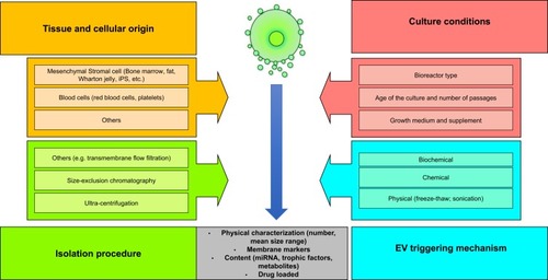
As with any complex biologicals that are very challenging to fully characterize only by analysis of the final product, it will be crucial in the case of EVs to ensure an optimal consistency of the various steps in their production process. We have assembled in some of the major production variables already known to influence EV properties. Those variables encompass (a) the cell type (such as the tissue source of MSCs); (b) the cell collection process and/or ex vivo processing or expansion methods including culture conditions (growth medium and supplement used, type of bioreactor, number of passages performed, etc.); (c) the mechanism to trigger EV release; and (d) the EV isolation and storage methods. Variations in these parameters can influence the EV population size, generation (yield/cell), membrane markers, EV contents including miRNAs, and ultimately the purity and safety profile of EVs.Citation15 Such a situation implies that these factors should be considered from the very early phases (lab-scale exploration) to the development process of EV-based therapeutic products. This also implies that EV manufacturing and scale-up processes should be robust, and thoroughly qualified and validated to identify their impacts on EV consistency, quality, and safety profiles. Key process parameters and their influence on EV products should be checked and monitored during routine production so that the end-product batches have characteristics that remain within pre-established specification criteria for release. Having well-defined process parameters is also important for tracing any deviation in the specifications of intermediates and final products. Specifications should be tailor-made to the cellular source, development parameters, and intended clinical use of EVs (targeted pathology, posology, drug loading parameters, storage conditions, and modes of in vivo administration).
Figure 1 Production variables known to influence extracellular vesicle (EV) properties such as the cell type, cellular origin, cell culture conditions, mechanisms used to trigger EV release, isolation procedure of EVs, and storage conditions.
EV Drug-Loading Approaches
Endogenous or exogenous approaches can be used to load therapeutic molecules into EVs.Citation3 The endogenous method first loads therapeutics into parent cells, followed by the generation and release of loaded EVs.Citation16 In the exogenous approach, therapeutics or imaging agents are incorporated into isolated EVs through an incubation process.Citation17,Citation18 Additionally, there are several other methods used for loading EVs, such as sonication, extrusion, and freeze-thaw cycles.Citation19–Citation21 All of the above approaches depend on various factors which should be taken into consideration to avoid any encapsulation method-based detrimental effects on the EV size, integrity, intrinsic contents, biological properties, or immunogenicity.Citation22,Citation23
Scalable EV Isolation Modes And Characterization Methods
Isolation methods and equipment processes considered for commercial-scale downstream isolation of EVs from conditioned medium or cell suspensions are most likely going to conservatively follow, at least for the time being, methodologies already well-established in the biotech industry for producing other therapeutic biological products.Citation24 The production sequence of EVs typically combines low-speed centrifugation to pelletize and remove cells/cell debris suspended in the starting raw material, followed by sequential centrifugation or filtration for clarification.Citation25 While earlier developed laboratory-scale processes typically used ultracentrifugation at 20,000~100,000 ×g for several hours to pelletize EVs, now concentration by tangential flow-filtration (TFF) followed by size-exclusion chromatography (SEC) is becoming the preferred approach to better preserve the functional activity of EVs.Citation26,Citation27 However, one drawback of SEC appears to be the lack of a commercial chromatographic resin best suited to separate the smallest EVs (eg, 20~70 nm) or subpopulation of EVs from high-molecular-weight proteins or lipoproteins originating from cells, biological fluids, or growth medium supplements.Citation28 This understandably has detrimental impacts on the recovery of the entire population of EVs. In addition, as SEC contributes to diluting the EVs by ca. 2~3-fold, a concentration step by TFF may be performed prior to formulation, aseptic filtration, and filling into the final storage containers.Citation15,Citation28 Furthermore, an isolation process based on SEC and TFF may be regarded as time-consuming on a commercial scale.Citation29–Citation32
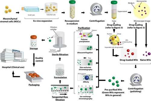
In general, the industrial development of EVs requires high-efficiency, scalable, and translatable isolation techniques compliant with good manufacturing practices (GMPs) and should be selected considering specifications of the biological raw materials and desired attributes of EVs. Detailed descriptions of various isolation techniques are reviewed elsewhere, including their advantages, limitations, and a discussion on involvement of several parameters,Citation3,Citation29,Citation30,Citation32 and a general scheme of EV production is shown in (reproduced with permission of Agrahari et alCitation3). Characterization of EVs is based on their origins and types. The current characterization, especially quantification methods, does not necessarily distinguish EVs on the basis of their origin or differentiate them from other similar types of vesicles/particles present in the same sample. Recently, various methods to determine the characterization parameters of EVs such as the size distribution, chemical composition, intrinsic content, and surface markers were reviewed and discussed.Citation3,Citation29,Citation33–Citation35
Figure 2 Illustration of manufacturing approaches considered for the production of extracellular vesicle (EV)-based therapeutics under good manufacturing practice-compliant, and scalable downstream processing methods to ensure the quality, safety, and consistency. Reprinted from of Trends Biotechnol. 37. Agrahari V, Agrahari V, Burnouf PA, Chew CH, Burnouf T. Extracellular microvesicles as new industrial therapeutic frontiers. 707–729. Copyright (2019).Citation3 with permission from Elsevier.
As the biotech and cell-therapy industries are increasingly involved in the research and development of therapeutic EV preparations, we believe that is very timely to suggest the strong and rapid involvement of industry suppliers in the development of scalable separation technologies and equipment best suited to EV isolation. Collaboration between academics, EV developers, and biotech industry suppliers should lead to the development of technologies offering optimal upstream and downstream processing approaches that provide good yields and optimal preservation of EVs. The application of scalable single-use technology and closed systems, as increasingly done at the upstream (cell culture) and downstream (isolation) stages for producing monoclonal antibodies,Citation36 should be of particular interest for processing EVs. Needless to say, as in all fields of the biopharmaceutical industry, all other components (raw materials, equipment, devices, and consumables) used in the manufacturing process or in contact with the product, should be compliant with GMP requirements.
Pathogen Safety
As EVs can be subjected to bacterial aseptic filtration, the most relevant pathogens potentially affecting the safety of EVs are viruses.Citation3 There are substantial similarities in the biophysical properties of EVs and viruses. Most human pathogenic viruses present in blood and other tissues, such as hepatitis A virus (HAV), parvovirus B19 (B19V), hepatitis C virus (HCV), hepatitis B virus (HBV), human immunodeficiency virus (HIV), cytomegalovirus (CMV), among many, have sizes ranging 20~200 nm, which overlap those of most EVs. In addition, like EVs, the most pathogenic human viruses (HIV, HCV, and HBV) have a lipid hydrophobic coating. Non-enveloped protein-coated viruses exhibit an electric charge that can also lead to co-purification and enrichment during EV isolation. For these reasons, there are major risks that any virus present or released by cells into the conditioned medium could be co-purified and co-enriched with EVs, making the final preparation potentially infectious.Citation37 For instance, processes based on centrifugation, ultracentrifugation, SEC, or TFF,Citation3 which are typically considered for extracting EVs, are unlikely to lead to partitioning from any viruses present. Dedicated purification methods, for instance those based on affinity or ion-exchange capture of EVs, may contribute to virus removal, but there are still limitations in their robustness to ensure consistent clearance of infectious viral particles, and virus removal may be hard to predict;Citation38 demonstrating or ensuring consistency of virus removal among batches is challenging when using techniques not tailor-made for virus removal.Citation39 The implementation of currently known virus-inactivation or -removal steps during downstream processing of EVs seems unrealistic and likely susceptible to affecting EV properties and functional performance. Virus-inactivation treatments based on solvent-detergent, caprylic acid, pasteurization (a heat-treatment typically done at 60 °C for 10 h),Citation39 or irradiation, which all are effective methods for inactivating at least some viral types possibly present in protein-based formulations, would most likely lead to detrimental alterations of EVs and/or their loaded contents.Citation37 Filtration on 15~35-nm-size membranes (nano-filtration) may lead to co-removal of viruses and at least part of the EVs,Citation40 but also potentially affect a product’s physiological properties. Final product virus testing, eg, by in vitro nucleic acid testing of targeted viruses or cellular assays, have limitations in their abilities to exclude contamination either by known or unknown viruses. Although “next-generation sequencing” may be capable of alleviating some of these technical limitations, for the time being, the best way to ensure virus safety relies on avoiding contamination in the upstream process and through implementation of GMPs.Citation3
It is currently thought that any allogeneic viral risk potentially associated with EVs is not greater than that of their cellular source, such as MSCs.Citation37 However, unless a strictly chemically defined medium, free of animal or human material, is used, one obvious potential source of possible pathogen contamination is fetal bovine serum (FBS) or human platelet lysate (HPL) supplements.Citation41–Citation43 For this reason, it appears mandatory that EV manufacturers and regulatory authorities scrutinize, through careful risk assessment, these supplements to ensure a sufficient margin of virus safety. In the case of HPL, for which no species barriers for viral transmission exist “vis à vis” expanded human cells or human EV recipients, important complementary safety measures include (a) epidemiological surveillance of the population at national and regional levels, (b) screening procedures of blood donors, (c) testing of donations, and (d) implementation of dedicated pathogen/virus-reduction procedures on starting platelet concentrates and/or during production.Citation43
In addition, adventitious viral contamination during cell culture or EV isolation cannot be excluded, as was realized in recent years with occasional episodes of virus contamination of biopharmaceuticals derived from genetically engineered mammalian cells.Citation44–Citation46 The causes of virus contamination were identified as originating from virus-infected/contaminated cell banks, operator-associated virus contamination, or contamination of raw materials. Preventative measures against adventitious contamination involve viral inactivation of growth media, by high-temperature short-time treatment and ultraviolet-C irradiation.Citation47 Alternative methods involve medium nano-filtration. Indeed, some nano-filters for virus-filtration processes are used as a medium filter, and some dedicated medium filters are on the market.Citation48,Citation49
EV Administration Strategies For Enhanced Targeting Potential And Biodistribution
EVs can be administered via intravenous (IV), subcutaneous, intraperitoneal (IP), oral, and intranasal routes to reach the desired in vivo target sites, depending on the intended therapeutic action and tissues to be targeted. Studies using specific labeling/imaging techniques showed that the biodistribution of EVs is determined by the cell source and route of administration, which greatly influence their therapeutic efficacy and safety.Citation50,Citation51 In addition, the biodistribution and targeting potential of EVs can be attributed to differences in EV sizes, surface markers, and their isolation methods. Moreover, various routes of EV administration have specific limitations as well as advantages, for example, the short half-life of EVs in circulation after systemic injection is one major limitation of this delivery approach. Imaging of systemically administered luciferase-loaded EVs (generated from HEK293T cells expressing Gluc or GlucB with sshBirA) revealed a half-life of <30 min in most tissues and almost complete clearance by 6 h.Citation51 In other work, EVs (exosomes generated from melanoma cells) loaded with the luciferase-lactadherin fusion protein disappeared very quickly from circulation with a half-life of ~2 min.Citation52 Moreover, the systemic administration of EVs resulted in preferential accumulation in organs such as the liver, kidneys, and spleen. The rapid clearance of EVs from circulation to mainly the liver and spleen was proposed as being driven by the action of macrophages.Citation53,Citation54 Still, IV injections of EVs were demonstrated to result in a crossing of the blood-brain barrier (BBB), mainly by transcytosis.Citation55 In addition, it was described that endothelial cells were able to internalize EVs by mechanisms such as micropinocytosis and clathrin-mediated endocytosis.Citation55
Other than IV, EVs can also be administered by an IP route, resulting in a lower accumulation in the liver and spleen.Citation50 IP administration allows the loading of larger EV doses, which may represent an advantage compared to other routes of administration, primarily IV.Citation56 But, this approach may be invasive and less desirable when repeated/frequent administration is needed. Therapeutic EVs can also be administered orally, which reduces the accumulation of EVs in the liver compared to IV injections.Citation57 However, orally administered EVs need to be designed to remain stable in the gastrointestinal (GI) environment of low pH, high enzyme activities, and the GI mucus barrier. The intestinal barrier and the microflora are further aspects that EVs need to overcome to reach their target tissues. In this respect, EVs derived from bovine milk were found to remain intact under GI conditionsCitation58 and were shown to escape the intestinal mucosal barrier.Citation59 The intranasal route is also usedCitation60,Citation61 and may reduce EV loss when targeting regions in the central nervous system, because it evades intestinal and hepatic metabolism. Using this route, curcumin loaded-EVs managed to induce a significant reduction in the number of activated microglial cells thus revealing this to be an effective carrier method for this anti-inflammatory compound.Citation61 In addition, the intranasal route is promising for Parkinson’s disease (PD) therapeutic approaches, using mice models.Citation62 Furthermore, compared to IV, intranasal administration of EVs led to enhanced brain accumulation.Citation63
Long-Term Preservation And Storage Stability Of EVs
A major challenge for the prolonged clinical applicability of EVs is to establish stable and reproducible storage conditions that do not compromise their therapeutic potential.Citation29,Citation64 This requires a systematic understanding of how to best preserve the functions of EVs during storage. Optimizing storage conditions of EVs is thus an area that deserves further investigations prior to moving into clinical phases. In particular, evaluating the effects of various temperature ranges on EV stability and biological activities, as was done in some studiesCitation65-68 to ensure preserved integrity for therapeutic applications, should be part of any industrial development.
A recent review summarized the recommended preservation and storage methods for EVs,Citation69 and some consensus is emerging towards using −80 °C for EV transportation and storage.Citation70,Citation71 However, temperatures of around −80 °C, may alter the functional activities, while storage at 4 °C causes EV damage and aggregation.Citation72 Although it was reported that EVs are relatively stable after several freeze/thaw cycles, the number of cycles should be as few as possible.Citation71,Citation73 Moreover, storage at −80 °C poses challenges in transportation and is a cost-ineffective approach.
Alternative methods such as lyophilization may be considered and have been suggested to improve EV stability during storage. However, freezing and dehydration steps may have obvious destructive effects on the morphology of EVs, their surface markers, and their contents. The addition of cryoprotectants (eg sugars, amino acid, etc.), may help preserve EVs and their cargoCitation69 with good effects found in particular using trehalose.Citation65,Citation74 Freeze-drying with a cryoprotectant does not seem to significantly affect EVs, and enzymes within the lyophilized EVs were found to exhibit activity comparable to that of EVs stored at −80 °C.Citation75 However, defining the optimal concentration of cryoprotectants is critical as excessively low concentrations may result in damage during the freezing process, whereas an excess can be toxic.Citation69 Consideration should also be given to the type of vials, as some materials may affect the integrity of stored EVs through irreversible binding and detrimental impacts on quality and concentration.Citation8 Use of siliconized vessels and containers throughout purification and storage can help prevent EV adherence and loss onto surfaces.Citation71
Possible Treatment Of Cancer And Neurodegenerative Disorders As Primary EV Therapeutic Targets
It is still somewhat premature to predict the primary clinical fields targeted for therapeutic applications by EVs as DDSs. One field of specific interest should be for the treatment of cancers. Interest in the applications of EVs in cancer treatment is supported by several objective reasons. EVs are regarded as being intimately involved into the pathology of cancers, due to their content in pro-oncogenic protein and nucleic acid components, and they may exert a prometastatic effect.Citation76 Thus, EVs are key physiologically active bystanders in the tumor microenvironment that can be exploited for designing DDSs using a “Trojan horse” drug delivery approach. Finally the development of EVs as a smart DDS is supported by limitations of currently licensed nanocarriers (such as liposomes), which exhibit a poor targeting capacity towards cancerous tissues,Citation77 leading to off-target toxicity of vital organs.Citation78,Citation79 Drug-loaded EVs physiologically decorated with functionally reactive membrane markers are expected to exert preferential tumor-homing capacities and thus to outperform synthetic nanocarriers with limited mean capacities (<1%)Citation80 as delivery packages to solid tumors.Citation5,Citation81 An essential question is the selection of parent cells best suited for clinical-grade EV generation and drug loading. So far, experimental, preclinical, and clinical evaluations involve diverse cellular sources, including dendritic cells, autologous tumor cells, cancer cell lines, ascites, macrophages, and plants.Citation5,Citation76,Citation81 Loaded drugs have included doxorubicin, paclitaxel, curcumin, cisplatin, and methotrexate.Citation76 Various options exist for drug loading in EVs, as recently reviewed.Citation3,Citation7,Citation82 It is however still unclear whether it will be achievable to achieve a drug loading efficiency equivalent to that obtained with synthetic nanoparticles. Nevertheless, the expected superior targeting ability of EVs might be able to provide advantages in efficacy and reduction in side effects over synthetic nanocarriers.Citation83
Another emerging area where EVs are being considered for therapeutic actions is neurological disorders.Citation84,Citation85 Within the nervous system, EVs can be secreted from one cell to target other cells and the circulatory system to perform multiple functions,Citation86 proving their ability to diffuse within the CNS. Other potential advantages include the fact that EVs are biocompatible and self-derived, and therefore immunologically transparent. Moreover, EVs such as exosomes have the ability to cross the BBB,Citation87–Citation89 and in a recent study, brain endothelial cell-derived exosomes were used as a drug carrier for treating brain cancer.Citation18 Further, to enhance the targeting ability of exosomes, their surfaces were functionalized with a peptide, which resulted in strong suppression of the inflammatory response and cellular apoptosis in the lesion region.Citation90 Despite the fact that EVs have enormous therapeutic potential in neurological disorders,Citation91 there are still challenges with drug-delivery approaches of EVs. The current state of EVs applications in the CNS is still in its early stages, unlike for treating cancer where significant developments have occurred. Unknowns remain with regard to loading doses and administration routes of EVs as DDSs of anticancer agents and therapeutics for neurological disorders, although the development paths traced by synthetic nanocarriers are likely to serve as an initial guide for both aspects. There should be clear guidelines as to the ratio and loading of therapeutics within EVs that are appropriate for these diseases.
Expert Opinion: Roadmap To Avoid EVs Being “Lost In Translation”
There are currently still many uncertainties as to whether or not EVs will turn into the promising advanced therapeutic medicinal products (ATMPs) for which we are hoping and aiming. Only a limited number of clinical trials focused on unveiling the therapeutic benefits of EVs are on-going.Citation92 Several important questions still need to systematically be addressed, for reliable translation, and the time needed for their resolution will depend upon both technical and regulatory factors, and the professionalism of manufacturers.
The range of human cells usable as parent cells for the generation of EVs is wide. It seems reasonable to believe that MSCs, which are currently developed for applications in the cell therapy field, constitute the most obvious candidates for the development of GMP-compliant EV preparations.Citation92 Such clinical-grade MSCs can already be cultured under upstream processing conditions meeting requirements for therapeutic use and transplantation into patients. As mentioned above, the second pragmatic class of candidates includes blood cells, erythrocytes (RBCs), and thrombocytes (platelets), as well as easily collected or producible biofluids, like plasma or serum, as the infrastructure for their collection is already in place for transfusion medicine. Using EVs derived from dendritic cells has also been clinically evaluated.Citation93 Regardless of the cellular origin or starting biofluid, conditions to promote sustainable release and productivity of EVs should be established, and robust downstream processes for the isolation of EVs need to be developed, validated, and operated under GMP conditions ensuring safety, quality, and consistency. One can anticipate that, at least initially, generic downstream EV isolation processes, potentially based on a combination of centrifugation, TFF, and SEC, will be proposed and established at a production scale, constituting the core isolation methodology that could be applied to many EV sources as first-generation products. Generic isolation platforms are already in place and approved for some biologicals, such as monoclonal antibodies, and continue to evolve as required for better productivity of advanced generation products.Citation94
As the understanding of the heterogeneity of EVs increases, and as characterization methods become increasingly sophisticated, accrued information on the relationship between EV structure and function will need to be researched. Whether this knowledge will justify the development of highly purified homogeneous EV sub-types for therapeutic use remains uncertain at this juncture. It is also questionable whether it will be clinically justified and economically feasible to implement scalable downstream technologies, likely relying on affinity-based isolation procedures, to isolate homogeneous EV preparations.
EV cell/tissue uptake mechanisms are highly diverse and vary according to the cell type and route of administration. Limitations influencing EV biodistribution through various routes of administration include transport through several specific physical barriers, non-target tissue uptake, immune cell-dependent phagocytosis, and rapid in vivo clearance. These necessitate higher administered doses of EVs to achieve the intended therapeutic effect at the targeted site, which creates a potential major safety issue in clinical applicability of EV-based therapeutics. One possible strategy to overcome this limitation is through functionalization of EVs with ligands specific to surface receptors of targeted tissues.Citation3,Citation95 Modifying EVs with CD47 (“don’t eat me” signals), may allow them to escape clearance by immune cells.Citation83,Citation96 However, detailed investigations are needed in these directions to further test EV stability and any induced damage to EVs or their contents when establishing the optimal route of administration for enhanced targeting potential and clinical translation.
Defining optimized storage conditions that do not affect EV characteristics still requires further research and sufficient quantities of EVs. While EV physical characteristics, such as particle size and protein content, have been focuses of investigation, information on their functional stability remains unexplored, largely due to a lack of appropriate functional assays. Moreover, EVs contain various proteins, lipids, carbohydrates, DNA, and RNA, and currently, optimal storage conditions to preserve all of these components in EVs are unknown. Overall, the eventual capacity to address these critical questions and thereby enhance therapeutic effectiveness will greatly influence the speed of translation of EV-based therapies.
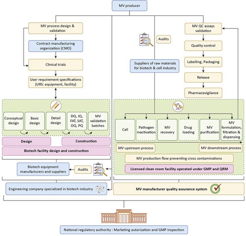
Regulators and other stakeholders will play key roles in establishing a reasonable framework defining the safety and quality criteria specifications of EVs for clinical trials and market approval. In the field of regenerative medicine, EVs should be regarded as being a likely safer alternative to cell therapies and a tool to hasten clinical applications for some indications.Citation97,Citation98 In contrast, using drug loaded-EVs as DDSs may take more time to establish themselves as realistic and efficient alternative therapeutic options to synthetic nanocarriers like liposomes. Pharmaceutical development phases (especially establishment of optimal drug loading and storage conditions, as well as stability studies) will be time-consuming, and in some pathologies like cancers, will require a large number of patients to establish superiority in targeting and safety profiles compared to existing licensed DDS nanoformulations. Careful design of EV manufacturing facilities and processing equipment is essential, which involves multiple stakeholders including EV manufacturers, equipment and raw material suppliers, and regulatory authorities (, reproduced with permission of Agrahari et alCitation3).
Figure 3 Phase-by-phase proposed development of an industrial microvesicle (MV) project and main stakeholders (identified in boxes with a light-blue background). After conducting pilot-scale design and validation of the MV production process at the research and development scale, the MV producer engages a contract manufacturing organization to produce MV batches for clinical trials under investigational new drug status, or equivalent, under supervision of a competent National Regulatory Authority (NRA). Once clinical data are conclusive, the MV producer hires an engineering company specializing in the biotechnology industry, which by closely following the user requirement specifications from the MV producer, can proceed with the conceptual, basic, and detailed design phases of the facility and select suitable equipment from qualified biotechnology industry suppliers. Next, the construction of the facility and the making of equipment go through the qualification phases: design qualification (DQ), installation qualification (IQ), factory acceptance tests (FAT), site acceptance tests (SATs), operational qualification (OQ), performance qualification (PQ), and production of consecutive MV validation batches meeting pre-established quality specifications. Following submission and approval of all the required validation and clinical documentation, the manufacturing site is licensed, and the MV product receives a marketing authorization by the relevant NRA. The MV producer operates the facility under good manufacturing practices (GMPs) and conducts quality audits of the suppliers of raw materials (eg, growth medium; fetal bovine serum, and buffer components) and excipients used during manufacture. The facility is operated under GMPs and is subjected to periodic NRA inspections to maintain manufacturing site license and MV marketing authorization. The MV product is subject to pharmacovigilance, in particular to monitor adverse reactions. Reprinted from of Trends Biotechnol. 37. Agrahari V, Agrahari V, Burnouf PA, Chew CH, Burnouf T. Extracellular microvesicles as new industrial therapeutic frontiers. 707–729. Copyright (2019)Citation3. With permission from Elsevier.
Abbreviation: QRM, quality risk management.
In conclusion, as the methodologies to characterize EVs and assess their safety and performance in preclinical models are at hand and their advantages and limits better understood, we think that clinical evaluation of naïve autologous or allogeneic EV preparations should be facilitated and encouraged. In order to maximize the intended therapeutic effects of EVs, the future may comprise more-complex delivery systems, such as their encapsulation in another nanoparticle-based delivery system, that are currently extensively developed and can be easily translatable. Though, the evaluation of biological fate of EVs-nanoparticle systems will be critical for their successful translation since nanoparticles in vivo journey can be significantly affected by their physicochemical properties which may also impact the EVs fate.Citation99,Citation100
All these evaluations should be made within the framework of a mature regulatory surveillance system that protects patients’ safety, and avoids deviations into unproven mercantile clinical applications that would jeopardize the potential and future of EVs as proven therapies.
Disclosure
The authors report no conflicts of interest in this work.
Acknowledgments
This study was supported by a grant (107-2314-B-038-084) from the Ministry of Science and Technology (MOST) of Taiwan to Thierry Burnouf, Taipei Medical University, Taiwan, and a Single Discipline Faculty Development Grant from Shenandoah University (Winchester, VA, USA) to Vibhuti Agrahari.
References
- Yanez-Mo M, Siljander PR, Andreu Z, et al. Biological properties of extracellular vesicles and their physiological functions. J Extracell Vesicles. 2015;4:27066. doi:10.3402/jev.v4.2706625979354
- van Niel G, D’Angelo G, Raposo G. Shedding light on the cell biology of extracellular vesicles. Nat Rev Mol Cell Biol. 2018;19(4):213–228. doi:10.1038/nrm.2017.12529339798
- Agrahari V, Agrahari V, Burnouf PA, Chew CH, Burnouf T. Extracellular microvesicles as new industrial therapeutic frontiers. Trends Biotechnol. 2019;37:707–729. doi:10.1016/j.tibtech.2018.11.01230638682
- Maas SLN, Breakefield XO, Weaver AM. Extracellular vesicles: unique intercellular delivery vehicles. Trends Cell Biol. 2017;27(3):172–188. doi:10.1016/j.tcb.2016.11.00327979573
- de Jong OG, Kooijmans SAA, Murphy DE, et al. Drug delivery with extracellular vesicles: from imagination to innovation. Acc Chem Res. 2019;52:1761–1770. doi:10.1021/acs.accounts.9b0010931181910
- Vader P, Mol EA, Pasterkamp G, Schiffelers RM. Extracellular vesicles for drug delivery. Adv Drug Deliv Rev. 2016;106(Pt A):148–156. doi:10.1016/j.addr.2016.02.00626928656
- Gudbergsson JM, Jonsson K, Simonsen JB, Johnsen KB. Systematic review of targeted extracellular vesicles for drug delivery - considerations on methodological and biological heterogeneity. J Control Release. 2019;306:108–120. doi:10.1016/j.jconrel.2019.06.00631175896
- Lener T, Gimona M, Aigner L, et al. Applying extracellular vesicles based therapeutics in clinical trials - an ISEV position paper. J Extracell Vesicles. 2015;4:30087. doi:10.3402/jev.v4.3008726725829
- Pachler K, Lener T, Streif D, et al. A good manufacturing practice–grade standard protocol for exclusively human mesenchymal stromal cell–derived extracellular vesicles. Cytotherapy. 2017;19(4):458–472. doi:10.1016/j.jcyt.2017.01.00128188071
- Keshtkar S, Azarpira N, Ghahremani MH. Mesenchymal stem cell-derived extracellular vesicles: novel frontiers in regenerative medicine. Stem Cell Res Ther. 2018;9(1):63. doi:10.1186/s13287-018-0791-729523213
- Burnouf T, Goubran HA, Chou ML, Devos D, Radosevic M. Platelet microparticles: detection and assessment of their paradoxical functional roles in disease and regenerative medicine. Blood Rev. 2014;28(4):155–166.24826991
- Tao SC, Guo SC, Zhang CQ. Platelet-derived extracellular vesicles: an emerging therapeutic approach. Int J Biol Sci. 2017;13(7):828–834. doi:10.7150/ijbs.1977628808416
- Burnouf T, Burnouf PA, Wu YW, Chuang EY, Lu LS, Goubran H. Circulatory-cell-mediated nanotherapeutic approaches in disease targeting. Drug Discov Today. 2018;23(5):934–943. doi:10.1016/j.drudis.2017.08.01228917821
- Palviainen M, Saari H, Karkkainen O, et al. Metabolic signature of extracellular vesicles depends on the cell culture conditions. J Extracell Vesicles. 2019;8(1):1596669. doi:10.1080/20013078.2018.156080831007875
- Gimona M, Pachler K, Laner-Plamberger S, Schallmoser K, Rohde E. Manufacturing of human extracellular vesicle-based therapeutics for clinical use. Int J Mol Sci. 2017;18(6). doi:10.3390/ijms18061190
- Pascucci L, Coccè V, Bonomi A, et al. Paclitaxel is incorporated by mesenchymal stromal cells and released in exosomes that inhibit in vitro tumor growth: a new approach for drug delivery. J Control Release. 2014;192:262–270. doi:10.1016/j.jconrel.2014.07.04225084218
- Tian Y, Li S, Song J, et al. A doxorubicin delivery platform using engineered natural membrane vesicle exosomes for targeted tumor therapy. Biomaterials. 2014;35(7):2383–2390. doi:10.1016/j.biomaterials.2013.11.08324345736
- Yang T, Martin P, Fogarty B, et al. Exosome delivered anticancer drugs across the blood-brain barrier for brain cancer therapy in Danio Rerio. Pharm Res. 2015;32(6):2003–2014. doi:10.1007/s11095-014-1593-y25609010
- Liao W, Du Y, Zhang C, et al. Exosomes: the next generation of endogenous nanomaterials for advanced drug delivery and therapy. Acta Biomater. 2019;86:1–14. doi:10.1016/j.actbio.2018.12.04530597259
- Kim MS, Haney MJ, Zhao Y, et al. Development of exosome-encapsulated paclitaxel to overcome MDR in cancer cells. Nanomedicine. 2016;12(3):655–664. doi:10.1016/j.nano.2015.10.01226586551
- Sato YT, Umezaki K, Sawada S, et al. Engineering hybrid exosomes by membrane fusion with liposomes. Sci Rep. 2016;6:21933. doi:10.1038/srep2193326911358
- Kim SM, Kim HS. Engineering of extracellular vesicles as drug delivery vehicles. Stem Cell Investig. 2017;4:74. doi:10.21037/sci.2017.08.07
- Tang TT, Lv LL, Lan HY, Liu BC. Extracellular vesicles: opportunities and challenges for the treatment of renal diseases. Front Physiol. 2019;10:226. doi:10.3389/fphys.2019.0022630941051
- Paganini C, Palmiero UC, Pocsfalvi G, Touzet N, Bongiovanni A, Arosio P. Scalable production and isolation of extracellular vesicles: available sources and lessons from current industrial bioprocesses. Biotechnol J. 2019;e1800528. doi:10.1002/biot.20180052831140717
- Konoshenko MY, Lekchnov EA, Vlassov AV, Laktionov PP. Isolation of extracellular vesicles: general methodologies and latest trends. Biomed Res Int. 2018;2018:8545347. doi:10.1155/2018/854534729662902
- Busatto S, Vilanilam G, Ticer T, et al. Tangential flow filtration for highly efficient concentration of extracellular vesicles from large volumes of fluid. Cells. 2018;7(12):273. doi:10.3390/cells7120273
- Watson DC, Yung BC, Bergamaschi C, et al. Scalable, cGMP-compatible purification of extracellular vesicles carrying bioactive human heterodimeric IL-15/lactadherin complexes. J Extracell Vesicles. 2018;7(1):1442088. doi:10.1080/20013078.2018.144208829535850
- Boing AN, van der Pol E, Grootemaat AE, Coumans FA, Sturk A, Nieuwland R. Single-step isolation of extracellular vesicles by size-exclusion chromatography. J Extracell Vesicles. 2014;3. doi:10.3402/jev.v3.24384
- Ramirez MI, Amorim MG, Gadelha C, et al. Technical challenges of working with extracellular vesicles. Nanoscale. 2018;10(3):881–906. doi:10.1039/c7nr08360b29265147
- Konoshenko MY, Lekchnov EA, Vlassov AV, Laktionov PP. Isolation of extracellular vesicles: general methodologies and latest trends. Biomed Res Int. 2018;2018:8545347. doi:10.1155/2018/854534729662902
- Liang K, Liu F, Fan J, et al. Nanoplasmonic quantification of tumor-derived extracellular vesicles in plasma microsamples for diagnosis and treatment monitoring. Nat Biomed Eng. 2017;1. doi:10.1038/s41551-016-0021
- Sunkara V, Woo HK, Cho YK. Emerging techniques in the isolation and characterization of extracellular vesicles and their roles in cancer diagnostics and prognostics. Analyst. 2016;141(2):371–381. doi:10.1039/c5an01775k26535415
- Hartjes TA, Mytnyk S, Jenster GW, van Steijn V, van Royen ME. Extracellular vesicle quantification and characterization: common methods and emerging approaches. Bioengineering. 2019;6(1):7. doi:10.3390/bioengineering6020029
- Rupert DLM, Claudio V, Lässer C, Bally M. Methods for the physical characterization and quantification of extracellular vesicles in biological samples. Biochim Biophys Acta. 2017;1861(1, Part A):3164–3179. doi:10.1016/j.bbagen.2016.07.028
- Szatanek R, Baj-Krzyworzeka M, Zimoch J, Lekka M, Siedlar M, Baran J. The methods of choice for extracellular vesicles (EVs) characterization. Int J Mol Sci. 2017;18(6):1153. doi:10.3390/ijms18061153
- Klutz S, Magnus J, Lobedann M, et al. Developing the biofacility of the future based on continuous processing and single-use technology. J Biotechnol. 2015;213:120–130. doi:10.1016/j.jbiotec.2015.06.38826091773
- Rohde E, Pachler K, Gimona M. Manufacturing and characterization of extracellular vesicles from umbilical cord-derived mesenchymal stromal cells for clinical testing. Cytotherapy. 2019;21:581–592. doi:10.1016/j.jcyt.2018.12.00630979664
- Cai K, Anderson J, Orchard JD, Afdahl CD, Dickson M, Li Y. Virus removal robustness of ion exchange chromatography. Biologicals. 2019;58:28–34. doi:10.1016/j.biologicals.2019.01.00430661901
- Burnouf T, Radosevich M. Reducing the risk of infection from plasma products: specific preventative strategies. Blood Rev. 2000;14(2):94–110. doi:10.1054/blre.2000.012911012252
- Chou ML, Lin LT, Devos D, Burnouf T. Nanofiltration to remove microparticles and decrease the thrombogenicity of plasma: in vitro feasibility assessment. Transfusion. 2015;55(10):2433–2444. doi:10.1111/trf.1316225988671
- Burnouf T, Strunk D, Koh MB, Schallmoser K. Human platelet lysate: replacing fetal bovine serum as a gold standard for human cell propagation? Biomaterials. 2016;76:371–387. doi:10.1016/j.biomaterials.2015.10.06526561934
- Henschler R, Gabriel C, Schallmoser K, Burnouf T, Koh MBC. Human platelet lysate current standards and future developments. Transfusion. 2019;59(4):1407–1413. doi:10.1111/trf.1517430741431
- Schallmoser K, Henschler R, Gabriel C, Koh MBC, Burnouf T. Production and quality requirements of human platelet lysate: a position statement from the working party on cellular therapies of the international society of blood transfusion. Trends Biotechnol. 2019. doi:10.1016/j.tibtech.2019.06.002
- Aranha H. Virus safety of biopharmaceuticals - absence of evidence is not evidence of absence. Contract Pharma. 2011;13:82–87.
- Moody M, Alvez W, Varghese J, Khan F. Mouse Minute Virus (MMV) contamination—a case study: detection, root cause determination, and corrective actions. PDA J Pharm Sci Technol. 2011;65(6):580–588. doi:10.5731/pdajpst.2011.0082422294580
- Kerr A, Nims R. Adventitious viruses detected in biopharmaceutical bulk harvest samples over a 10 year period. PDA J Pharm Sci Technol. 2010;64(5):481–485.21502056
- Roush JD. Integrated viral clearance strategies — reflecting on the present, projecting to the future. Curr Opin Biotechnol. 2018;53:137–143. doi:10.1016/j.copbio.2018.01.00329367164
- Carbrello C, Nhiem D, Priest M, Mann K, Greenhalgh P. Supplement: upstream virus safety: protect your bioreactor by media filtration. Genet Eng Biotechnol News. 2017;37(17).
- Mann K, Royce J, Carbrello C, et al. Protection of bioreactor culture from virus contamination by use of a virus barrier filter. BMC Proc. 2015;9(9):22. doi:10.1186/1753-6561-9-S9-P22
- Wiklander OP, Nordin JZ, O’Loughlin A, et al. Extracellular vesicle in vivo biodistribution is determined by cell source, route of administration and targeting. J Extracell Vesicles. 2015;4:26316. doi:10.3402/jev.v4.2631625899407
- Lai CP, Mardini O, Ericsson M, et al. Dynamic biodistribution of extracellular vesicles in vivo using a multimodal imaging reporter. ACS Nano. 2014;8(1):483–494. doi:10.1021/nn404945r24383518
- Takahashi Y, Nishikawa M, Shinotsuka H, et al. Visualization and in vivo tracking of the exosomes of murine melanoma B16-BL6 cells in mice after intravenous injection. J Biotechnol. 2013;165(2):77–84. doi:10.1016/j.jbiotec.2013.03.01323562828
- Matsumoto A, Takahashi Y, Nishikawa M, et al. Role of phosphatidylserine-derived negative surface charges in the recognition and uptake of intravenously injected B16BL6-derived exosomes by macrophages. J Pharm Sci. 2017;106(1):168–175. doi:10.1016/j.xphs.2016.07.02227649887
- Imai T, Takahashi Y, Nishikawa M, et al. Macrophage-dependent clearance of systemically administered B16BL6-derived exosomes from the blood circulation in mice. J Extracell Vesicles. 2015;4:26238. doi:10.3402/jev.v4.2623825669322
- Matsumoto J, Stewart T, Banks WA, Zhang J. The transport mechanism of extracellular vesicles at the blood-brain barrier. Curr Pharm Des. 2017;23(40):6206–6214. doi:10.2174/138161282366617091316473828914201
- Pinheiro A, Silva AM, Teixeira JH, et al. Extracellular vesicles: intelligent delivery strategies for therapeutic applications. J Control Release. 2018;289:56–69. doi:10.1016/j.jconrel.2018.09.01930261205
- Munagala R, Aqil F, Jeyabalan J, Gupta RC. Bovine milk-derived exosomes for drug delivery. Cancer Lett. 2016;371(1):48–61. doi:10.1016/j.canlet.2015.10.02026604130
- Pieters BC, Arntz OJ, Bennink MB, et al. Commercial cow milk contains physically stable extracellular vesicles expressing immunoregulatory TGF-beta. PLoS One. 2015;10(3):e0121123. doi:10.1371/journal.pone.012112325822997
- Manca S, Giraud D, Zempleni J. Bioavailability and biodistribution of fluorophore-labeled exosomes from cow’s milk after intravenous and oral administration in C57Bl/6J mice. Faseb J. 2016;30(1_supplement):690.698.
- Narbute K, Pilipenko V, Pupure J, et al. Intranasal administration of extracellular vesicles derived from human teeth stem cells improves motor symptoms and normalizes tyrosine hydroxylase expression in the substantia nigra and striatum of the 6-hydroxydopamine-treated rats. Stem Cells Transl Med. 2019;8(5):490–499. doi:10.1002/sctm.18-016230706999
- Zhuang X, Xiang X, Grizzle W, et al. Treatment of brain inflammatory diseases by delivering exosome encapsulated anti-inflammatory drugs from the nasal region to the brain. Mol Ther. 2011;19(10):1769–1779. doi:10.1038/mt.2011.16421915101
- Haney MJ, Klyachko NL, Zhao Y, et al. Exosomes as drug delivery vehicles for Parkinson’s disease therapy. J Control Release. 2015;207:18–30. doi:10.1016/j.jconrel.2015.03.03325836593
- Betzer O, Perets N, Angel A, et al. In vivo neuroimaging of exosomes using gold nanoparticles. ACS Nano. 2017;11(11):10883–10893. doi:10.1021/acsnano.7b0449528960957
- Ingato D, Lee JU, Sim SJ, Kwon YJ. Good things come in small packages: overcoming challenges to harness extracellular vesicles for therapeutic delivery. J Control Release. 2016;241:174–185. doi:10.1016/j.jconrel.2016.09.01627667180
- Jeyaram A, Jay SM. Preservation and storage stability of extracellular vesicles for therapeutic applications. Aaps J. 2017;20(1):1. doi:10.1208/s12248-017-0160-y29181730
- Charoenviriyakul C, Takahashi Y, Nishikawa M, Takakura Y. Preservation of exosomes at room temperature using lyophilization. Int J Pharm. 2018;553(1–2):1–7. doi:10.1016/j.ijpharm.2018.10.03230316791
- Park SJ, Jeon H, Yoo SM, Lee MS. The effect of storage temperature on the biological activity of extracellular vesicles for the complement system. In Vitro Cell Dev Biol Anim. 2018;54(6):423–429. doi:10.1007/s11626-018-0261-729748909
- Richter M, Fuhrmann K, Fuhrmann G. Evaluation of the storage stability of extracellular vesicles. J Vis Exp. 2019;(147). doi:10.3791/59584
- Kusuma GD, Barabadi M, Tan JL, Morton DAV, Frith JE, Lim R. To protect and to preserve: novel preservation strategies for extracellular vesicles. Front Pharmacol. 2018;9(1199). doi:10.3389/fphar.2018.01199
- Valkonen S, van der Pol E, Boing A, et al. Biological reference materials for extracellular vesicle studies. Eur J Pharm Sci. 2017;98:4–16. doi:10.1016/j.ejps.2016.09.00827622921
- Witwer KW, Buzas EI, Bemis LT, et al. Standardization of sample collection, isolation and analysis methods in extracellular vesicle research. J Extracell Vesicles. 2013;2:20360.
- Lorincz AM, Timar CI, Marosvari KA, et al. Effect of storage on physical and functional properties of extracellular vesicles derived from neutrophilic granulocytes. J Extracell Vesicles. 2014;3:25465. doi:10.3402/jev.v3.2438425536933
- Jin Y, Chen K, Wang Z, et al. DNA in serum extracellular vesicles is stable under different storage conditions. BMC Cancer. 2016;16(1):753. doi:10.1186/s12885-016-2783-227662833
- Bosch S, de Beaurepaire L, Allard M, et al. Trehalose prevents aggregation of exosomes and cryodamage. Sci Rep. 2016;6:36162. doi:10.1038/srep3616227824088
- Frank J, Richter M, de Rossi C, Lehr CM, Fuhrmann K, Fuhrmann G. Extracellular vesicles protect glucuronidase model enzymes during freeze-drying. Sci Rep. 2018;8(1):12377. doi:10.1038/s41598-018-30786-y30120298
- Xu R, Rai A, Chen M, Suwakulsiri W, Greening DW, Simpson RJ. Extracellular vesicles in cancer - implications for future improvements in cancer care. Nat Rev Clin Oncol. 2018;15(10):617–638. doi:10.1038/s41571-018-0036-929795272
- Hinde E, Thammasiraphop K, Duong HT, et al. Pair correlation microscopy reveals the role of nanoparticle shape in intracellular transport and site of drug release. Nat Nanotechnol. 2017;12(1):81. doi:10.1038/nnano.2016.16027618255
- Erkoc P, Yasa IC, Ceylan H, Yasa O, Alapan Y, Sitti M. Mobile microrobots for active therapeutic delivery. Adv Ther. 2019;2(1):1800064. doi:10.1002/adtp.v2.1
- Brambilla D, Luciani P, Leroux JC. Breakthrough discoveries in drug delivery technologies: the next 30 years. J Control Release. 2014;190:9–14. doi:10.1016/j.jconrel.2014.03.05624794899
- Wilhelm S, Tavares AJ, Dai Q, et al. Analysis of nanoparticle delivery to tumours. Nat Rev Mater. 2016;1(5):16014. doi:10.1038/natrevmats.2016.14
- Kibria G, Ramos EK, Wan Y, Gius DR, Liu H. Exosomes as a drug delivery system in cancer therapy: potential and challenges. Mol Pharm. 2018;15(9):3625–3633. doi:10.1021/acs.molpharmaceut.8b0027729771531
- Armstrong JPK, Stevens MM. Strategic design of extracellular vesicle drug delivery systems. Adv Drug Deliv Rev. 2018;130:12–16. doi:10.1016/j.addr.2018.06.01729959959
- Kamerkar S, LeBleu VS, Sugimoto H, et al. Exosomes facilitate therapeutic targeting of oncogenic KRAS in pancreatic cancer. Nature. 2017;546(7659):498–503. doi:10.1038/nature2234128607485
- You Y, Ikezu T. Emerging roles of extracellular vesicles in neurodegenerative disorders. Neurobiol Dis. 2019;130:104512. doi:10.1016/j.nbd.2019.10451231229685
- Hall J, Prabhakar S, Balaj L, Lai CP, Cerione RA, Breakefield XO. Delivery of therapeutic proteins via extracellular vesicles: review and potential treatments for Parkinson’s disease, Glioma, and Schwannoma. Cell Mol Neurobiol. 2016;36(3):417–427. doi:10.1007/s10571-015-0309-027017608
- Budnik V, Ruiz-Cañada C, Wendler F. Extracellular vesicles round off communication in the nervous system. Nat Rev Neurosci. 2016;17(3):160–172. doi:10.1038/nrn.2015.2926891626
- Rufino-Ramos D, Albuquerque PR, Carmona V, Perfeito R, Nobre RJ, Pereira de Almeida L. Extracellular vesicles: novel promising delivery systems for therapy of brain diseases. J Control Release. 2017;262:247–258. doi:10.1016/j.jconrel.2017.07.00128687495
- Ha D, Yang N, Nadithe V. Exosomes as therapeutic drug carriers and delivery vehicles across biological membranes: current perspectives and future challenges. Acta Pharm Sin B. 2016;6(4):287–296. doi:10.1016/j.apsb.2016.02.00127471669
- Qu M, Lin Q, Huang L, et al. Dopamine-loaded blood exosomes targeted to brain for better treatment of Parkinson’s disease. J Control Release. 2018;287:156–166. doi:10.1016/j.jconrel.2018.08.03530165139
- Tian T, Zhang H-X, He C-P, et al. Surface functionalized exosomes as targeted drug delivery vehicles for cerebral ischemia therapy. Biomaterials. 2018;150:137–149. doi:10.1016/j.biomaterials.2017.10.01229040874
- Vinaiphat A, Sze SK. Clinical implications of extracellular vesicles in neurodegenerative diseases. Expert Rev Mol Diagn. 2019;1–12.
- Rayyan M, Zheutlin A, Byrd JB. Clinical research using extracellular vesicles: insights from the International Society for Extracellular Vesicles 2018 annual meeting. J Extracell Vesicles. 2018;7(1):1535744. doi:10.1080/20013078.2018.153574431162489
- Besse B, Charrier M, Lapierre V, et al. Dendritic cell-derived exosomes as maintenance immunotherapy after first line chemotherapy in NSCLC. Oncoimmunology. 2016;5(4):e1071008. doi:10.1080/2162402X.2015.107100827141373
- Shukla AA, Thommes J. Recent advances in large-scale production of monoclonal antibodies and related proteins. Trends Biotechnol. 2010;28(5):253–261. doi:10.1016/j.tibtech.2010.02.00120304511
- Armstrong JPK, Holme MN, Stevens MM. Re-engineering extracellular vesicles as smart nanoscale therapeutics. ACS Nano. 2017;11(1):69–83. doi:10.1021/acsnano.6b0760728068069
- Koh E, Lee EJ, Nam GH, et al. Exosome-SIRPalpha, a CD47 blockade increases cancer cell phagocytosis. Biomaterials. 2017;121:121–129. doi:10.1016/j.biomaterials.2017.01.00428086180
- Riazifar M, Pone EJ, Lotvall J, Zhao W. Stem Cell Extracellular Vesicles: extended Messages of Regeneration. Annu Rev Pharmacol Toxicol. 2017;57:125–154. doi:10.1146/annurev-pharmtox-061616-03014627814025
- Akyurekli C, Le Y, Richardson RB, Fergusson D, Tay J, Allan DS. A systematic review of preclinical studies on the therapeutic potential of mesenchymal stromal cell-derived microvesicles. Stem Cell Rev. 2015;11(1):150–160. doi:10.1007/s12015-014-9545-9
- Agrahari V, Burnouf PA, Burnouf T, Agrahari V. Nanoformulation properties, characterization, and behavior in complex biological matrices: Challenges and opportunities for brain-targeted drug delivery applications and enhanced translational potential. Adv Drug Deliv Rev. doi:10.1016/j.addr.2019.02.008 Epub 2019 Feb 22.
- Agrahari V, Hiremath P. Challenges associated and approaches for successful translation of nanomedicines into commercial products. Nanomedicine (Lond). 2017;12(8):819–823. doi:10.2217/nnm-2017-0039