Abstract
Several reports have demonstrated the anticancer activities of plumbagin, a naphthoquinone derivative isolated from plants belonging to Plumbaginaceae family. However, to the best of our knowledge, there are no reports which describe gold nanoconjugation with plumbagin, even though plumbagin is considered to be a promising therapeutic agent. In this report, we demonstrate the fabrication and characterization of gold nanoparticles conjugated with plumbagin (AuPB) that can reduce the toxicity of the latter, and their capacity for cellular localization and generation of reactive oxygen species. The anticancer activity and ability of plumbagin to produce reactive oxygen species was studied and compared with that of bromoderivatives of 1,4 naphthoquinones such as 2-bromo-1,4-naphthoquinone (2-BNQ) and 2,3-dibromo-1, 4-naphthoquinone (2,3-DBNQ) and their gold nanoconjugates. Plumbagin and bromoderivatives of 1,4 naphthoquinones in the form of gold nanoconjugates showed reduced cytotoxicity and apoptosis compared with the pristine compounds, ie, plumbagin, 2-BNQ, and 2,3-DBNQ. Interestingly, we observed that the gold nanoparticles could quench the reactive oxygen species-generating capacity of plumbagin, 2-BNQ, and 2,3-BNQ, which is one of the main mechanisms of action of the naphthoquinones. Therefore, it can be concluded that conjugation with gold nanoparticles can reduce the cytotoxicity of these compounds.
Introduction
In Indian Ayurvedic medicine, plants belonging to Plumbaginaceae family, which are a rich source of plumbagin (5-hydroxy-2-methyl-1,4-naphthoquinone), have been used to treat a wide variety of ailments. Research in the last two decades has suggested that plumbagin is associated with a range of potential therapeutic or preventive effects.Citation1–Citation4 Though several studies have reported the anticancer activity of plumbagin, it is relatively nontoxic to normal cells.Citation5–Citation9 The mechanism of action of plumbagin involves generation of reactive oxygen species, such as superoxide radicals (O2−) and hydrogen peroxide (H2O2), in the system.Citation10,Citation11 Thus, plumbagin has also been widely used as a model for studies of oxidative damage.Citation12 The hydroxyl group of plumbagin increases its electrophilic properties and makes it a more potent cytotoxic molecule via its ability to accept electrons, which increases its capacity to generate reactive oxygen species. Although plumbagin has been shown to be more toxic to cancer cells, it can also be toxic to normal cells at high concentrations.Citation13 In this context, nanotechnologies offer an exciting range of new therapeutic possibilities for cancer treatment.
In cancer nanotechnology, inorganic nanoparticles, especially gold nanoparticle-mediated drug delivery, might significantly reduce the drug doses needed, with high specificity, low toxicity, and better bioavailability. Recently, several groupsCitation14–Citation16 including ours, have demonstrated that inorganic nanoparticles, especially gold nanoparticles (AuNP), possess enormous potential to improve the efficacy of several cancer treatments.Citation17–Citation19 Colloidal AuNP have been used widely for biomedical diagnostics, drug delivery, and therapeutic applications in cancer. AuNP were chosen for our study because they have several advantages, including ease of synthesis, easy characterization due to the presence of a surface plasmon resonance band, a very high surface area, stability, biocompatibility, and surface functionalization. Gold is chemically inert but is highly reactive and catalytic at the nanoscale level due to its high surface to volume ratio and quantum size effect. Spherical AuNP (size 5 nm) shows a single strong surface plasmon resonance absorption band in the visible region around 512 nm, which has significant biological importance.Citation18–Citation20
In the present study, we demonstrated conjugation and characterization of gold nanoconjugates with plumbagin (Au-PB) and bromoderivatives of naphthoquinone, their cytotoxicity, localization, and capacity to generate reactive oxygen species. We also showed that the pristine compounds could induce apoptosis more effectively than the gold conjugates. We also observed that plumbagin, but not gold nanoconjugates with plumbagin and polyethylene glycol (PEG)-amine (Au-PAM-PB), could inhibit p53 protein expression because plumbagin specifically inhibits acetylation of p53, which makes it susceptible to ubiquitination. We also analyzed bromoderivatives of 1,4 naphthoquinones (2-bromo-1,4-naphthoquinone [2-BNQ] and 2,3-bibromo-1,4-naphthoquinone [2,3-DBNQ]) to see if they could have a better conjugation effect with AuNP or show any change in cytotoxicity on AuNP conjugation when compared with Au-PB. To the best of our knowledge, there are no reports that describe gold nanoconjugation with plumbagin for use as a therapeutic agent in cancer.
Materials and methods
Synthesis of AuNP
AuNP were synthesized by wet chemical methods using tetrachloroauric acid (HAuCl4, Aldrich Chemicals, St Louis, MO) and sodium borohydride (NaBH4, Aldrich Chemicals) as reducing agents, as described elsewhere.Citation17,Citation18 Briefly, an aqueous solution of HAuCl4 was mixed with an aqueous solution of NaBH4 and mixed under vigorous stirring for about 16 hours. AuNP were then characterized by several physicochemical techniques, including ultraviolet-visible spectroscopy and transmission electron microscopy.
Conjugation of plumbagin, 2-BNQ, and 2,3-DBNQ with AuNP
Initially we tried to conjugate plumbagin, 2-BNQ, and 2,3-DBNQ (Sigma-Aldrich, St Louis, MO) with colloidal AuNP directly by a single incubation step. Different concentrations of these compounds were added to the AuNP solution and stirred vigorously for one hour. The solutions were ultracentrifuged at 18,000 rpm for one hour at 10°C in an OTD80B Sorvall ultracentrifuge (Thermofisher Scientific Inc, Waltham, MA) using a 50 2Ti rotor. The resulting loose pellets were used for further characterization and finally for in vitro cell culture experiments.
Conjugation of plumbagin with AuNP via PEG
Several PEG backbones of sulfhydryl and PAM chemicals, like PEG disulfhydryl 2000, PEG tetrasulfhydryl 20,000, PEG disulfhydryl 10,000, methyl PEG sulfhydryl 2000, PEG-amine 10,000, and PEG-diamine 2000 (SunBio Corporation, Orinda, CA) were used to make Au-PEG-PB nanoconjugates. In the first step, the PEG molecules were incubated with AuNP at room temperature for one hour to prepare AuNP-PEG, and subsequently incubated with plumbagin for another hour under similar conditions to make AuNP-PEG-PB in line with our previously published work.Citation20 Finally, the nanoconjugates were centrifuged at 18,000 rpm for one hour at 10°C, and the loose pellets and supernatant were analyzed for further characterization and in vitro cell culture experiments.
Ultraviolet spectroscopy
Absorption of the AuNP was measured using a Shimadzu spectrophotometer (Shimadzu Scientific Instruments Inc, Columbia, MD) to identify their ultraviolet-visible spectra (from 800 to 200 nm with a resolution of 1 nm).
Release of plumbagin from nanoconjugates
The release study for the gold nanoconjugates was done as described elsewhere.Citation20 Briefly, the original pellets of gold nanoconjugates (Au-PEG-PB) were divided into several equal fractions, diluted with equal volumes of phosphate-buffered solution (pH 7.4), and incubated for different time periods (0, 0.30, 1, 2, 4, and 6 hours) at 37°C. All the fractions were centrifuged at 15,000 rpm for one hour at 4°C after incubation. The supernatant was collected from each fraction and we then measured the absorbance of plumbagin using ultraviolet spectroscopy.
In vitro cytotoxicity studies with MDA-MB-231 cells
In this study, we used MDA-MB-231 cells (American Type Culture Collection) as a model of breast cancer cells. Cells were maintained in Dulbecco’s modified Eagle’s medium with 10% fetal calf serum and 1% antibiotics in a humidified 5% CO2 incubator at 37°C. In order to check the functional activity of plumbagin and its derivatives, the cells were incubated for different periods of time and with different concentrations of the study compounds, as appropriate for such experiments.
MTS and thymidine incorporation assays
An in vitro cell proliferation assay was done using a colorimetric 3-(4-5-dimethylthiazol-2-yl)-5-(3-carboxymethoxyphenyl)-2-(4-sulfophenyl)-2H-tetrazolium salt (MTS) assay (Promega, Madison, WI). Briefly, the cells were treated with 100 nM of the gold nanoconjugates or the pristine compounds (plumbagin, 2-BNQ, and 2,3-DBNQ) in a 96-well plate (10,000 cells/well) for 48 hours, and the experiment was performed as described previously.Citation21 Thymidine incorporation was also done to measure the growth proficiency of the cells. Briefly, after 48 hours of treatment with the compounds, 2.4 × 10⁵ cells in a 24-well plate were treated with 3H thymidine for four hours (0.5 μCi, in RPMI with 10% fetal bovine serum per well). After washing with ice-cold phosphate-buffered solution, the proteins were fixed by adding 1 mL of ice-cold methanol into each well and incubated for 15 minutes at room temperature. The cells were then washed with water, and 300 μL of 0.2 N NaOH was then added to each well and incubated for 30 minutes. Two milliliters of scintillation fluid was added, and the radioactivity was counted using a liquid scintillation counter. The extent of thymidine incorporation was measured as the growth proficiency of the cells.
Apoptosis by Annexin V binding assay
After treating the cells for 24 hours with 100 nM of the pristine compounds (plumbagin, 2-BNQ, or 2,3-DBNQ), Au-2-BNQ, Au-2,3-DBNQ), Au-PEG-PB, or AuNP alone were dispersed by trypsin-ethylenediamine tetra-acetic acid and washed further with 1× phosphate-buffered solution. Early apoptotic events were then determined using the ApoAlert Annexin V kit (Clontech, Palo Alto, CA) as described by the manufacturer. Briefly, five million cells were resuspended in 200 μL of 1× binding buffer containing Annexin V 0.1 μg/mL and propidium iodide 0.5 μg/mL. The cells were analyzed by flow cytometry. Flow cytometric quantification of DNA was performed using a FACScan (Becton Dickinson, San Jose, CA) and data analysis was carried out using Modfit software.
Western blot analysis
The cells were incubated with the 100 nM conjugates for 24 hours. The whole cell lysate, including the floating cells (total eight million cells), was collected in RIPA buffer supplemented with a protease inhibitor cocktail. Protein 10 μg was loaded for blotting. Mouse monoclonal antibodies, p53 (DO-1, SC-126, Santa Cruz Biotechnology, Santa Cruz, CA) and β-actin (Sigma) were incubated overnight at 4°C and used for immunodetection. After washing the membrane with 1× phosphate-buffered solution, the membrane was incubated with horseradish peroxidase-conjugated secondary antibody (antimouse IgG, Santa Cruz Biotechnology) for 45 minutes. The bands were visualized as per the protocols given in the West Pico chemiluminescence detection kit and the Super Signal West Pico substrate (Pierce Biotechnology, Rockford, IL).
Transmission electron microscopy
About one million cells were incubated with 1 μM Au-PAM-PB for 24 hours. After treatment, the cells were trypsinized and resuspended in Trump’s fixative containing 1% glutaraldehyde and 4% formaldehyde in 0.1 M phosphate buffer at pH 7.2 and processed as already described in the literature.Citation19,Citation22 Thin (90 nm) sections were cut using a Reichert Ultra cut E ultramicrotome, placed on 200 mesh copper grids, and stained with lead citrate. Micrographs were taken on a TECNAI 12 operating at 120 kV.
Spectrofluorometric analysis of reactive oxygen species
The cells were seeded at a concentration of 10,000 cells per well in a 96-well plate. After overnight incubation, the cells were treated with 100 nM of different AuNP/compounds/H2O2 10 μM in serum-free Dulbecco’s modified Eagle’s medium for 30 minutes. Cells were then washed and resuspended in phosphate-buffered solution and incubated with 5 μM 2′, 7′-dichlorofluorescin diacetate H2 (Molecular Probes, Invitrogen, Carlsbad, CA) in ethanol for 30 minutes in the dark. Readings were taken on a Shimadzu UV PC2101 (Shimadzu Scientific Instruments Inc) at 495 nm excitation and 523 nm emission.
Results and discussion
Plumbagin conjugated with PEG-diamine 2000-coated AuNP
Characterizations of AuNP formed by the NaBH4 reduction method were performed using ultraviolet-visible spectroscopy which showed a characteristic surface plasmon resonance band at 512 nm, suggesting the formation of spherical AuNP. Plumbagin, 2-BNQ, and 2,3-DBNQ showed absorbance peaks at 420, 350, and 360 nm, respectively. To analyze whether the plumbagin, 2-BNQ, and 2,3-DBNQ had conjugated with AuNP, the loose pellets collected after centrifugation were analyzed by ultraviolet spectroscopy. We found that 2-BNQ and 2,3-DBNQ conjugated directly with AuNP. This may have been due to the bromine side chain in naphthoquinone where the bromo group can make a slight positive charge in the benzene ring, enabling weak bond formation between the positively charged benzene ring and the slight negatively charged AuNP. Therefore, 2-BNQ and 2,3-DBNQ can conjugate with AuNP. Another report suggests that this may be due to the bromine side chain which can bond with AuNP directly, because bromide ions of bromoderivatives can replace the chloride ions in AuCl4.Citation23 A 2-BNQ concentration of 20 μg/mL was used for further studies because maximum absorbance at 350 nm was observed at this concentration (). The same concentration was chosen for 2,3-DBNQ because the absorption at 520 nm was maximum at 20 μg/mL (). On the other hand, AuNP with plumbagin did not show any absorbance at 420 nm, indicating that direct conjugation of plumbagin with AuNP had not taken place. The maximum number of naphthoquinones was attached to the AuNP at 20 μg/mL, beyond which aggregation took place instead of binding more molecules to the AuNP. The decrease in absorbance indicates aggregation of AuNP in line with our previously published work.Citation19,Citation20
Figure 1 Ultraviolet-visible spectra of Au-2-BNQ, Au-2,3-DBNQ, and Au-PAM-PB nanoconjugates. Using different concentrations of conjugates (10 μg/mL of AuNP solution), an increase in absorbance of (A) Au-2-BNQ at 350 nm and (B) Au-2,3-DBNQ at 360 nm is observed with increasing concentrations of 2-BNQ (10, 20, 30, and 50 μg/mL) and 2,3-DBNQ (10, 15, 20, 25 and 30 μg/mL), respectively, up to 20 μg/mL is seen, and then a decrease. Absorbance increases up to 20 μg/mL and then the absorbance decreases with increasing concentrations of both 2-BNQ and 2,3-DBNQ. (C) Similarly, imilarly, for Au-PAM-PB (5, 10, 15, 20, 30, 40, and 50 μg/mL), the absorbance at 420 nm increases to a maximum at 30 μg/mL and then decreases with an increase in concentration of plumbagin. All experiments were performed three times with similar results.
Abbreviations: Au, gold; 2-BNQ, 2-bromo-1,4-naphthoquinone; 2,3-DBNQ, 2,3-dibromo-1,4-naphthoquinone; PB, plumbagin; PAM, polyethylene glycol-amine.
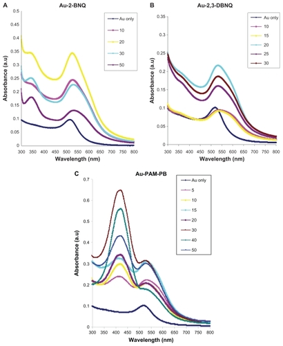
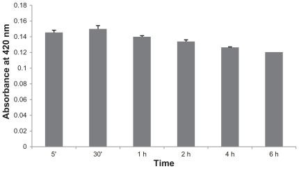
In this context, we tried to conjugate plumbagin with AuNP through various sulfhydryl and amine derivatives of PEG molecules, whereby PEG was used as a linker because plumbagin did not conjugate directly with AuNP. Among several PEG molecules (discussed in the Material and methods section), PEG-diamine 2000 was found to be conjugated with AuNP, so we used PAM for surface functionalization of AuNP, after which plumbagin was conjugated with AuNP without any aggregation and showed absorbance at 420 nm (), where 15 μg/mL of PAM was used for conjugation with AuNP.Citation20 The dependence on absorption of the conjugates at 420 nm indicated that a concentration ratio of 30 μg/mL (with respect to 1 mL of AuNP-PAM solution) of plumbagin could be used for further experiments. The change of absorption maxima (λmax) of AuNP upon addition of PEG-PB, 2-BNQ, or 2,3-DBNQ, as compared with the spectrum for AuNP alone (λmax = 512 nm) clearly demonstrates a red shift in the surface plasmon resonance band maxima in Au-PEG-PB (λmax = 536 nm), Au-2-BNQ (λmax = 542), and Au-2,3-DBNQ (λmax = 535), respectively (). Addition of PAM compounds to AuNP caused red shifts in the λmax values, suggesting interaction and binding of the individual components with the surface of the negatively charged AuNP. Therefore, further experiments were done with the Au-2-BNQ, Au-2,3-DBNQ, and Au-PAM-PB complexes. According to the published literature, we hypothesized that the naphthoquinone-derived AuNP conjugate concept is based on a combination of the favorable properties of the AuNP and the naphthoquinone molecules when forming a naphthoquinone-gold nanoparticle assembly.Citation24–Citation26 A predicted schematic diagram of the Au-PAM-PB complex is shown in . According to our earlier work, the absorption peak at 512 nm indicates the spherical nature of the nanoparticles. The high wavelength indicates an association with AuNP, which increases the stability of the nanoconjugate and the hydrodynamic radius of the particles.Citation18 The release kinetics of plumbagin from the Au-PAM-PB nanoconjugates was also analyzed, and a constant release of plumbagin was observed for up to 24 hours ().
Figure 2 Ultraviolet-visible spectral readings of AuNP and nanoconjugates indicating the change in peak of wavelength (shift in λmax) of gold nanoparticles with increasing concentrations of (A) 2-BNQ, (B) 2,3-DBNQ, and (C) Au-PAM-PB. A predicted schematic diagram of Au-PAM-PB complex is shown in (D). Au-PAM may conjugate with plumbagin by amide bonding.
Abbreviations: Au, gold; 2-BNQ, 2-bromo-1,4-naphthoquinone; 2,3-DBNQ, 2,3-dibromo-1,4-naphthoquinone; PB, plumbagin; PAM, polyethylene glycol-diamine.
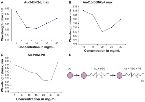
Figure 3 Release profiles for plumbagin from Au-PAM-PB nanoconjugates. There was a constant release of plumbagin from Au-PAM-PB for up to six hours in phosphate-buffered solution at pH 7.4.
Abbreviations: Au, gold; PB, plumbagin; PAM, polyethylene glycol-amine.
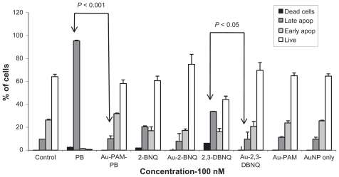
AuNP-conjugated compounds may reduce cytotoxicity and apoptosis
MTS assay showed that AuNP-conjugated plumbagin, 2-BNQ, and 2,3-DBNQ (Au-2-BNQ, Au-2,3-DBNQ, and Au-PAM-PB) could reduce antiproliferative effects to the same extent as the pristine compounds (). Analysis of the number of cells in the S phase using the thymidine incorporation assay showed similar results. Changes in the phospholipid bilayer of the cell membrane were also observed early in apoptosis. The phosphatidylserine component of the phospholipid bilayer was externalized and could be detected by fluorescence labeling. Annexin V has a high affinity for the phosphatidylserine-containing phospholipid bilayer. Staining with FITC-conjugated Annexin V and propidium iodide can identify subpopulations of cells with membrane changes and any associated loss of membrane integrity. In this study, further apoptotic induction studies by Annexin-propidium iodide staining confirmed that the AuNP-conjugated molecules could reduce apoptosis more than the pristine compounds alone (). Similar results showing the inhibitory effect of anisamide-targeted liposomal doxorubicin on the growth of DU-145 tumors were reported by Banerjee et al, who showed that despite the improved targeting, doxorubicin formulated in liposomes containing distearoylglycerol phosphatidylethanolamine polyethylene glycol(3400)- -[2-(4-methoxybenzamido)] ethylamine were less potent than the free drug. This might be due to the fact that the tumor cells were exposed to the free drug for an extended period of time.Citation27 In vivo, this condition might not occur as the free drug is rapidly cleared after administration.
Figure 4 (A) MTS cell proliferation assay with naphthoquinones in MDA-MB-231 cells. Concentrations of all free compounds and compounds with AuNP conjugates were constant (100 nM) as indicated. The concentration was calculated from the standard curve drawn using the absorbance obtained at 420, 350, and 360 nm for plumbagin, 2-BNQ, and 2,3-DBNQ, respectively. MTS reading was taken after 48 hours of incubation with the compounds and AuNP conjugates. (B) Thymidine incorporation assay also showed fewer S phase cells in those treated with the pristine compound compared than the AuNP conjugates. The experiment was repeated at least three times, with similar results.
Abbreviations: Au, gold; 2-BNQ, 2-bromo-1,4-naphthoquinone; 2,3-DBNQ, 2,3-dibromo-1,4-naphthoquinone; PB, plumbagin; NP, nanoparticles.
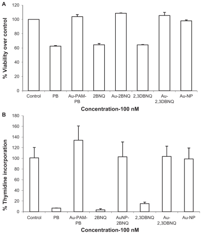
Figure 5 Cells were counted after Annexin-propidium iodide staining. Cells with bound Annexin V show green staining in the plasma membrane. Cells that have lost membrane integrity show red staining (propidium iodide) throughout the cytoplasm. Flow cytometric analysis showed live (no stain), early apoptotic cells (only annexin staining), late apoptotic cells (both annexin and propidium iodide staining), and dead (propidium iodide staining) cells. Most of the plumbagin-treated cells were in the late apoptotic stage, whereas the AuNP conjugates had fewer numbers of late apoptotic cells. More live cells are observed in AuNP conjugates than in samples treated with the pristine compound. A t-test showed a statistically significant difference between the percentage of late apoptotic cells induced by plumbagin versus Au-PAM-PB and 2,3-DBNQ versus Au-2,3-DBNQ. The experiment was repeated at least three times with similar results.
Abbreviations: Au, gold; 2-BNQ, 2-bromo-1,4-naphthoquinone; 2,3-DBNQ, 2,3-dibromo-1,4-naphthoquinone; PB, plumbagin; PAM, polyethylene glycol-amine; NP, nanoparticles.
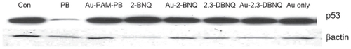
p53 and apoptosis
Apoptosis can be p53-dependent or p53-independent. Plumbagin is reported specifically to inhibit p300-mediated acetylation of p53, making it susceptible to ubiquitination. The chemical entity in plumbagin responsible for inhibition of acetyltransferase activity is the hydroxyl group.Citation28 Here we observed that treatment with plumbagin but not with Au-PAM-PB could inhibit p53 protein expression (). 2-BNQ and 2,3-DBNQ and their gold conjugates did not inhibit p53. This might be due to the lack of a hydroxyl group in these quinones. This indirectly shows that AuNP can change the electrophilicity of the hydroxyl group on plumbagin.
Figure 6 Western blot analysis of p53 protein expression showed that only plumbagin-treated cells had reduced expression of p53 at 100 nM concentration.
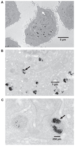
Au-PAM-PB aggregates in lysosomes
MDA-MB-231 cells were treated with 1 μM Au-PAM-PB for 24 hours, and their localization inside the cells was checked by transmission electron microscopy, revealing uptake of synthesized AuNP in vacuoles and lysosomes in the cytoplasmic compartment, as shown in . AuNP are reported to be internalized in the cell and to accumulate inside lysosomes.Citation29 However, in this study, it was not analyzed whether the AuNP found in the lysosomes contained bound plumbagin or not.
Figure 7 Cellular uptake of Au-PAM-PB. Transmission electron microscopic analysis of MDA-MB-231 cells treated with Au-PAM-PB 100 nM for 24 hours showed internalization of Au-PAM-PB by lysosomes in the cells. Magnifications of the figures shown as (A) 6000×, (B) 20,000×, and (C) 80,000×. The experiment was repeated at least three times with similar results.
Abbreviations: Au, gold; PB, plumbagin; PAM, polyethylene glycol-amine.
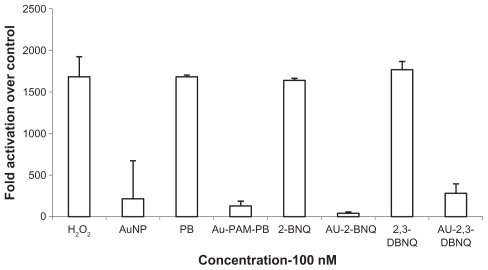
AuNP quenches production of reactive oxygen species by naphthoquinones
The ability of AuNP-conjugated plumbagin to generate reactive oxygen species was analyzed because plumbagin is a well known generator of reactive oxygen species.Citation10,Citation11 There is extensive electron delocalization between the C1 to C4 carbon atoms, which is responsible for the reversible redox cycling observed for plumbagin (by electrochemical measurement) whereby it generates successive semiquinone and catecholic forms. The intermediate semiquinone formed is highly reactive and interacts with molecular dioxygen species present in biological systems to oxidize itself back to the original quinone form, while the molecular oxygen gets reduced to superoxide species. This is what is commonly known as “redox cycling” of quinones, and is responsible for most of their biological activity. As we observed quenching of the redox cycling behavior of plumbagin upon making an AuNP conjugate, such conjugation does not allow or favor addition of one electron in the lowest unoccupied molecular orbits. This could be due to substitution of electrons, withdrawing a chloro/bromo substituent at the C3/C2 position or perturbation by the molecular orbits of gold in the quinone reduction. It has been reported that AuNP can cause an approximately 1.5-fold increase in reactive oxygen species levels in human hepatoma cells.Citation30 Another report by Ito et alCitation31 showed that AuNP increase the yield of reactive oxygen species and that the antioxidant enzymes, catalase and superoxide dismutase, are both effective in reducing the total amount of reactive oxygen species.Citation31 This analysis was done using electron paramagnetic resonance spectroscopy in cell-free media. Other reports also show that AuNP can cause elevation of reactive oxygen species levels without cytotoxicity.Citation32,Citation33 On the other hand, quinones can undergo intracellular electron reduction to the hydroquinone or to the semiquinone.Citation34,Citation35 The semiquinone or hydroquinone may regenerate quinone through redox cycling by reacting with molecular oxygen to produce superoxide radicals. This may culminate in damage to macromolecules and cytotoxicity.Citation36 The production of oxygen radicals and the cytotoxic damage vary for different quinones and in different environments.Citation37,Citation38 Thus, both AuNP and naphthoquinones generate reactive oxygen species. However, AuNP-conjugated naphthoquinones might exhibit a relative antioxidant potential compared with the pristine compounds. It is known that antioxidant capacity is a relative phenomenon. A pro-oxidant may become antioxidant when it encounters a stronger pro-oxidant. Similarly, an antioxidant may become pro-oxidant depending on the redox potential. Therefore, interpretation of the antioxidant/pro-oxidant status of chemical entities should be done only after considering these issues. We had shown earlier that pretreatment of cells with N-acetylcysteine, an antioxidant, protected cells from undergoing apoptosis, confirming the role of reactive oxygen species in plumbagin-mediated apoptosis.Citation39 Here we have found that the capacity of plumbagin to generate reactive oxygen species, which is the major mechanism of action, can be quenched by AuNP conjugation through PEG, which shows that cytotoxicity can be reduced by gold nanoconjugation (). However, further studies need to be done to confirm this in vivo.
Figure 8 Reactive oxygen species generation study with DCFH-DAH2 staining. H2O2 production was detected using nonfluorescent DCFH-DAH2 dye, which when oxidized is converted into fluorescent 2′,7′-dichlorofluorescein (excitation 488 nm, emission 540 nm). The AuNP conjugates and AuNP alone could not induce reactive oxygen species production, while the positive control (10 μM H2O2) and the pristine compounds generated reactive oxygen species. The experiment was repeated at least three times with similar results.
Abbreviations: Au, gold; NP, nanoparticles.
Conclusion
We have synthesized AuNP and their conjugates with plumbagin, 2-BNQ, and 2,3-DBNQ molecules and characterized them by several physicochemical techniques. We have also studied the anticancer activity of the free and gold nanoconjugated compounds. We have shown that the cytotoxicity of the free compounds can be reduced by conjugation with AuNP. A plausible mechanism might be less production of reactive oxygen species when these compounds are conjugated with AuNP. The outcome of targeted chemotherapy depends mainly on two factors, ie, the ability of the carrier molecule to recognize malignant cells selectively and the nature of the chemical linkage used for coupling the cytotoxic agent to the carrier. Chemical bonding between plumbagin and AuNP-PAM might affect the hydroxyl group of plumbagin, ie, the electrophilic moiety, making it a less toxic molecule, resulting in an ability to quench reactive oxygen species. Further studies of these AuNP-naphthoquinone conjugates with targeting antibodies or liposomes might enable these compounds to be administered with targeted cytotoxicity in a safe manner.
Acknowledgment
This study was supported by an overseas associateship grant to PS by the Department of Biotechnology, Government of India (San No. 102/IFD/SAN/415/2009-10). The authors are grateful to J Charlesworth from the Mayo Clinic for the transmission electron microscopic analysis. We acknowledge Dr Priyabrata Mukherjee, Department of Biochemistry and Molecular Biology, Mayo Clinic College of Medicine, and Professor Subhash Padhye from the Center for Drug Design and Molecular Medicine, Department of Environmental Sciences, University of Pune, India, for their kind suggestions for the study and preparation of the manuscript. CRP is grateful to the Department of Science and Technology, Govt of India, New Delhi for the award of Ramanujan Fellowship (SR/S2/RJN-04/2010, GAP 0305\DST\CP). This work was also supported by a generous gift to DM from Bruce and Martha Atwater.
Disclosure
The authors report no conflicts of interest in this work.
References
- BhargavaSKEffects of plumbagin on reproductive function of male dogIndian J Exp Biol1984221531566083981
- de PaivaSRFigueiredoMRAragãoTVKaplanMAAntimicrobial activity in vitro of plumbagin isolated from Plumbago speciesMem Inst Oswaldo Cruz20039895996114762525
- HsuYLChoCYKuoPLHuangYTLinCCPlumbagin (5-hydroxy-2-methyl-1,4-naphthoquinone) induces apoptosis and cell cycle arrest in A549 cells through p53 accumulation via c-Jun NH2-terminal kinase-mediated phosphorylation at serine 15 in vitro and in vivoJ Pharmacol Exp Ther200631848449416632641
- ItoigawaMTakeyaKFurukawaHCardiotonic action of plumbagin on guinea-pig papillary musclePlanta Med1991573173191775570
- ChenCAChangHHKaoCYTsaiTHChenYJPlumbagin, isolated from Plumbago zeylanica, induces cell death through apoptosis in human pancreatic cancer cellsPancreatology2009979780920110748
- DemmaJHallbergKHellmanBGenotoxicity of plumbagin and its effects on catechol and NQNO-induced DNA damage in mouse lymphoma cellsToxicol In Vitro20092326627119124069
- JacksonJKHigoTHunterWLBurtHMTopoisomerase inhibitors as anti-arthritic agentsInflamm Res20085712613418301866
- SrinivasPGopinathGBanerjiADinakarASrinivasGPlumbagin induces reactive oxygen species, which mediate apoptosis in human cervical cancer cellsMol Carcinog20044020121115264212
- ThasniKARakeshSRojiniGRatheeshkumarTSrinivasGPriyaSEstrogen-dependent cell signaling and apoptosis in BRCA1-blocked BG1 ovarian cancer cells in response to plumbagin and other chemotherapeutic agentsAnn Oncol20081969670518187487
- XuKHLuDPPlumbagin induces ROS-mediated apoptosis in human promyelocytic leukemia cells in vivoLeuk Res20103465866519748668
- InbarajJJChignellCFCytotoxic action of juglone and plumbagin: a mechanistic study using HaCaT keratinocytesChem Res Toxicol200417556214727919
- BurgerRMDrlicaKSuperoxide protects Escherichia coli from bleomycin mediated lethalityJ Inorg Biochem20091031273127719679357
- AhmadABanerjeeSWangZKongDSarkarFHPlumbagin-induced apoptosis of human breast cancer cells is mediated by inactivation of NF-κB and Bcl-2J Cell Biochem20081051461147118980240
- BrownSDNativoPSmithJAGold nanoparticles for the improved anticancer drug delivery of the active component of oxaliplatinJ Am Chem Soc20101324678468420225865
- PaciottiGFMyerLWeinreichDColloidal gold: a novel nanoparticle vector for tumor directed drug deliveryDrug Deliv20041116918315204636
- LiJWangXWangCThe enhancement effect of gold nanoparticles in drug delivery and as biomarkers of drug-resistant cancer cellsChem Med Chem2007237437817206735
- PatraCRBhattacharyaRWangETargeted delivery of gemcitabine to pancreatic adenocarcinoma using cetuximab as a targeting agentCancer Res2008681970197818339879
- PatraCRBhattacharyaRMukherjeePFabrication and functional characterization of goldnanoconjugates for potential application in ovarian cancerJ Mater Chem20102054755420436942
- DasAMukherjeePSinglaSKFabrication and characterization of an inorganic gold and silica nanoparticle mediated drug delivery system for nitric oxideNanotechnology20102130510220610873
- BhattacharyaRPatraCREarlAAttaching folic acid on gold nanoparticles using noncovalent interaction via different polyethylene glycol backbones and targeting of cancer cellsNanomedicine20073224238
- WangLLauJSPatraCRRGS-GAIP-interacting protein controls breast cancer progressionMol Cancer Res201081591160021047775
- PatraCRBhattacharyaRPatraSProangiogenic properties of europium (III) hydroxide nanorodsAdv Mater200820753756
- EustisSEl-SayedMAMolecular mechanism of the photochemical generation of gold nanoparticles in ethylene glycol: support for the disproportionation mechanismJ Phys Chem20061101401414019
- MiyachiMYamanoiYShibataYA photosensing system composed of photosystem I, molecular wire, gold nanoparticle, and double surfactants in waterChem Commun20104625572559
- TerasakiNYamamotoNTamadaKBio-photosensor: Cyanobacterial photosystem I coupled with transistor via molecular wireBiochim Biophys Acta200717665365917184727
- QuartaADi CoratoRMannaLMultifunctional nanostructures based on inorganic nanoparticles and oligothiophenes and their exploitation for cellular studiesJ Am Chem Soc2008130105451055518627147
- BanerjeeRTyagiPLiSHuangLAnisamide-targeted stealth liposomes: a potent carrier for targeting doxorubicin to human prostate cancer cellsInt J Cancer200411269370015382053
- RavindraKCSelviBRArifMInhibition of lysine acetyltransferase KAT3B/p300 activity by a naturally occurring hydroxynaphthoquinone, plumbaginJ Biol Chem2009284244532446419570987
- HostaLPla-RocaMArbiolJConjugation of Kahalalide F with gold nanoparticles to enhance in vitro antitumoral activityBioconjug Chem20092013814619072538
- FanJHHungWILiWTYehJBiocompatibility study of gold nanoparticles to human cellsIFMBE Proc200923870873
- ItoSMiyoshiNDegraffWGNagashimaKKirschenbaumLJRieszPEnhancement of 5-aminolevulinic acid-induced oxidative stress on two cancer cell lines by gold nanoparticlesFree Radic Res2009431214122419905984
- FarkasJChristianPUrreaJARoosNEffects of silver and gold nanoparticles on rainbow trout (Oncorhynchus mykiss) hepatocytesAquat Toxicol200996445219853932
- PanYLeifertARuauDGold nanoparticles of diameter 1.4 nm trigger necrosis by oxidative stress and mitochondrial damageSmall200952067207619642089
- O’BrienPJMolecular mechanisms of quinone cytotoxicityChem Biol Interact1991801411913977
- MorganWAHartleyJACohenGMQuinone-induced DNA single strand breaks in rat hepatocytes and human chronic myelogenous leukaemic K562 cellsBiochem Pharmacol1992442152211642637
- NgoEOSunTPChangJYMenadione-induced DNA damage in a human tumor cell lineBiochem Pharmacol199142196119681741774
- RockwellSSartorelliACTomaszMKennedyKACellular pharmacology of quinone bioreductive alkylating agentsCancer Metastasis Rev1993121651768375019
- GibsonNWHartleyJAButlerJSiegelDRossDRelationship between DT-diaphorase-mediated metabolism of a series of aziridinylbenzoquinones and DNA damage and cytotoxicityMol Pharmacol1992425315361406604
- SrinivasGAnnabLAGopinathGBanerjiASrinivasPAntisense blocking of BRCA1 enhances sensitivity to plumbagin but not tamoxifen in BG-1 ovarian cancer cellsMol Carcinog200439152514694444