Abstract
Background
Worldwide, oral squamous cell carcinoma (potentially mediated by HER2) is recognized as the most commonly occurring malignant neoplasm of the oral cavity. Anti-HER2 nanobodies conjugated to gold-silica nanoshells and used as photothermal treatment for oral squamous cell carcinoma may provide a novel therapeutic alternative to current treatment for this disease.
Methods
KB epithelial or HeLaS3 cell cultures (controls) were exposed to these immunonanoshells, and plasmon resonance electron initiation specific to gold was employed to burn the tumor cells.
Results
Following this treatment, significant cell death occurred in the KB tumor cell cultures while there was no evidence of cellular damage or death in the HeLaS3 cell cultures.
Conclusion
These findings suggest that photothermal treatment of oral squamous cell carcinoma has considerable advantages.
Introduction
Oral squamous cell carcinoma is histopathologically characterized by invasive islands of malignant squamous epithelial cells, and accounts for more than 90% of all malignant neoplasms of the oral cavity. The majority of these cancers are located on the tongue, mainly at the lateral posterior border.Citation1,Citation2 Current treatments are based on the clinical stage of the disease and may include surgical excision, radiation therapy, chemotherapy, or combination therapy, as well as new investigational methods such as immunotherapy or gene therapy.Citation1,Citation3,Citation4 Surgical excision can result in complications, and the use of conventional radiotherapy may cause an oral cavity disorder, resulting in reduced patient survival time.Citation5 Because local carcinoma recurrences are common and current treatments can result in significant problems, alternative therapeutic approaches are warranted.Citation6
Thermal therapy is emerging as a promising alternative strategy that is simple to perform and is advantageous as an alternative treatment for certain malignancies. The use of hyperthermia in the treatment of a variety of solid tumors has been under investigation for quite some time.Citation7–Citation13
When generating nanoshells, it is possible to manipulate the size and layer of their composition and to conjugate antibodies with the nanoshells to target cancerous cells specifically.Citation14–Citation16 The nanoshells neither absorb nor scatter light emission, thereby allowing local destruction of tumor tissue.Citation14,Citation17,Citation18 It is also possible to construct gold nanoshells that preferentially absorb light in the near infrared region of the spectrum (700–1100 nm) so that optical transmission through tissue is optimal.Citation19 This property could be used for noninvasive excitation of nanoshells within tissue. Using antibody conjugation to target nanoparticle-encapsulated drugs to tumor sites has increasingly been incorporated as a therapeutic approach in cancer.Citation20 A relatively high incidence of endothelial growth factor receptor and HER2 overexpression has been reported in oral squamous cell carcinoma.Citation21–Citation23
A previous study demonstrated that nanoshells injected into mice could accumulate at tumor sites because of the small size of the nanoparticles and enhanced permeability and retention effects.Citation24 Following near-infrared laser irradiation to introduce cellular hyperthermia, tumors were completely eliminated. Neutron activation analysis has been used to detect and qualify gold (nanoshells) in blood, bone, and other tissues, and has been used to determine nanoshell concentrations in tumors.Citation25 Further, the nanoshell is composed of substances (silica, gold, and targeting antibodies) that have also been used previously as nontoxic pharmacological agents. In this study, for the first time, the efficacy of photothermal treatment/near-infrared laser light with anti-HER2 nanobody-conjugated nanoshells was investigated in an in vitro model of oral squamous cell carcinoma.
Materials and methods
Cell lines and cell culture
All cell lines were obtained from the cell bank at the Pasteur Institute of Iran. HER2-positive KB cells were grown in RPMI 1640 medium (Gibco-Invitrogen Corporation, Auckland, New Zealand). HER2-negative HeLaS3 cells were grown in HAM F12 medium (Gibco-Invitrogen Corporation). All media were supplemented with 10% fetal bovine serum and 1% antibiotics (90 U/mL penicillin and 0.9 μg/mL streptomycin). The cell cultures were maintained at 37°C in a humidified incubator under 5% CO2.
Gold-silica nanoshell fabrication
The nanoshells were fabricated as previously described.Citation26,Citation27 All chemicals were purchased from Merck, Hohenbrunn, Germany. Three milliliters of 30% ammonia solution was mixed with 50 mL of absolute ethanol. After this, 1.5 mL of tetraethyl orthosilicate 6.7 mM was added, and the solution was stirred overnight. The silica nanoparticles produced during this process were 100 nm in diameter. Treatment with organosilane produced surface-containing terminal amine groups. To make the nanoparticles functional, we mixed 50 μL of 0.28 mM organosilane with the silica nanoparticles. Following an overnight reaction, the solution was kept at a low boil for one hour to facilitate covalent bonding of organosilane to the surface of the silica nanoparticles. We then added dry ethanol gradually to maintain a constant volume. The solution was centrifuged at 2000 rpm per minute at 4°C for 30 minutes and redispersed in ethanol at least five times to remove excess materials.
For preparation of the gold nanoparticles, we rapidly mixed 45 mL of high-pressure liquid chromatography grade water with 0.5 mL NaOH, 1 mL tetrakis (hydroxymethyl) phosphonium chloride and 1.5 mL HAuCL4 into a 1% H2O solution with vigorous stirring. The gold nanoparticles produced were approximately 3 nm in diameter, and were prepared as previously described by Duff et al and stored for two weeks at 6°C.Citation28 Following a 2-week incubation, 25 mg of K2CO3 dissolved in 100 mL water was added, and 1.5 mL of 1% 2-chloroauric acid was then vigorously mixed into the solution. Four milliliters of this solution was mixed with 100 mL of silica-gold nanoparticles. Finally, 10 μL of 0.36 mM formaldehyde was added, turning the solution dark blue. This solution was then centrifuged at 2000 rpm per minute at 4°C for 30 minutes and redispersed in high-pressure liquid chromatography grade water at least twice to remove excess materials. The final estimation of the nanoshells was determined by optical absorption profiling using an ultraviolet-visible spectrophotometer (3100; Shimadzu, Tokyo, Japan). The dimensions of the nanoshells were assessed by electron microscopy (EM-900, 800 Kev; Zeiss, Oberkochen, Germany). The nanoshells used in this study were approximately 100 nm in diameter with a 10 nm thick gold shell and displayed an extinction peak at 820 nm.
Nanoshells anti-HER2 nanobody conjugation
Approximately 8 × 10⁸ nanoshells were suspended in a K2CO3 solution (0.2 M, pH 8.2–8.8) at a final concentration of 8 μg/mL of SR-86 nanobody and then stirred gently using a stirrer magnet bar at room temperature. Next, 20 μL of 1% polyethylene glycol (molecular weight 20,000) was added per mL of solution and stirred gently for 5 minutes at room temperature. A 20 μL sample of 10% bovine serum albumin solution in phosphate-buffered saline (pH 7.2–7.4) was added for each milliliter of suspension and gently stirred for 5 minutes. The solution was poured into a tube (Sigma 3K30 Laborzentrifugen, Germany) and centrifuged at 14,000 g for 45 minutes at 4°C to remove excess reagents, such as unbound nanoshells or nanobodies. Following centrifugation, we removed 4.5 mL of supernatant and resolved the pellet in 20 μL of 1% bovine serum albumin in phosphate-buffered saline (pH 7.2–7.4) per mL of solution with gentle stirring for 5 minutes. The solution was then centrifuged at 14,000 g for 45 minutes at 4°C. This step was then repeated, with the centrifugation step reduced to 30 minutes. The pellet was resuspended in 1% bovine serum albumin and stored at 4°C.
Molecular imaging of HER2 expression and in vitro photothermal therapy
Cells were seeded onto 96-well plates at a density of 5 × 10³ cells/cm2 and grown until nearly confluent. Images were taken with a Zeiss fluorescence microscope before and after laser irradiation. Cells were washed with phosphate-buffered saline twice, and 8 × 10⁸ nanoshells/mL were mixed with cell culture media without fetal bovine serum at an 8:1 ratio. The culture medium was removed from each well, and replaced with 100 μL of the nanoshell solution. After one hour of incubation at 37°C under 5% CO2, the cells were washed three times with phosphate-buffered saline to remove the unbound nanoshells. Next, a 4 mm diameter spot in each well was exposed to laser light (Med Art, Hvidovre, Denmark) at 820 nm and 4 W/cm2 for two minutes. Eight hours later, the cells were examined using the MTT assay.Citation29 A 50 μL sample of MTT dye (Merck, 10 mg/mL in phosphatebuffered saline) was added to each well. The plates were incubated at 37°C for three hours and then centrifuged at 800 g for 10 minutes. Finally, the supernatant was aspirated. Formazan production was determined one hour after addition of 100 μL of dimethyl sulfoxide (Merck) using an enzyme-linked immunosorbent assay microplate reader (Labsystem, Multiskan MS, England) at 575 nm.
Results
Production of gold-silica nanoshells
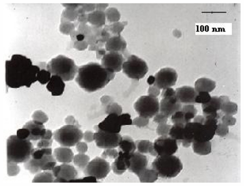
The gold-silica nanoshells were produced as previously described, and aliquots were conjugated to a targeting antibody. The extra sites on the nanoshells were blocked by adding a solution of bovine serum albumin. indicates that the absorption spectra of the bare nanoshells were nearly identical. The antibody did not have any detectable adsorption in the near-infrared region, indicating that the optical properties of the nanoshells must originate from the bare nanoshells. This finding suggests that the properties of the nanoshells were not altered by antibody conjugation or addition of bovine serum albumin. We visualized the gold-silica nanoshells using transmission electron microscopy ().
Figure 1 Spectral characteristics of near-infrared-absorbing nanoshells. The absorption spectrum shows the absorbing near-infrared nature (820 nm) of nanoshells with dimensions consisting of a silica core of 100 nm in diameter and shells approximately 10 nm thick. Predicted optical properties were confirmed using ultraviolet-visible spectrophotometry.
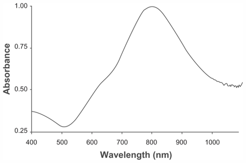
Figure 2 Transmission electron microscopic image of gold-silica nanoshells with an overall diameter of 111 ± 3 nm.
Note: Scale bar = 100 nm.
HER2-targeted nanoshells in KB and HeLaS3 cell lines
As expected, bare nanoshells could be absorbed nonspecifically to the cell surface in both cell lines. Nonspecific attachment of the bare nanoshells could induce cell death in the area treated with laser, but cell mortality was low in the KB and HeLaS3 cells.
HER2-targeted nanobody-conjugated nanoshells in KB and HeLaS3 cells
The nanoshells conjugated to nanobodies were able to induce cell death effectively in KB cells overexpressing HER2 on their surface. The specificity and affinity of binding was confirmed previously by antibodies and antigen-based studies.Citation22 A comparison of the images demonstrated the relationship between nanoshell absorption and cell cytotoxicity following laser treatment ( and B versus ).
Figure 3 (A) HER2-positive KB cells exposed to anti-HER2 immunonanoshells (nanobody-conjugated nanoshells). (B) Cytotoxicity was observed in cells treated with near-infrared laser. Images represent cells targeted with anti-HER2 nanoshells only.
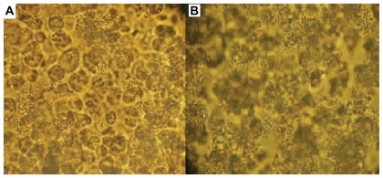
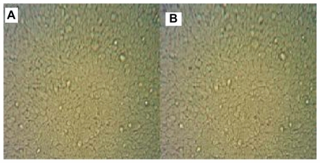
Figure 4 (A) HER2-negative HeLaS3 cells treated with anti-HER2 immunonanoshells. (B) No cytotoxicity was observed in HeLaS3 cells following near-infrared laser treatment.
Viability staining
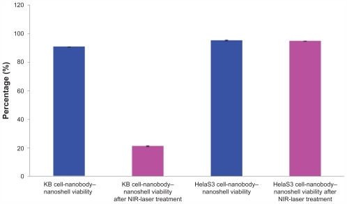
A cell viability staining experimentCitation29 was performed in all the experimental groups. These assays were used to evaluate the number of living cells following near-infrared radiation. Each cell line was divided in four groups, and the viability of each group of cells was evaluated by the MTT assay (–). Nonspecific binding was responsible for 0.6%–1.5% of cell death in KB cells while this number was slightly lower at 0.3%–0.9% in HeLaS3 cells ( and ). The highest mortality was observed in KB cells (69.4%) following addition of the nanobody-nanoshell conjugates and subsequent exposure to laser radiation. Mortality was much lower in the HeLaS3 cells, at approximately 4% (). It should be noted that there was loss of both viable and dead cells due to reagent removal and washing during the experiment. It should be noted that there was a significant difference between all experimental groups.
Figure 5 MTT assay results for normal HER2-positive (KB) and HER2-negative (HeLaS3) cells before and after near-infrared laser irradiation.
Abbreviation: NIR, near-infrared.
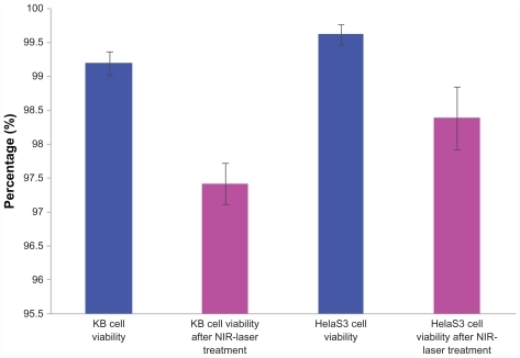
Figure 6 MTT assay results for KB and HeLaS3 cells following treatment with HER2-targeted nanobodies and near-infrared laser.
Abbreviation: NIR, near-infrared.
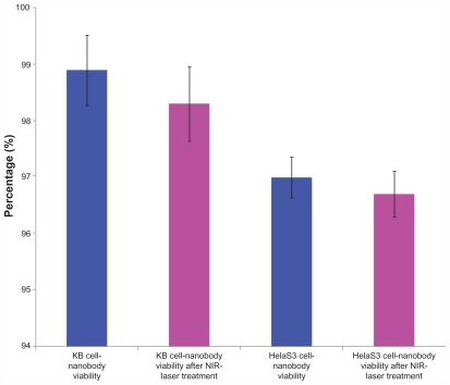
Figure 7 MTT assay results for KB and HeLaS3 cells exposed to bare nanoshells. Both cell types were treated with a near-infrared laser.
Abbreviation: NIR, near-infrared.
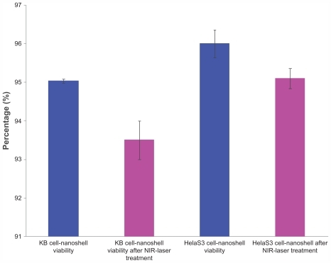
Figure 8 Anti-HER2 immunonanoshells (nanobody-conjugated nanoshells) were added to KB and HeLaS3 cells, and the cells were treated with an near-infrared laser. An MTT assay was performed on both cell types and our results indicated that immunotargeted nanoshells can selectively induce specific cell death in vitro.
Abbreviation: NIR, near-infrared.
Discussion
Improved therapies for oral squamous cell carcinoma, including surgery, chemotherapy, radiotherapy, and immunotherapy, are currently being evaluated to determine their efficacy in promoting long-term, disease-free survival. Photothermal therapy is a novel noninvasive treatment approach and is rapidly being recognized as one of the most effective therapeutic strategies for targeting tumor cells specifically.
In this study, anti-HER2 nanobody-tagged gold-silica nanoshells were generated, and their photothermal potency was examined in KB and HeLaS3 cells. Our data demonstrated selective tumor destruction of more than 69.4% of KB (HER2-positive) cells with no mortality in HeLaS3 (HER2-negative) cells. It should be noted that nonspecific binding of conjugated nanoshells and extra reagents was minimized by washing to prevent cell death following near-infrared laser radiation.
Our current data highlight several advantages of nanobody-conjugated nanoshells compared with current treatments for oral squamous cell carcinoma. Nanobody labeling of nanoshells serves to increase nanoparticle specificity and therapeutic efficiency in vitro and is rapidly emerging as a new therapeutic class of immunonanoshells. For use in vivo, increasing nanoparticle specificity would be beneficial in that it would result in a higher accumulation of nanoparticles in the target tissue. It should be noted that there is no resistance to photothermal application for use in vivo. It has been observed that current tumor therapies can induce cancer cells to alter their cell surface marker expression; however, nanoparticles could localize to target cancer cells without causing any detectable cellular alterations. Furthermore, our toxicology results indicate that the anticancer effects of our immunonanoconjugate are limited to the areas exposed to near-infrared laser irradiation. The requirement for the presence of both nanoshells and laser light to induce cell death is promising for use in tumors in which local recurrence is often the most serious risk and the surrounding normal tissues perform critical functions. In conclusion, we demonstrate for the first time that immunonanoshells containing nanobody conjugates can be used to induce selective cancer cell death in vitro. In vivo studies are currently underway in mouse models of oral squamous cell carcinoma to determine the therapeutic efficacy of gold–silica nanobody immunonanoshells.
Acknowledgment
We thank the staff of the Biotechnology Department of Medical Sciences, Tarbiat Modares University, for their technical assistance with this research.
Disclosure
The authors report no conflicts of interest in this work.
References
- NevilleBWDammDDAllenCMOral and Maxillofacial Pathology3rd edSt Louis, MOSaunders2009365370
- HirotaSKMigliariDASugayaNNOral squamous cell carcinoma in a young patient Case report and literature reviewA Bras Dermatol2006813251254
- SpencerKRFergusonJWWiesenfeldDCurrent concepts in the management of oral squamous cell carcinomaAust Dent J200247428428912587762
- MéndezEChengCFarwellDGTranscriptional expression profiles of oral squamous cell carcinomasCancer20029571482149412237917
- YaoMChangKFunkGFThe failure patterns of oral cavity squamous cell carcinoma after intensity-modulated radiotherapy – the University of Iowa experienceInt J Radiat Oncol Biol Phys20076751332134117276613
- RobergKJonssonACGrenmanRNorberg-SpaakLRadiotherapy responses in oral squamous carcinoma cell lines: evaluation of apoptotic proteins as prognostic factorsHead Neck200729432533417163470
- O’NealDPHirschLRHalasNJPayneJDWestJLPhoto-thermal tumor ablation in mice using near infrared-absorbing nanoparticlesCancer Lett2004209217117615159019
- PhilippCMRohdeEBerlienHPNd:YAG laser procedures in tumor treatmentSemin Surg Oncol19951142902987481366
- PrudhommeMTangJRouySInterstitial diode laser hyperthermia in the treatment of subcutaneous tumorLasers Surg Med19961944454508983005
- ChenWRAdamsRLHigginsAKBartelsKENordquistREPhotothermal effects on murine mammary tumors using indocyanine green and an 808 nm diode laser: an in vivo efficacy studyCancer Lett19969821691738556705
- JoleszFAHynenKMagnetic resonance image-guided focused ultrasound surgeryCancer J20028Suppl 1S10011212075696
- GazelleGSGoldbergSNSolbiatiLLivraghiTTumor ablation with radio-frequency energyRadiology2000217363364611110923
- HirschLRGobinAMLoweryARMetal nanoshellsAnn Biomed Eng2006341152216528617
- LoweryARGobinAMDayESHalasNJWestJLImmunonanoshells for targeted photothermal ablation of tumor cellsInt J Nanomedicine20061214915417722530
- WuXLiuHLiuJImmunofluorescent labeling of cancer marker HER-2 and other cellular targets with semiconductor quantum dotsNat Biotechnol2003211414612459735
- VerelIHeiderKHSiegmundMTumor targeting properties of monoclonal antibodies with different affinity for target antigen CD44V6 in nude mice bearing head and neck cancer xenograftsInt J Cancer200299339640211992408
- BernardiRJLoweryARThompsonPABlaneySMWestJLImmunonanoshells for targeted photothermal ablation in medulloblastoma and glioma: an in vitro evaluation using human cell linesJ Neurooncol200886216517217805488
- HirschLRStaffordRJBanksonJANanoshell-mediated near infrared thermal therapy of tumors under magnetic resonance guidanceProc Natl Acad Sci U S A200310023135491355414597719
- WeisslederRA clearer vision for in vivo imagingNature Biotechnol200119431631711283581
- ReynoldsARMoghimiSMHodivala-DilkeKNanoparticle mediated gene delivery to tumor neovasculatureTrend Mol Med20039124
- XiaWLauYKZhangHZCombination of EGFR, HER-2/neu, and HER-3 are a stronger predictor for the outcome of oral squamous cell carcinoma than any individual family membersClin Cancer Res19995124164417410632356
- SheikholeslamiFRasaeeMJShokrgozarMAIsolation of a novel nanobody against HER2/neu using phage display technologyLabMedicine20104126975
- BernardeVFGleber-NettoFOSousaSFSilvaTAAguiarMCClinical significance of EGFR, Her-2 and EGF in oral squamous cell carcinoma: a case control studyExp Clin Cancer Res201029140
- MaedaHFangJInutsukaTKitamotoYVascular permeability enhancement in solid tumor: various factors, mechanisms involved and its implicationsInt Immunopharmacol20033331932812639809
- JamesWDHirschLRWestJLO’NealPDPayneJDApplication of INAA to the build-up and clearance of gold nanoshells in clinical studies in miceJRNC20072712455459
- OldenburgSJAverittRDWestcottSLHalasNJNanoengineering of optical resonancesChem Phys Lett19982882–4243247
- StöberWFinkABohnEControlled growth of monodisperse silica spheres in the micron size rangeJ Colloid Interface Sci1968266269
- DuffDGBaikerAEdwardsPA new hydrosol of gold clusters. 1. Formation and Particle size variationLangmuir1993923012309
- MosmannTRapid colorimetric assay for cellular growth and survival: application to proliferation and cytotoxicity assaysJ Immunol Methods1983651–2624