Abstract
Background
Realizing the therapeutic benefits of quercetin is mostly hampered by its low water solubility and poor absorption. In light of the advantages of nanovehicles in the delivery of flavanoids, we aimed to deliver quercetin perorally with nanomicelles made from the diblock copolymer, polyethylene glycol (PEG)-derivatized phosphatidylethanolamine (PE).
Methods
Quercetin-loaded nanomicelles were prepared by using the film casting method, and were evaluated in terms of drug incorporation efficiency, micelle size, interaction with Caco-2 cells, and anticancer activity in the A549 lung cancer cell line and murine xenograft model.
Results
The incorporation efficiency into the nanomicelles was ≥88.9% when the content of quercetin was up to 4% w/w, with sizes of 15.4–18.5 nm and polydispersity indices of <0.250. Solubilization of quercetin by the nanomicelles increased its aqueous concentration by 110-fold. The quercetin nanomicelles were stable when tested in simulated gastric (pH 1.2) and intestinal (pH 7.4) fluids, and were non-toxic to the Caco-2 cells as reflected by reversible reduction in transepithelial electrical resistance and ≤25% lactose dehydrogenase release. The anticancer activity of quercetin could be significantly improved over the free drug through the nanomicellar formulation when tested using the A549 cancer cell line and murine xenograft model. The nanomicellar quercetin formulation was well tolerated by the tumor-bearing animals, with no significant weight loss observed at the end of the 10-week study period.
Conclusion
A stable PEG-PE nanomicellar formulation of quercetin was developed with enhanced peroral anticancer activity and no apparent toxicity to the intestinal epithelium.
Acknowledgments
The authors would like to thank Dr Marcel Bally, Dr Dawn Waterhouse, and Ms Hong Yan (Department of Experimental Therapeutics, BC Cancer Research Center, Canada) for providing the facilities and technical support of the animal experiment. The research project is funded by Singapore Economic Development Board Biomedical Sciences Proof-of- Concept Scheme (COY-15-POC/N197-9).
Disclosure
The authors declare no conflicting interests in connection with this article.
Supplementary data
Figure S1 MTT viability of free and nanomicellar quercetin in MDA-MB-231 human breast cancer cell line upon 72 hours of exposure. The concentrations of empty micelle were 96 μM and 192 μM for 30 μM and 60 μM nanomicellar quercetin, respectively.
Notes: Data represent mean ± SEM from three independent experiments. *P < 0.05 as compared to free quercetin group.
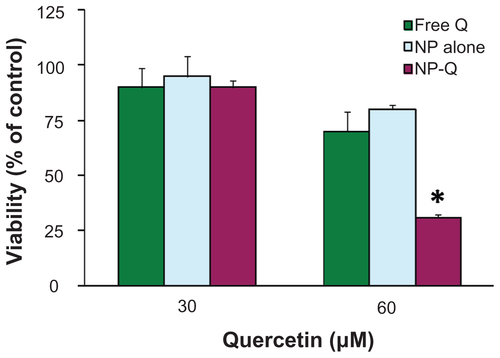
Figure S2 Cellular accumulation of FITC-labeled nanomicelles over 24 hours of incubation under various conditions: (A) at 4°C, (B) at 37°C, (C) without cyclosporine A, (D) with cyclosporine A, (E) without 0.45 M sucrose, (F) with 0.45 M sucrose, (G) without brefeldine A, (H) with brefeldine A.
Notes: Representative images from three independent studies are shown.
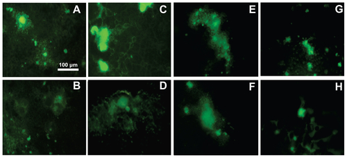
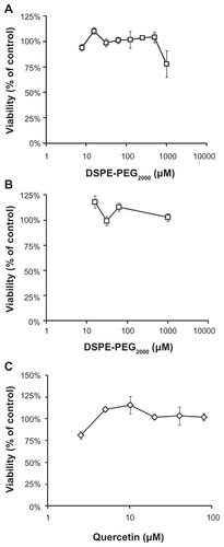
Figure S3 MTT viability of (A) empty, non-drug loaded nanomicelles, (B) quercetin-loaded nanomicelles (showing micelle concentrations), and (C) free quercetin in Caco-2 cell monolayer upon 24 h of exposure.