Abstract
Chitosan, a natural polymer, is a promising system for the therapeutic delivery of both plasmid DNA and synthetic small interfering RNA. Reports attempting to identify the optimal parameters of chitosan for synthetic small interfering RNA delivery were inconclusive with high molecular weight at high amine-to-phosphate (N:P) ratios apparently required for efficient transfection. Here we show, for the first time, that low molecular weight chitosan (LMW-CS) formulations at low N:P ratios are suitable for the in vitro delivery of small interfering RNA. LMW-CS nanoparticles at low N:P ratios were positively charged (ζ-potential ~20 mV) with an average size below 100 nm as demonstrated by dynamic light scattering and environmental scanning electron microscopy, respectively. Nanoparticles were spherical, a shape promoting decreased cytotoxicity and enhanced cellular uptake. Nanoparticle stability was effective for at least 20 hours at N:P ratios above two in a slightly acidic pH of 6.5. At a higher basic pH of 8, these nanoparticles were unravelled due to chitosan neutralization, exposing their polynucleotide cargo. Cellular uptake ranged from 50% to 95% in six different cell lines as measured by cytometry. Increasing chitosan molecular weight improved nanoparticle stability as well as the ability of nanoparticles to protect the oligonucleotide cargo from nucleases at supraphysiological concentrations. The highest knockdown efficiency was obtained with the specific formulation 92-10-5 that combines sufficient nuclease protection with effective intracellular release. This system attained >70% knockdown of the messenger RNA, similar to commercially available lipoplexes, without apparent cytotoxicity. Contrary to previous reports, our data demonstrate that LMW-CS at low N:P ratios are efficient and nontoxic polynucleotide delivery systems capable of transfecting a plethora of cell lines.
Introduction
RNA interference (RNAi), an evolutionary endogenous gene regulation mechanism based on double-stranded RNA (short hairpin RNA, microRNA, Piwi-interacting RNA, and small interfering RNA [siRNA]), has provided a potential new class of therapeutics.Citation1 Since its discovery in Caenorhabditis elegans,Citation2 RNAi has been proven effective in mammalian cellsCitation1,Citation3–Citation11 and has reached clinical trials.Citation1,Citation12–Citation14 However, direct delivery of RNAi-inducing entities such as synthetic siRNA or short hairpin RNA continues to be problematic owing to their rapid extracellular/intracellular degradation by nucleases (ie, RNAse and DNAse), limited blood stability, poor cellular uptake, and nonspecific targeting.Citation15–Citation17 As a consequence, the translation of RNAi into a clinical therapeutic reality is still pending resolution of these issues.
Chemical modification of synthetic siRNAs has provided resistance to nuclease degradation and improved blood stability.Citation18–Citation22 For example, selective addition of a phosphorothioate linkage or substitution with 2′-O-methyl on the C2 position of specific riboses increases nuclease resistance of siRNAs without compromising activity.Citation14,Citation19,Citation20 Nevertheless, some chemical modifications can increase cytotoxicity, off-target effects and reduce messenger RNA (mRNA) hybridization.Citation23–Citation27 Despite progress achieved through chemical modification to increase siRNA half-life, transfection efficiency, cellular targeting, and uptake remain as obstacles to effective delivery. Therefore, packaging systems which can both protect and transport chemically unmodified/modified siRNA to target cells are required.
Liposomes/Lipoplexes have been extensively used as nonviral vehicles for plasmid and RNAi entities and pose toxicity concerns. For example, the repeated administration of lipid-based delivery vehicles caused phospholipidosis.Citation28 Intravenous injection of stable nucleic acid-lipid particles has successfully targeted the liver to silence the apolipoprotein B (ApoB) gene in mice and nonhuman primates.Citation10 However, a significant 20-fold transient elevation in serum transaminases (aspartate transaminase, alanine transaminase) indicative of hepatocellular necrosis was identified at the effective dose. Liposomal formulations of nucleic acids are known inducers of inflammatory cytokines including tumor necrosis factor-alpha, interferon-gamma, and interleukin-6 which may be related to liver damage.Citation29 Polyethylene glycol (PEG) modification of liposomes (PEGylation), for the purpose of reducing their toxicity, was also demonstrated to elicit acute hypersensitivity after repeated dosing.Citation30–Citation32 Similarly, the highly studied cationic family of polymers such as polyethylenimine demonstrated high gene transfer efficiency but was also associated with significant toxicity issuesCitation1,Citation33 limiting their broad use in clinical trials. Polyethylenimine cytotoxicity was characterized as a two-phase process where the polycation-cell interaction induces loss of cell membrane integrity and the induction of programmed cell death. Insights into polyethylenimine toxicity highlight the importance of polycation/organelle interactions – ie, mitochondria and lysosomes – on the induction of toxicity.Citation34,Citation35 In general, cationic polymers display less toxicity associated with cytokine induction – immune activation – compared to their cationic lipid counterparts.Citation36
Chitosan, a family of cationic polymers of β-1-4 N-acetyl-glucosamine and D-glucosamine residues, has been extensively studied for the delivery of plasmid DNA (pDNA) and siRNA both in vitro and in vivo.Citation3,Citation8,Citation17,Citation37–Citation43 Chitosan properties include mucoadhesivity,Citation44 biocompatibility, biodegradability,Citation45 nontoxicity, and low cost of production. Primary amine residues confer a polycationic nature to chitosan at pH values below its pKa (~6.5) thus enabling it to condense polyanionic compounds such as nucleic acids. Electrostatic interaction between chitosan and nucleic acids leads to the spontaneous formation of nanoparticles of different sizes and shapes.Citation46 The ability of chitosan-based nanoparticles to transfect cells efficiently depends on several parameters such as: (1) the degree of deacetylation (DDA), which represents the fraction of ionizable monomers; (2) the average molecular weight (Mn), proportional to chain length, and (3) the amine-to-phosphate (N:P) charge ratio represented by the amine-(chitosan)-to-phosphate (DNA or RNA) ratio used to form nanoparticles.
We have previously demonstrated that maximization of in vitro transfection efficiency for the delivery of pDNA depends on a fine balance between these tunable parameters of chitosanCitation38–Citation40 and found maximum transgene expression for DDA:Mn values that run along a diagonal from high DDA/ low Mn to low DDA/high Mn.Citation38 We have also demonstrated that specific chitosan formulations [DDA, Mn, and N:P ratio] efficiently express transgene in vivo.Citation37,Citation41
We also demonstrated that specific formulations are able to trigger an anti-transgene immune response;Citation37 therefore, nanoparticles can be designed based on the fine-tuning of chitosan parameters for application-specific purposes such as genetic vaccination or gene therapy.
The structural differences between pDNA and siRNA are believed to affect the complexation/stability of nanoparticles and optimal parameters required for effective delivery. Chitosan has also been used for siRNA delivery both in vitro and in vivo.Citation1,Citation8,Citation10,Citation17,Citation43 However, and despite attempts to identify optimal physicochemical parameters for siRNA delivery,Citation43 inconclusive results have been observed in the literature due to experimental discrepancies.Citation8,Citation17 For example, it was reported that intermediate DDA (80%) and high Mw (64–170 kDa) chitosan were more efficient than low molecular weight chitosan (LMW-CS) (10 kDa) in delivering siRNA.Citation17,Citation43 However, high molecular weight chitosans are found to be cytotoxic,Citation47–Citation49 thus potentially limiting their use in future clinical trials. Additionally, most of the reports evaluating the physicochemical parameters of chitosan/siRNA nanoparticles were performed at high N:P ratios (N:P >25).Citation8,Citation17,Citation43 Such formulations bring significant practical problems including limited dosing due to aggregation and the nonspecific effects of large quantities of soluble chitosan.Citation50 Here, we investigate, for the first time, the ability of specific LMW-CS formulations (92-10-5, 80-80-10, 80-40-5, and 80-10-10) [DDA, Mn, and N:P ratio] at low N:P ratios to in vitro deliver siRNA targeting: (1) the RecQL1 DNA helicase mRNA in the colon adenocarcinoma RecQL1 overexpressing cell line (LS174T) and (2) ApoB mRNA in the hepatocarcinoma-derived cell line (HepG2). The choice of these two targets resides in their relevance to cancer and atherosclerosis, respectively.Citation6,Citation7,Citation9,Citation51,Citation52 We also explored the ability of these formulations to transfect multiple cell lines such as A549, AsPC1, HEK293, and Raw264.7 without apparent toxicity. In this study, we hypothesized that, contrary to previous literature,Citation8,Citation17,Citation42,Citation43 low Mw chitosans (LMW-CS) complexed at low N:P ratios represent suitable formulations for siRNA delivery and gene knockdown; similar to our observations with pDNA.Citation37–Citation41 Additionally, we hypothesized that low N:P ratios assure sufficient protection and efficient delivery of the siRNA cargo. Moreover, we explore the physicochemical properties of these specific formulations with the prospect of optimizing nanoparticle transfection and silencing efficiencies. Our results demonstrate, for the first time, that LMW-CSs at low N:P ratios are effective and nontoxic delivery systems for polynucleotide and siRNA delivery for in vitro gene silencing.
Materials and methods
Synthesis of siRNAs and dsODNs
siRNAs targeting the RecQL1 DNA helicase and ApoB mRNAs were synthesized using a novel RNA synthesis chemistry, the 5′-silyl-2′-orthoester protecting groups (2′-ACE)Citation54 combined with a standard phosphoramitide solid-phase technology by Dharmacon (Thermo Scientific, Dharmacon RNAi Technologies, Lafayette, CO). RecQL1 mRNA-specific siRNA (siRNA-RecQL1) contains the sense sequence of 5′-GUUCAGACCACUUCAGCUUdTdT-3′ and antisense 5′-AAGCUGAAGUGGUCUGAACdTdT-3′ whereas ApoB mRNA-specific siRNA (siRNA-ApoB) contains the sense sequence of 5′-GUCAUCACACUGAAUACCAAU-3′ and antisense 5′-AUUGGUAUUCAGUGUGAUGACAC-3′. Mock siRNA was also used as a negative control. Mock siRNA is a nontargeting siRNA (Dharmacon, D-001710-01-05) designed to have minimal targeting of known genes in human, mouse, and rat cells.
Double-stranded oligodeoxynucleotides (dsODNs, 21 bp) encoding the same sequences and mimicking siRNA physicochemical proper ties were used for nanoparticle characterization. The double-stranded oligodeoxynucleotide (dsODN) sequences were synthesized using the phosphoramidite chemistry (Integrated DNA Technologies Inc, Coralville, IO) and used for size and zeta potential determination, nanoparticle stability, and nuclease protection assays. For confocal microscopy and flow cytometry analysis, 6-carboxyfluorescein (6FAM) 5′-labeled dsODNs were used (Integrated DNA Technologies Inc). The rationale for using dsODN for chitosan nanoparticle physicochemical characterization is their siRNA-mimicking properties. These mimicking properties are due to similarities at the structural level (double-stranded structure, length, and nucleotide overhangs) between siRNA and dsODNs. Additionally, charge densities are similar between siRNA and dsODNs due to identical phosphate residue numbers on their backbone. The main differences between siRNA and dsODNs lie in the substitution of uracil to thymine (U → T) in the dsODN sequences, and in the deoxyribosilation of the dsODN sugar backbone.
Preparation and characterization of depolymerized chitosan
Clinical-grade chitosan at different DDAs was obtained from BioSynthec Inc, (Laval, QC, Canada) and depolymerized using nitrous acid to achieve specific number-average molecular weight targets (Mn) of 80, 40, and 10 kDa. Chitosan number- and weight-average molecular weights (Mn and Mw) were determined by gel permeation chromatography using a Shimadzu LC-20AD isocratic pump, autosampler SIL-20AC HT, oven CTO-20AC coupled with a Dawn HELEOS II multiangle laser light scattering detector (Wyatt Technology Co, Santa Barbara, CA), a Viscostar II (Wyatt Technology Co), an Optilab rEX interferometric refractometer (Wyatt Technology Co), and two Shodex OHpak (SB-806M HQ and SB-805 HQ; Showa Denko America, Inc, New York, NY) columns eluted with a pH 4.5 acetic acid (0.15 M)/sodium acetate (0.1 M)/sodium azide (4 mM) buffer.Citation54,Citation55 The injection volume was 100 μL, the flow rate 0.8 mL min−1 and the temperature 25°C. The dn/dc value was previously calculated for chitosan with a DDA of 92% (for a laser’s wavelength of 658 nm) and is equal to 0.208 and 0.201 for chitosan with 80% DDA. The degree of deacetylation was determined by 1H NMR according to our previous reports.Citation38,Citation56
Preparation of chitosan nanoparticles
Chitosans with specific Mn and DDA () were dissolved overnight on a rotary mixer at 0.5% (w/v) in hydrochloric acid using a glucosamine:HCl ratio of 1:1 at a final concentration of 5 mg/mL. Sterile filtered solutions were then diluted with deionized water to obtain the desired ratio (N:P) of amine (chitosan deacetylated groups) to phosphate (dsODNs/siRNA nucleic acids). Nanoparticles (92-10-5, 80-10-10, 80-40-5, and 80-80-5) were then prepared by rapid mixing (pipetting) of 100 μL of diluted chitosan solution to 100 μL of dsODNs or siRNA at a concentration of 0.05 μg/μL or 100 nM.
Table 1 Physicochemical characteristics of bulk chitosans
Nanoparticle size and ζ-potential analysis
The size of chitosan/dsODN-RecQL1 and chitosan/dsODN-ApoB nanoparticles – intensity average diameter – was determined by dynamic light scattering at an angle of 173° at room temperature using the Malvern Zetasizer Nano ZS (Malvern, Worcestershire, UK). Following nanoparticle formation, samples were diluted in 10 mM NaCl at a ratio of 1:10 and measured in triplicate. The ζ-potential was measured in triplicate using laser Doppler velocimetry at 25°C on the same instrument with the viscosity and dielectric constant of pure water used for calculations.
Environmental scanning electron microscopy (ESEM)
Chitosan/dsODN-RecQL1 and chitosan/dsODN-ApoB nanoparticles were sprayed on silicon wafer substrate then sputter-coated with gold (Agar Manual Sputter Coater; Marivac Inc, Montreal, QC, Canada) and imaged using a Quanta 200 FEG Environmental Scanning Electron Microscope (FEI Inc, Hillsboro, OR). Observations were performed at 20 kV using the high-vacuum mode. The average particle diameter (± standard deviation) was determined using the XT Docu image analysis software (FEI Inc).
Nanoparticle stability assessment by polyacrylamide gel electrophoresis
The stability of chitosan/dsODN nanoparticles at different pHs (6.5 and 8) and for different incubation times (0.5, 4, and 24 hours) was assessed using polyacrylamide gel electrophoresis. Upon formation, nanoparticles were mixed at a ratio of 1:1 with 2-(N-morpholino)ethanesulfonic acid buffer (MES 1X) (20 mM MES, 8 mM sodium acetate, pH 6.5) or Tris- acetate (TAE)-EDTA buffer (TAE 1X) (2 M Tris-acetate, 50 mM EDTA, pH 8). The samples were then migrated on a 13% polyacrylamide gel (BioRad Laboratories, Mississauga, ON, Canada) for 2 hours at 100 mV in either MES or TAE buffer. Gels were stained with 0.5 μg/mL ethidium bromide solution (BioRad Laboratories) to visualize dsODNs. Gel documentation and image analysis were performed using a Bio-Vision 3000 (Vilbert Lourmat, Marne-la-Vallée, France) and the Vision-Capt software, respectively.
Nuclease protection assay
The level of protection against nuclease attack offered by chitosan formulations (92-10-5, 80-80-10, 80-40-5, and 80-80-5) was assessed electrophoretically on a 5% agarose gel. Chitosan/dsODN-RecQL1 and chitosan/dsODN-ApoB nanoparticles at different DDA, Mw, and N:P ratios were incubated with 0.5, 1, 2, 5, or 10 units of DNAse I (Sigma-Aldrich, Oakville, ON, Canada) per μg of dsODNs in 20 μL of MES-MgCl2 buffer (20 mM MES, 1 mM MgCl2, pH 6.5) for 30 minutes at 37°C. The reaction was stopped by adding 2 μL of EDTA (50 mM) (Sigma-Aldrich). To ensure proper migration of the nondigested dsODNs, samples were treated with Streptomyces griseus type III chitosanase (Sigma-Aldrich) at 10 mU/μL for 1.5 hours at 37°C and stopped by placing the samples at −20°C for 15 minutes as previously described.Citation3 Samples were migrated at 90 V during 1 hour then stained with 0.5 μg/mL ethidium bromide solution before visualization. Captured images were analyzed using Vision-Capt software (v 15.06; Vilber Lourmat, Paris, France). Relative amounts of dsODN-RecQL1 or dsODN-ApoB (%) were determined by comparison of the integrated signal intensity of nuclease-treated samples versus nontreated samples.
In vitro cell transfection
Cell culture
All cell lines were purchased from American Type Cell Culture (Manassas, VA). The HepG2 cell line was cultured in minimal essential medium (MEM). The HEK293, Raw294.7, and LS174T cell lines were cultured in high-glucose Dulbecco’s modified eagle’s media (DMEM-HG). The A549 and AsPC1 cell lines were cultured in F12-K and Roswell Park Memorial Institute medium media, respectively. All cell culture media contained 1.85 g/L of sodium bicarbonate (NaHCO3) and were supplemented with 10% fetal bovine serum (Cedarlane Laboratories, Burlington, ON, Canada). All cell lines were cultured at 37°C in a 5% CO2 incubator. For transfection, cells were plated in 96-well or 24-well culture plates (Corning, Lowell, MA) to obtain a ~50% confluence the day of transfection using 100 μL/well or 500 μL/well, respectively, of complete culture medium.
Cell transfection
For in vitro transfection, DMEM-HG was prepared with 0.976 g/L of MES and 0.84 g/L of NaHCO3 at a pH of 6.5. Transfection media containing 10% fetal bovine serum was equilibrated overnight at 37°C in a 5% CO2 incubator and the pH was adjusted to 6.5 using sterile HCl (1N) prior to transfection. For siRNA transfection performed in a 96-well plate, chitosan/siRNA nanoparticles were prepared as described above, 30 minutes before use. A 100 μL siRNA solution at a concentration of 0.05 μg/μL was used for siRNA complexation with chitosan at a 1:1 ratio (v/v). Following complexation, nanoparticles were incubated in a ghost plate containing the transfection media (DMEM-HG + fetal bovine serum) at a final concentration of 1.35 ng/μL; equivalent to 10 pmol per well of siRNA. For dsODN transfection performed in a 24-well plate, nanoparticles were complexed as described above and incubated at a final concentration of 8.07 ng/μL, equivalent to 60 pmol per well of dsODNs. Plates containing nanoparticles were equilibrated for 10 minutes at 37°C, 5% CO2. Medium over cells was aspirated and replenished with either 500 μL (24-well plate) or 100 μL per well (96-well plate) of the transfection medium containing dsODN- or siRNA-based nanoparticles. Cells were incubated with chitosan/siRNA or chitosan/dsODN nanoparticles until analysis 24 hours post transfection. The commercially available liposome, DharmaFECT™ (Dharmacon RNAi Technologies), was used as a positive control and both untreated cells and uncomplexed siRNA/dsODN-treated cells were used as negative controls.
Transfection with DharmaFECT
DharmaFECT was used as a positive control for transfection efficiency in all tested cell lines. DharmaFECT/dsODN (flow cytometry and confocal microscopy) or DharmaFECT/ siRNA (qPCR and viability assay) lipoplexes (1:2 [w/v] ratio) were prepared following the manufacturer’s protocol.
In vitro cell viability assay
Nanoparticle toxicity was evaluated using the alamarBlue® proliferation assay (Invitrogen, Carlsbad, CA). The principle of the assay is based on the natural reducing power of viable cells to convert resazurin, a blue and nonfluorescent compound, into resofurin; a red and fluorescent molecule. Viable cells continuously convert resazurin to resofurin, thereby providing a quantitative measure of viability. Transfection was performed as described above using chitosan-siRNA nanoparticles. Five thousand cells/well were seeded 24 hours before transfection. To alleviate the experimental bias from the effect of RecQL1 gene silencing on cell viability, nontargeting siRNA (siRNA mock) was used instead. Twenty-four hours post transfection with chitosan-based nanoparticles, 20 μL of alamarBlue reagent, pre-warmed at 37°C was added to each well and incubated for another 4 hours. At the end of the incubation 100 μL of media containing reduced alamarBlue dye was transferred to a black Corning 96-well plate and read on an infinite 200 fluorescence plate reader (Tecan Systems, San Jose, CA) with excitation 560 nm, emission 590 nm and a cut-off of 570 nm. Cells without the addition of alamarBlue were used as blank and dimethyl sulfoxide was used as a positive control of toxicity. The viability of nontransfected control cells was arbitrarily defined as 100%. The relative cell viability was calculated using the following formula: (fluorescence intensitysample/fluorescence intensitycontrol) × 100.
Uptake analysis by flow cytometry and confocal microscopy
Fluorescence-activated cell sorting (FACS) analysis
The cellular uptake of dsODNs was determined by transfecting AsPC1, A549, LS174T, HepG2, HEK293, and Raw264.7 cell lines with nanoparticles formed with (6FAM) 5′labeled dsODNs. Twenty-four hours post-transfection, cells were chitosanase treated for 60 minutes to eliminate any cell surface-associated nanoparticles left from the transfection as described previously.Citation3 Afterward, cells were washed twice with phosphate-buffered saline, trypsinized, and resuspended in phosphate-buffered saline. The analysis of cell uptake was made using a BD Canto flow cytometer (Becton Dickinson, San Jose, CA). For each sample, 20,000 events were counted and to exclude cell debris, dead cells, and aggregated cells, a collection gate was established using a dot plot of the forward light scatter against the side scatter. Nontransfected cells were used as negative controls to discriminate (6FAM) positive cells from auto-fluorescence.
Confocal microscopy
For nanoparticle internalization analysis, the LS174T, HepG2, HEK293, and Raw264.7 cell lines were seeded on 35 mm glass-bottom culture dishes (MatTek, Ashland, MA) at 40,000 cells/dish using 500 μL of complete culture medium. Nanoparticles were formed with fluorescent rhodamine B isothiocyanate-labeled chitosan and dsODNs labeled with 6FAM on their 5′ extremities (Integrated DNA Technologies). Prior to imaging, cell membranes were stained with 5 μg/mL of Cell Mask™ Deep Red (Invitrogen, Burlington, ON, Canada). Images were taken in multitrack mode using a Zeiss LSM 510 META confocal Axioplan 200 microscope (Carl Zeiss AG, Feldbach, Switzerland). Chitosan and dsODNs were visualized as red and green pseudocolors, respectively. The spatial overlap of these two colors produced yellow which permitted a qualitative assessment of colocalization.
Quantitative PCR (qPCR) analysis of RecQL1 and ApoB mRNA knockdown
RNA extraction and assessment methods (yield, purity, and integrity)
RNA extraction was performed using the RNA XS® extraction kit from Macherey-Nagel (Biolynx, Montréal, QC, Canada) according to the manufacturer’s protocol following chitosanase treatment, as described previously.Citation3 Total RNA was quantified and RNA integrity was measured using the Agilent BioAnalyzer 2100 (Agilent Technologies, Mississauga, ON, Canada) following the manufacturer’s protocol. RNA integrity was evaluated by the ratio of 28S/18S ribosomal RNACitation57 and the RNA integrity number (RIN). The Agilent 2100 BioAnalyzer uses automated microfluidics, capillary electrophoresis, and fluorescence to evaluate RNA integrity. The RIN is a relative measure of RNA quality that is based largely on electrophoretic trace analysis. The BioAnalyzer 2100 automatically computes RIN, where an ideal nondegraded RNA sample has RIN = 10.
Reverse transcription
Total RNA was reverse transcribed in a final volume of 20 μL using the First Strand cDNA Transcriptor Kit (Roche Diagnostics, Laval, QC, Canada) with oligodT primers as described by the manufacturer’s protocol. Samples were stored at −20°C.
Gene expression assays
The RecQL1 and ApoB mRNA expression level was determined using assays designed with the Universal Probe Library (UPL) from Roche (Roche Applied Science, Laval, QC, Canada). Endogenous control (hypoxanthine guanine phosphoribosyl transferase) and glyceraldehyde 3-phosphate dehydrogenase expression levels were determined using pre-validated TaqMan® Gene Expression Assays (Applied Biosystems, Carlsbad, CA). RecQL1 and ApoB mRNA (target detection) reactions for 384-well plate formats were performed using 1.5 μL of cDNA samples (25–50 ng), 5 μL of the Fast Universal qPCR MasterMix (Applied Biosystems) 2 μM of each primer, and 1 μM of a Universal Probe Library probe (RecQL1 [probe #29]/ApoB [probe #55]) in a total volume of 10 μL. For endogenous control assessment, reactions were performed using identical volumes of cDNA and, Fast Universal qPCR Master Mix, 0.5 μL of the TaqMan Gene Expression Assay (20×) and 2.5 μL of water in a total volume of 10 μL.
Detection and analysis
The ABI PRISM® 7900HT Sequence Detection System (Applied Biosystems) was used to detect the amplification level and was programmed with an initial step of 3 minutes at 95°C, followed by 45 cycles of 5 seconds at 95°C and 30 seconds at 60°C. All reactions were run in triplicate and the average values of Cts (threshold cycle) were used for quantification. Glyceraldehyde 3-phosphate dehydrogenase and hypoxanthine guanine phosphoribosyl transferase were used as endogenous controls. The relative quantification of target genes was determined using the ρρCT method. Briefly, the Ct values of target genes were normalized to an endogenous control gene (endogenous control) (ΔCT = Cttarget − CtendoC) and compared with a calibrator: ΔΔCT = ΔCttarget − ΔCtcalibrator. Relative expression (RQ) was calculated using the Sequence Detection System 2.2.2 software using the RQ = 2−ΔΔCT formula.
Statistical analysis
The statistical analysis was performed using Statistica 9.0 Software (STATSOFT; Statistica, Tulsa, OK). Data are expressed as mean ± standard deviation. Statistical significance was determined with one-way analysis of variance, followed by Tukey’s post hoc test. The results were considered significant and highly significant (P < 0.05 and P < 0.01, respectively).
Results
Size and ζ-potential of chitosan nanoparticles
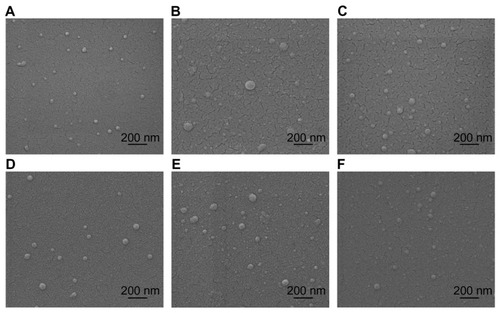
All formulations of chitosan/dsODN nanoparticles were in the range of 41–109 nm as measured by environmental scanning electron microscopy (ESEM) and dynamic light scattering ( and ). Chitosan/dsODN nanoparticles showed higher size values with increasing Mn. No statistically significant differences were observed when comparing DDAs for these specific formulations. The excess chitosan in all formulations resulted in positively charged nanoparticles as shown by ζ-potential measurements ().
Table 2 Size and zeta potential values obtained by dynamic light scattering for chitosan/dsODN-RecQL1 and chitosan/dsODN-ApoB nanoparticles
Figure 1 Environmental scanning electron microscopy images of spherical chitosan/dsODN nanoparticles. (A) 92-10-5 chitosan/dsODN-RecQL1 nanoparticles; (B) 80-40-5 chitosan/dsODN-RecQL1 nanoparticles; (C) 80-10-10 chitosan/dsODN-RecQL1 nanoparticles; (D) 92-10-5 chitosan/dsODN-ApoB nanoparticles; (E) 80-80-5 chitosan/ dsODN-ApoB nanoparticles, and (F) 80-10-10 chitosan/dsODN-ApoB nanoparticles.
Abbreviations: ApoB, apolipoprotein B; dsODN, double-stranded oligodeoxynucleotide.
Chitosan/dsODN nanoparticle stability
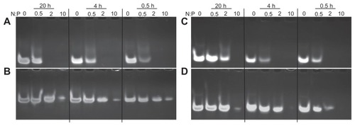
Chitosan-based nanoparticles were incubated for 0.5, 4, and 20 hours in two different buffers (pH 6.5 and 8) to assess the effect of time and pH on nanoparticle stability (). Nanoparticles were stable up to 20 hours at an N:P ratio above 2 in slightly acidic buffers (pH 6.5). At 4 hours following nanoparticle formation, and under slightly acidic conditions, no detectable dsODNs were observed at N:P ratios of 2 or higher (). On the contrary, dsODN release was observed for the same N:P ratios at a pH of 8 (). Longer exposure time – 20 hours – at a pH of 6.5 resulted in increased dsODN-ApoB release at an N:P ratio of 2. This pattern was not observed for the dsODN-RecQL1 sequence. This may be due to sequence/structural differences between the two dsODNs. Furthermore, our results at a pH of 8 show a rapid partial-to-complete dsODN release after 0.5 hour at an N:P ratio of 2 (). At N:P ratio 10 and for the same pH of 8, chitosan showed a partial release of dsODNs indicating the effect of excess chitosan on preserving stability. Overall, our specific chitosan formulations assured nanoparticle stability for a minimum period of 20 hours at an N:P ratio above 2 in slightly acidic near-neutral pH environments.
Figure 2 Chitosan nanoparticle temporal stability. Stability was assessed at 0.5, 4, and 24 hours after complex formation using polyacrylamide gel electrophoresis at a pH of 6.5 (MES 1X) and pH8 (TAE 1X). Chitosan 92-10 at different N:P ratios (0.5, 2, and 10) was complexed with: (A) dsODN-RecQL1 at a pH of 6.5; (B) dsODN-RecQL1 at a pH of 8; (C) dsODN-ApoB at a pH of 6.5, and (D) dsODN-ApoB at a pH of 8. Unstable nanoparticles release dsODNs which become visible following EtBr staining on polyacrylamide gel following ethidium bromide staining of the polyacrylamide gel.
Abbreviations: ApoB, apolipoprotein B; dsODN, double-stranded oligodeoxynucleotide; N:P, amine to phosphate.
Nanoparticle protection assay
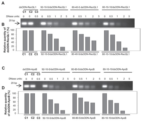
For effective gene expression and/or inhibition, nucleic acids entrapped in the delivery vehicle must be protected from degradation by enzymes such as serum nucleases.Citation58 The ability of chitosan-based nanoparticles to protect siRNA mimicking dsODN sequences was assessed using a DNAse I protection assay against different chitosan formulations complexed with dsODN-RecQL1 or dsODN-ApoB. Upon incubation with DNAse I, naked dsODN-RecQL1 and dsODN-ApoB (controls) were completely degraded (, lane 3). In contrast, DNAse I protection assay showed that all chitosans tested protected dsODNs from degradation at DNAse I concentrations <2 units DNAse I per μg dsODN (). Chitosan formulations demonstrated an average of ~80% protection of dsODNs at DNAse I concentrations of 0.5 U/μg (). The ability of LMW-CS (92-10, 80-40, 80-80, and 80-10) to protect dsODNs from nuclease degradation decreased with increased concentrations of DNAse I. Our results show that protection decreased from ~50% at a DNAse I concentration of 1 U/μg to less than ~20% at 2 U/μg (92-10 and 80-10). Moreover, our results suggest that higher Mn chitosan (80-40 and 80-80) offers a slightly better protection of dsODNs as compared to lower Mn chitosan (92-10 and 80-10) at high DNAse I concentrations (2 U/μg) (). The enhanced cargo protection observed with higher molecular weight chitosans is consistent with previous studies where higher binding affinities between high Mw chitosans and nucleic acids was demonstrated.Citation59 Altogether, our results show that DNAse I protection is considerable when using intermediate to low DDA/Mn and preserves approximately 60% of nucleic acid when using 1 unit of DNAse I per μg of dsODNs.
Figure 3 Nuclease protection assays of chitosan/dsODN nanocomplexes. (A) Chitosan (92-10-5, 80-40-5 or 80-10-10) complexed with dsODN-RecQL1. (B) dsODN-RecQL1 remaining after the DNAse I digestion was assessed using the signal intensity of the treated samples with the control (ie, 0 U DNAse I = 100% intensity). This comparison was made between the samples of the same chitosan formulation. (C) Chitosans (92-10-5, 80-80-5 or 80-10-10) complexed with dsODN-ApoB. (D) dsODN-ApoB remaining after the DNAse I digestion was similarly assessed as in (B).
Abbreviations: ApoB, apolipoprotein B; dsODN, double-stranded oligodeoxynucleotide.
In vitro cell uptake analysis by flow cytometry and confocal microscopy
Nanoparticle internalization into cells can be another rate-limiting step for effective drug delivery systems. In general, efficient nanoparticle internalization depends on several factors, such as the cell type, the physicochemical surface properties of the nanoparticles, and the bio–nano interface. Citation60 The internalization of RecQL1- and ApoB-bearing nanoparticles was assessed in two different sets of relevant cell lines using flow cytometry (FACS). For the assessment of (6FAM) dsODN-RecQL uptake, transfection and FACS analysis were performed on AsPC1, A549, and LS174T cancer cell lines whereas (6FAM) dsODN-ApoB uptake was performed on HEK293, HepG2, and Raw269.7 cell lines. Our FACS results show that cell uptake using chitosan/(6FAM) dsODN-ApoB nanoparticles achieved levels comparable to the commercially used lipoplex (DharmaFECT) ( and ), demonstrating the internalization efficiency of LMW-CS formulations in different cell lines. Moreover, our results indicate that different chitosan formulations show statistically significant differences in their cell uptake efficiency, with LMW-CSs 92-10-5 and 80-10-10 more easily internalized compared to the higher molecular weight 80-80-5 and 80-40-5, in a cell-line-dependent manner. Interestingly, the A549 and HEK293 cell lines demonstrated no statistical differences in uptake efficiency between the different chitosan formulations ( and ). However, the A549 and HEK293 cell lines showed statistically significant increases in uptake when compared to the LS174T and Raw264.7 cell lines, again highlighting some important cell-type dependencies.
Figure 4 Cellular uptake of dsODN-RecQL1 nanoparticles 24 hours post transfection in AspC1, A549, and LS174T cancer cell lines. Chitosan formulations 92-10-5, 80-40-5, and 80-10-10 were complexed to (6FAM) 5′ labeled dsODN-RecQL1 and transfected at 60 pmol/well 24 hours prior to fluorescence-activated cell sorting analysis. DharmaFECT™ was used as the positive uptake control.
Notes: Values are mean ± SD; n = 3; **P > 0.01.
Abbreviations: dsODN, double-stranded oligodeoxynucleotide; SD, standard deviation.
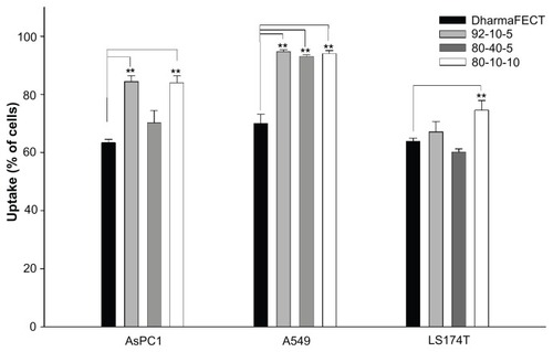
Figure 5 Cellular uptake of dsODN-ApoB nanoparticles 24 hours post transfection in HEK293, Raw269.7, and HepG2 cell lines. Chitosan formulations 92-10-5, 80-80-5, and 80-10-10 were complexed to (6FAM) 5′ labeled dsODN-ApoB and transfected at 60 pmol/well 24 hours prior to fluorescence-activated cell sorting analysis. (A) Uptake efficiency of dsODN-ApoB in percentage (%). (B) Uptake efficiency of dsODN-ApoB in HepG2 cells at different passage numbers. DharmaFECT™ was used as the positive uptake control.
Notes: Values are mean ± SD; n = 3; *P > 0.05; **P > 0.01.
Abbreviations: ApoB, apolipoprotein B; dsODN, double-stranded oligodeoxynucleotide; SD, standard deviation.
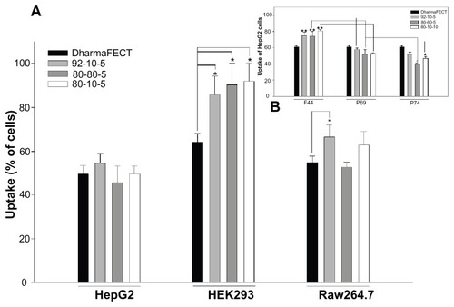
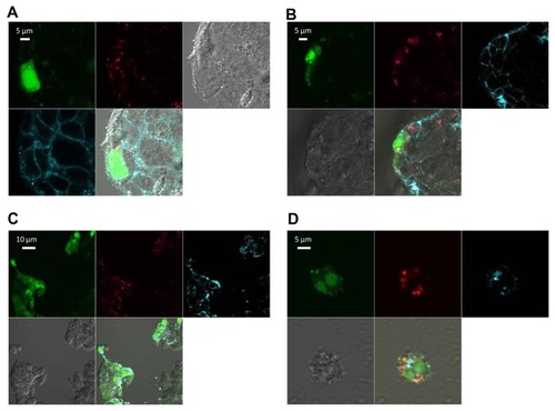
In general, LMW-CS (92-10-5 and 80-10-10) showed higher uptake efficiency, ranging from approximately 65% to 95% depending on the transfected cell line ( and ). These results are in accordance with confocal microscopy data, where images representative of the whole population show that the vast majority of cells for each of the four cell types imaged show nanoparticle internalization (). The lack of colocalization at 24 hours between dsODNs and chitosan indicates that complete release of the dsODN cargo was achieved 24 hours post transfection. Furthermore, the diffuse staining pattern of dsODNs seen in most transfected cells suggests that complexes have escaped endocytic vesicles (), consistent with previous live cell imaging work using chitosan–plasmid DNA nanoparticles.Citation40
Figure 6 Confocal imaging of chitosan/dsODN nanocomplex uptake 24 hours post transfection. Chitosan 92-10 (DDA, Mn) was labeled with rhodamine (red) and dsODNs were 5′ labeled with (6FAM) (green). Chitosan 92-10 was complexed to dsODNs at an N:P ratio of 5. Cell membranes were stained prior to imaging with CellMask™ (blue) to differentiate between internalized and membrane-bound nanoparticles. Images shown represent each separate channel, with dsODNs in green, chitosan in red, membrane in blue, differential interference contrast image in grey, and the merged images shown on the bottom left quadrant. (A) LS174T cells transfected with chitosan/dsODNRecQL1 nanoparticles. (B) HepG2 cells transfected with chitosan/dsODN-ApoB nanoparticles. (C) HEK293 cells transfected with chitosan/dsODN-ApoB nanoparticles. (D) Raw 294.7 cells transfected with chitosan/dsODN-ApoB nanoparticles.
Abbreviations: ApoB, apolipoprotein B; dsODN, double-stranded oligodeoxynucleotide; N:P, amine to phosphate.
Specific gene silencing and cell cytotoxicity evaluation of chitosan nanoparticles in different cell lines
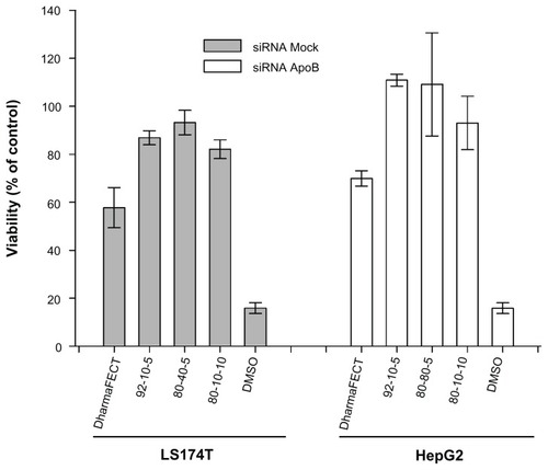
Gene silencing occurs when complementarity is achieved between the siRNA seed region and target mRNA.Citation1 Chitosan- specific formulations (92-10-5, 80-40-5, 80-10-10, and 80-80-5) were assessed for mRNA knock-down in two different cell lines relevant to cancer and atherosclerosis, targeted by RecQL1 and ApoB siRNA, respectively. qPCR analysis revealed inhibition of RecQL1 and ApoB since their coding mRNAs were downregulated more than twofold (). More specifically, in LS174T cells, chitosan 92-10-5 showed a high level of silencing (~80%) of RecQL1, similar to the current commercial gold standard liposomal formulation (~80%), used here as a positive control. Formulations 80-40-5 and 80-10-10 also induced significant silencing but to a lower degree than 92-10-5 and also with an increase of nonspecific mock silencing, especially for formulation 80-10-10, for reasons that remain to be elucidated. For the HepG2 cell line, only the best performing 92-10-5 was tested and induced significant silencing (~55% versus ~80% for positive control) of ApoB but slightly lower than RecQL1 for LS174T. Importantly, our results showed that silencing efficiency with chitosan reached similar levels to the positive control, with a markedly reduced cytotoxicity from the delivery system as assessed using the alamarBlue assay ().
Figure 7 Real-time polymerase chain reaction analysis of the inhibition of RecQL1 and ApoB gene expression in specific cell lines. LS174T cells were transfected with chitosan (92-10-5, 80-40-5, and 80-10-10)/siRNA-RecQL1 nanoparticles, whereas HepG2 cells were transfected with chitosan (92-10-5)/siRNA-ApoB nanoparticles. The inhibition percentage was obtained by comparing the transfected and nontransfected cells, using the ΔΔCT method.
Notes: Values are mean ± SD; n = 3; **P > 0.01.
Abbreviations: ApoB, apolipoprotein B; siRNA, small interfering RNA; SD, standard deviation.
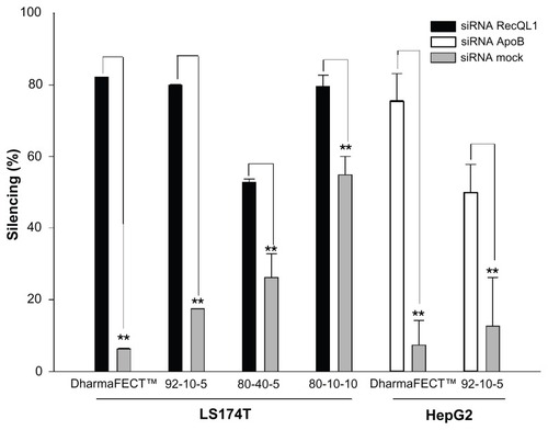
Figure 8 Cell viability assessment using the alamarBlue® assay 24 hours post transfection with different chitosan/siRNA formulations. To alleviate the apoptotic effect of RecQL1 gene silencing for a proper assessment of chitosan-siRNA toxicity, mock siRNA was used for transfection in the LS174T cell line. The HepG2 cell line was transfected with ApoB siRNA. DharmaFECT™ was used for comparison purposes whereas dimethyl sulfoxide was used as a positive control of toxicity.
Notes: Values are presented as mean ± SD; n = 3.
Abbreviations: ApoB, apolipoprotein B; siRNA, small interfering RNA.
Discussion
In this study, we evaluated the efficiency of specific low molecular weight chitosan (LMW-CS) formulations at low N:P ratios for the in vitro delivery of siRNA targeting either RecQL1 or ApoB genes. RecQL1 is a DNA helicase playing a major role in homologous recombination, maintenance of genomic stability, and DNA repair at damaged replication forks.Citation52,Citation61 Overexpression of RecQL1 has been implicated in cancer by preventing cell apoptosis.Citation6,Citation7,Citation62 As for ApoB, it is a major gene involved in atherosclerosis through its essential role in the formation of very low density lipoprotein which will therefore generate low density lipoproteins following triacylglycerol hydrolyzation in the circulation.Citation51,Citation63,Citation64
Nanoparticle size is one parameter affecting uptake and intracellular trafficking, both considered as potential rate-limiting steps for effective gene therapy.Citation41,Citation42 For instance, nano-sized particles have been shown to be internalized more efficiently than micro-sized particles.Citation65–Citation67 In this study, LMW-CS- based nanoparticles ranged in size from 41–110 nm, a size range promoting uptake, prolonged blood circulation, higher tissue penetration, and a relatively free passage from the mononuclear phagocyte system.Citation48,Citation68–Citation70 Therefore, our results show that these specific LMW-CS nanoparticles at low N:P ratios meet performance criteria () and are potentially relevant for in vivo administration. The different chitosan parameters – DDA, Mn, and N:P ratios – used in this study did not significantly affect nanoparticle size, with higher molecular weight chitosan promoting a slightly increased size (). Our results are in contrast to previously published reports where the authors found increased nanoparticle size for lower molecular weight chitosan.Citation43 This discrepancy may be due to differences in experimental conditions and to the high N:P ratio used in Liu et al and Howard et al reportsCitation8,Citation43 versus low N:P ratios reported in our study. ESEM analysis revealed that these small nanoparticles were of spherical shape consistent with previous findings for pDNA,Citation37 siRNA,Citation8 and dsODNs.Citation71 The effect of nanoparticle shape on internalization efficiency showed spherical particles of similar size being internalized 500% more efficiently than rod-shaped particles.Citation72–Citation74 This is mainly explained by increased membrane-wrapping time required for elongated particles and greater thermodynamic forces required for their engulfment.Citation60,Citation74 It was previously demonstrated that the morphology of chitosan-pDNA nanoparticles is strongly dependent upon their charge ratios, and the variation of the latter resulted in nanoparticles with different topological conformations including spherical,Citation75 toroidal,Citation76,Citation77 and globular morphologies.Citation76,Citation78 Chitosan-based nanoparticle shape may also seem to be affected by the type of nucleic acid – pDNA or siRNA/dsODN – used for complexation and the process of nanoparticle formation; ie, ionic gelation. The fact that these LMW-CS nanoparticles demonstrated a reproducible pattern of spherical particles at low N:P ratios may be indicative of higher internalization efficiency than nanoparticles of different topological conformations.
Table 3 Safety and performance criteria for the development of effective nonviral gene delivery systems
Nanoparticle stability and nucleic acid protection are important parameters for efficient nucleic acid delivery. Our results of nuclease protection indicate that all LMW-CS formulations tested were able to protect dsODNs at supraphysiological concentrations of nucleases. Nuclease protection is of great importance for nucleic acid delivery systems through maintenance of cargo bioavailability and improved pharmacokinetic profile, thereby increasing the therapeutic potential of these nanoparticles. Increasing chitosan molecular weight resulted in an enhanced cargo protection () in agreement with previous findings.Citation17,Citation38,Citation42,Citation43,Citation46 Nevertheless, enhancing the ability of nanoparticles to protect their siRNA from degradation may render their intracellular disassembly more difficult, as demonstrated with high molecular weight chitosan-pDNA nanoparticles.Citation40 Further characterization of nanoparticle stability by gel retardation assays show that LMW-CS used at low N:P ratios can effectively complex and compact dsODNs into stable particles. We found LMW-CS nanoparticles at N:P ratios above 2 to be stable in slightly acidic buffers for at least 20 hours. These interesting findings are in contrast with most previous studies using chitosan-siRNA nanoparticles, where high Mn and high N:P ratios are usually required to achieve particle stability.Citation8,Citation17,Citation42,Citation43,Citation79 This discrepancy can be explained by the lower pH (pH 6.5) of the electrophoresis buffer in our study compared to the commonly used TAE buffer at a pH of 8 for chitosan-based nanoparticle characterization,Citation8,Citation17,Citation42,Citation43,Citation80 a difference that was clearly highlighted by our gel retardation assay performed at both pHs (). The use of a lower pH in the electrophoresis buffer results in higher degrees of chitosan ionization which translates to stronger electrostatic attraction to the polyanionic nucleic acid and hence more stable nanoparticles. This simple modification of the pH permits lower N:P ratios than those observed previouslyCitation8,Citation17,Citation42,Citation43,Citation80 to achieve nanoparticle stability. A direct consequence of this modification translates into reduced dosing, aggregation, and other undesirable nonspecific effects of large quantities of soluble chitosan for in vivo delivery where nanoparticles are to be injected at physiological pH values close to the chitosan pKa of 6.5.
In general, efficient nanoparticle internalization depends on factors such as cell type, physicochemical surface properties of the nanoparticles, and the bio–nano interface.Citation60 In this report, we demonstrated that LMW-CS nanoparticles were efficiently internalized in multiple cell lines. The uptake efficiency as measured by flow cytometry ranged from 50% (Raw269.7) to 95% (A549 and HEK293), depending on the cell line. Statistical analysis of uptake efficiency intercell lines showed meaningful differences when comparing the A549 and HEK293 (high uptake) to the LS174T and Raw269.7 (medium uptake), indicating a cell-line dependency of chitosan uptake. The cell-line dependency of chitosan nanoparticles uptake was previously suggested to be associated with different endocytic pathways.Citation81,Citation82 Flow cytometry data showed LMW-CS nanoparticles to be efficiently internalized to levels similar or higher than commercially available liposomal systems such as DharmaFECT.
Finally, the transfection efficiency of LMW-CS nanoparticles as measured by gene-silencing efficacy was evaluated in two different cell lines: RecQL1 in LS174T cells and ApoB in HepG2 cells. The ability of these chitosan formulations to efficiently silence gene expression reached more than a twofold specific mRNA knockdown; with chitosan 92-10-5 being the most efficient and specific in the LS174T cell line. Other low molecular weight formulations also achieved good levels of gene silencing in the LS174T cell line. Interestingly, chitosan 80-10-10 achieved a high level of silencing with a concomitant increase in silencing when delivering mock siRNA. This intriguing observation is currently under investigation in our laboratory. The chitosan formulation 92-10-5 complexed to ApoB siRNA showed lower target mRNA knockdown in HepG2 when compared to the LS174T cell lines targeted with the RecQL1 siRNA. The silencing efficiency correlated well with uptake efficiency as observed by flow cytometry where chitosan 92-10-5 showed both high uptake and high silencing efficiencies. Despite structural differences between pDNA and siRNA,Citation46 the chitosan formulation 92-10-5 has shown the highest transfection efficiencies for both siRNA and pDNA to date.Citation37–Citation41 Taken together, our results show that LWM-CS nanoparticles at low N:P ratios can achieve efficient uptake and gene silencing in vitro, serving as a proof of concept for their use as efficient siRNA delivery vectors in cancer and atherosclerotic animal models. Although in vitro and in vivo performance criteria differ, no consensus on such performances has been established. For in vivo performance, safety remains the major issue, with guidance available from the US Food and Drug Administration for the development of gene and cell therapy products.Citation83 Therefore, the development of nonviral drug delivery systems for in vivo use should take into account physicochemical criteria, cell-based criteria, and, most importantly, in vivo performance and safety criteria (). The low molecular weight, low N:P system presented here meets many of these criteria and has already been demonstrated as efficient in vivo for plasmid DNA delivery.Citation37,Citation41 Thus a complete characterization of the safety and in vivo performance of our LMW-CS system delivering RecQL1 and ApoB targeting siRNA is currently under investigation in animal models of cancer and atherosclerosis.
Acknowledgments
This work was supported by the National Science and Engineering Research Council (NSERC) and by the Groupe de Recherche en Sciences et Technologies Biomédicales of the Fonds de la Recherche en Santé Quebec. We are grateful to Dr Monica Nelea for the ESEM analyses.
Disclosure
The authors declare no conflicts of interest in this work.
References
- de FougerollesAVornlocherHPMaraganoreJLiebermanJInterfering with disease: a progress report on siRNA-based therapeuticsNat Rev Drug Discov20076644345317541417
- FireAXuSMontgomeryMKKostasSADriverSEMelloCCPotent and specific genetic interference by double-stranded RNA in Caenorhabditis elegansNature199839166698068119486653
- AlamehMJeanMDeJesusDBuschmannMDMerzoukiAChitosanase-based method for RNA isolation from cells transfected with chitosan/siRNA nanocomplexes for real-time RT-PCR in gene silencingInt J Nanomedicine2010547320957169
- BantounasIPhylactouLAUneyJBRNA interference and the use of small interfering RNA to study gene function in mammalian systemsJ Mol Endocrinol200433354555715591019
- CastanottoDRossiJJThe promises and pitfalls of RNA-interference-based therapeuticsNature2009457722842643319158789
- FutamiKKumagaiEMakinoHInduction of mitotic cell death in cancer cells by small interference RNA suppressing the expression of RecQL1 helicaseCancer Sci2008991718017953710
- FutamiKKumagaiEMakinoHAnticancer activity of RecQL1 helicase siRNA in mouse xenograft modelsCancer Sci20089961227123618422747
- HowardKARahbekULLiuXRNA interference in vitro and in vivo using a novel chitosan/siRNA nanoparticle systemMol Ther200614447648416829204
- SoutschekJAkincABramlageBTherapeutic silencing of an endogenous gene by systemic administration of modified siRNAsNature2004432701417317815538359
- ZimmermannTSLeeACAkincARNAi-mediated gene silencing in non-human primatesNature2006441708911111416565705
- ElbashirSMHarborthJLendeckelWYalcinAWeberKTuschlTDuplexes of 21-nucleotide RNAs mediate RNA interference in cultured mammalian cellsNature2001411683649449811373684
- WhelanJFirst clinical data on RNAiDrug Discov Today200510151014101516055013
- CoreyDRRNA learns from antisenseNat Chem Biol20073181117173018
- CoreyDRChemical modification: the key to clinical application of RNA interference?J Clin Invest2007117123615362218060019
- SteinCAPhosphorothioate antisense oligodeoxynucleotides: questions of specificityTrends Biotechnol19961451471498645447
- Urban-KleinBWerthSAbuharbeidSCzubaykoFAignerARNAi-mediated gene-targeting throught systemic application of polyethylenimine (PEI)-complexed siRNA in vivoGene Ther200412546146615616603
- KatasHAlparHODevelopment and characterisation of chitosan nanoparticles for siRNA deliveryJ Control Release2006115221622516959358
- ElmenJThonbergHLjungbergKLocked nucleic acid (LNA) mediated improvements in siRNA stability and functionalityNucleic Acids Res200533143944715653644
- JudgeADBolaGLeeACMacLachlanIDesign of noninflammatory synthetic siRNA mediating potent gene silencing in vivoMol Ther200613349450516343994
- WhiteheadKALangerRAndersonDGKnocking down barriers: advances in siRNA deliveryNat Rev Drug Discov20098212913819180106
- BramsenJBLaursenMBNielsenAFA large-scale chemical modification screen identifies design rules to generate siRNAs with high activity, high stability and low toxicityNucleic Acids Res20093792867288119282453
- LayzerJMMcCaffreyAPTannerAKHuangZKayMASullengerBAIn vivo activity of nuclease-resistant siRNAsRNA200410576677115100431
- WeyermannJLochmannDGeorgensCZimmerAAlbumin-protamine- oligonucleotide-nanoparticles as a new antisense delivery system. Part 2: cellular uptake and effectEur J Pharm Biopharm200559343143815760723
- AmarzguiouiMHolenTBabaieEPrydzHTolerance for mutations and chemical modifications in a siRNANucleic Acids Res200331258959512527766
- ParrishSFleenorJXuSMelloCFireAFunctional anatomy of a dsRNA trigger: differential requirement for the two trigger strands in RNA interferenceMol Cell2000651077108711106747
- BraaschDAJensenSLiuYRNA interference in mammalian cells by chemically-modified RNABiochemistry200342267967797512834349
- HarborthJElbashirSMVandenburghKSequence, chemical, and structural variation of small interfering RNAs and short hairpin RNAs and the effect on mammalian gene silencingAntisense Nucleic Acid Drug Dev20031328310512804036
- ReasorMJKacewSDrug-induced phospholipidosis: are there functional consequences?Exp Biol Med (Maywood)2001226982583011568304
- TousignantJDGatesALIngramLAComprehensive analysis of the acute toxicities induced by systemic administration of cationic lipid:plasmid DNA complexes in miceHum Gene Ther200011182493251311119421
- IshidaTIchiharaMWangXInjection of PEGylated liposomes in rats elicits PEG-specific IgM, which is responsible for rapid elimination of a second dose of PEGylated liposomesJ Control Release20061121152516515818
- JudgeAMcClintockKPhelpsJRMacLachlanIHypersensitivity and loss of disease site targeting caused by antibody responses to PEGylated liposomesMol Ther2006132328337 Epub November 7, 200516275098
- SempleSCHarasymTOClowKAAnsellSMKlimukSKHopeMJImmunogenicity and rapid blood clearance of liposomes containing polyethylene glycol-lipid conjugates and nucleic acidJ Pharmacol Exp Ther2005312310201026 Epub November 3, 200415525796
- BoeckleSvon GersdorffKvan der PiepenSCulmseeCWagnerEOgrisMPurification of polyethylenimine polyplexes highlights the role of free polycations in gene transferJ Gene Med20046101102111115386739
- MoghimiSMSymondsPMurrayJCHunterACDebskaGSzewczykAA two-stage poly(ethylenimine)-mediated cytotoxicity: implications for gene transfer/therapyMol Ther200511699099515922971
- HunterACMoghimiSMCationic carriers of genetic material and cell death: a mitochondrial taleBiochim Biophys Acta201017976–71203120920381448
- Al-DosariMSGaoXNonviral gene delivery: principle, limitations, and recent progressAAPS J20094671681 Epub October 16, 200919834816
- JeanMSmaouiFLavertuMChitosan-plasmid nanoparticle formulations for IM and SC delivery of recombinant FGF-2 and PDGF-BB or generation of antibodiesGene Ther20091691097111019440230
- LavertuMMéthotSTran-KhanhNBuschmannMDHigh efficiency gene transfer using chitosan/DNA nanoparticles with specific combinations of molecular weight and degree of deacetylationBiomaterials200627274815482416725196
- NimeshSThibaultMMLavertuMBuschmannMDEnhanced gene delivery mediated by low molecular weight chitosan/DNA complexes: effect of pH and serumMol Biotechnol201046218219620454872
- ThibaultMNimeshSLavertuMBuschmannMDIntracellular trafficking and decondensation kinetics of chitosan-pDNA polyplexesMol Ther201018101787179520628361
- JeanMAlamehMBuschmannMDMerzoukiAEffective and safe gene-based delivery of GLP-1 using chitosan/plasmid-DNA therapeutic nanocomplexes in an animal model of Type 2 DiabetesGene Ther201118880781621412280
- HowardKAKjemsJPolycation-based nanoparticle delivery for improved RNA interference therapeuticsExpert Opin Biol Ther20077121811182218034647
- LiuXHowardKADongMThe influence of polymeric properties on chitosan/siRNA nanoparticle formulation and gene silencingBiomaterials20072861280128817126901
- de CamposAMDieboldYCarvalhoELSanchezAAlonsoMJChitosan nanoparticles as new ocular drug delivery systems: in vitro stability, in vivo fate, and cellular toxicityPharm Res200421580381015180338
- OnishiHMachidaYBiodegradation and distribution of water-soluble chitosan in miceBiomaterials199920217518210022787
- MaoSSunWKisselTChitosan-based formulations for delivery of DNA and siRNAAdv Drug Deliv Rev2010621122719796660
- RichardsonSCKolbeHVDuncanRPotential of low molecular mass chitosan as a DNA delivery system: biocompatibility, body distribution and ability to complex and protect DNAInt J Pharm1999178223124310205643
- HuangMKhorELimLYUptake and cytotoxicity of chitosan molecules and nanoparticles: effects of molecular weight and degree of deacetylationPharm Res200421234435315032318
- WiegandCWinterDHiplerUCMolecular-weight-dependent toxic effects of chitosans on the human keratinocyte cell line HaCaTSkin Pharmacol Physiol201023316417020110767
- MaPLBuschmannMDWinnikFMOne-step analysis of DNA/ chitosan complexes by field-flow fractionation reveals particle size and free chitosan contentBiomacromolecules201011354955420158194
- OlofssonSOBorenJApolipoprotein B: a clinically important apolipoprotein which assembles atherogenic lipoproteins and promotes the development of atherosclerosisJ Intern Med2005258539541016238675
- SharmaSDohertyKMBroshRMJrMechanisms of RecQ helicases in pathways of DNA metabolism and maintenance of genomic stabilityBiochem J2006398331933716925525
- ScaringeSAWincottFECaruthersMHNovel RNA synthesis method using 5′-silyl-2′-orthoester protecting groupsJ Am Chem Soc19981201182011821
- NguyenSHisigerSJolicoeurMWinnikFMBuschmannMDFractionation and characterization of chitosan by analytical SEC and 1H-NMR after semi-preparative SECCarbohydr Polym200975636646
- NguyenSWinnikFMBuschmannMDImproved reproducibility in the determination of the molecular weight of chitosan by analytical size exclusion chromatographyCarbohydr Polym200975528533
- LavertuMXiaZSerreqiANA validated 1H-NMR method for the determination of the degree of deacetylation of chitosanJ Pharm Biomed Anal20033261149115812907258
- SkrypinaNATimofeevaAVKhaspekovGLSavochkinaLPBeabealashvilliRTotal RNA suitable for molecular biology analysisJ Biotechnol20031051–21914511905
- QuongDNeufeldRJDNA protection from extracapsular nucleases, within chitosan- or poly-L-lysine-coated alginate beadsBiotechnol Bioeng199860112413410099413
- MaPLLavertuMWinnikFMBuschmannMDNew insights into chitosan-DNA interactions using isothermal titration microcalorimetryBiomacromolecules20091061490149919419142
- NelAEMadlerLVelegolDUnderstanding biophysicochemical interactions at the nano-bio interfaceNat Mater20098754355719525947
- WuLHicksonIDDNA helicases required for homologous recombination and repair of damaged replication forksAnnu Rev Genet20064027930616856806
- KawabeTTsuyamaNKitaoSDifferential regulation of human RecQ family helicases in cell transformation and cell cycleOncogene200019414764477211032027
- SchaeferJRScharnaglHBaumstarkMWHomozygous familial defective apolipoprotein B-100. Enhanced removal of apolipoprotein E-containing VLDLs and decreased production of LDLsArterioscler Thromb Vasc Biol19971723483539081691
- ItabeHOxidative modification of LDL: its pathological role in atherosclerosisClin Rev Allergy Immunol200937141118987785
- GrefRDombAQuellecPThe controlled intravenous delivery of drugs using PEG-coated sterically stabilized nanospheresAdv Drug Deliv Rev199516215233
- Bivas-BenitaMRomeijnSJungingerHEBorchardGPLGA-PEI nanoparticles for gene delivery to pulmonary epitheliumEur J Pharm Biopharm20045811615207531
- PanyamJLabhasetwarVBiodegradable nanoparticles for drug and gene delivery to cells and tissueAdv Drug Deliv Rev200355332934712628320
- GuyJDrabekDAntoniouMDelivery of DNA into mammalian cells by receptor-mediated endocytosis and gene therapyMol Biotechnol1995332372487552693
- DesaiMPLabhasetwarVAmidonGLLevyRJGastrointestinal uptake of biodegradable microparticles: effect of particle sizePharm Res19961312183818458987081
- SeymourLWPassive tumor targeting of soluble macromolecules and drug conjugatesCrit Rev Ther Drug Carrier Syst1992921351871386002
- JeanMAlamehMDe JesusDChitosan-based therapeutic nanocomplexes for combination gene therapy and gene silencing of in vitro cell lines relevant to type 2 diabetesEur J Pharm Sci2011451–2138149 Epub November 9, 201122085632
- ChithraniBDChanWCElucidating the mechanism of cellular uptake and removal of protein-coated gold nanoparticles of different sizes and shapesNano Lett2007761542155017465586
- ChithraniBDGhazaniAAChanWCDetermining the size and shape dependence of gold nanoparticle uptake into mammalian cellsNano Lett20066466266816608261
- VermaAStellacciFEffect of surface properties on nanoparticle-cell interactionsSmall201061122119844908
- LiuWSunSCaoZAn investigation on the physicochemical properties of chitosan/DNA polyelectrolyte complexesBiomaterials200526152705271115585274
- ErbacherPZouSBettingerTSteffanAMRemyJSChitosan-based vector/DNA complexes for gene delivery: biophysical characteristics and transfection abilityPharm Res1998159133213399755882
- DanielsenSStrandSde Lange DaviesCStokkeBTGlycosaminoglycan destabilization of DNA-chitosan polyplexes for gene delivery depends on chitosan chain length and GAG propertiesBiochim Biophys Acta200517211–34454 Epub November 2, 200415652178
- Köping-HöggårdMMel’nikovaYSVårumKMLindmanBArturssonPRelationship between the physical shape and the efficiency of oligomeric chitosan as a gene delivery system in vitro and in vivoJ Gene Med20035213014112539151
- GaoSDagnaes-HansenFNielsenEJThe effect of chemical modification and nanoparticle formulation on stability and biodistribution of siRNA in miceMol Ther20091771225123319401674
- JiAMSuDCheOFunctional gene silencing mediated by chitosan/ siRNA nanocomplexesNanotechnology2009204040510319752491
- BishopNEAn Update on Non-clathrin-coated EndocytosisRev Med Virol19977419920910398484
- HuangMMaZKhorELimLYUptake of FITC-chitosan nanoparticles by A549 cellsPharm Res200219101488149412425466
- Center for Biologics Evaluation and Research, US Food and Drug AdministrationGuidance for Industry: Considerations for Plasmid DNA Vaccines for Infectious Disease IndicationsRockville, MD2007 Available from: http://www.fda.gov/BiologicsBloodVaccines/GuidanceComplianceRegulatoryInformation/Guidances/Vaccines/ucm074770.htmAccessed January 1, 2012
- Center for Biologics Evaluation and ResearchGuidance for Industry: Considerations for Plasmid DNA Vaccines for Infectious Disease IndicationsRockville, MDUS FDA2007 Available from: http://iccvam.niehs.nih.gov/SuppDocs/FedDocs/FDA/FDA_plasdnavac.pdfAccessed February 23, 2012
- Center for Biologics Evaluation and ResearchGuidance for FDA Reviewers and Sponsors: Content and Review of Chemistry, Manufacturing, and Control (CMC) Information for Human Gene Therapy Investigational New Drug Applications (INDs)Rockville, MDUS FDA2008 Available from: http://www.fda.gov/downloads/BiologicsBloodVaccines/GuidanceComplianceRegulatoryInformation/Guidances/CellularandGeneTherapy/ucm078694.pdfAccessed February 23, 2012
- US Pharmacopeial ConventionGene therapy productsSecond Supplement to USP 34-NF 29Rockville, MDUnited States Pharmacopeial201151355158