Abstract
Using nanoparticles to deliver chemotherapeutics offers new opportunities for cancer therapy, but challenges still remain when they are used for the delivery of multiple drugs, especially for the synchronous delivery of hydrophilic and hydrophobic drugs in combination therapies. In this paper, we developed an approach to deliver hydrophilic–hydrophobic anticancer drug pairs by employing magnetic mesoporous silica nanoparticles (MMSNs). We prepared 50 nm-sized MMSNs with uniform pore size and evaluated their capability for the loading of two combinations of chemotherapeutics, namely doxorubicin–paclitaxel and doxorubicin–rapamycin, by means of sequential adsorption from the aqueous solution of doxorubicin and nonaqueous solutions of paclitaxel or rapamycin. Experimental results showed that the present strategy successfully realized the co-loading of hydrophilic and hydrophobic drugs with high-loading content and widely tunable ratio range. We elaborate on the theory behind the molecular interaction between the silica hydroxyl groups and drug molecules, which underlie the controllable loading, and the subsequent release of the drug pairs. Then we demonstrate that the multidrug-loaded MMSNs could be easily internalized by A549 human pulmonary adenocarcinoma cells, and produce enhanced tumor cell apoptosis and growth inhibition as compared to single-drug loaded MMSNs. Our study thus realized simultaneous and dose-tunable delivery of hydrophilic and hydrophobic drugs, which were endowed with improved anticancer efficacy. This strategy could be readily extended to other chemotherapeutic combinations and might have clinically translatable significance.
Acknowledgments
This work was supported by National 863 High-Tech Program (2009AA03Z333), National Natural Science Foundation China (30900756), “Rising Star” Grant from Science and Technology Commission of Shanghai (09QA1403400), a start-up grant from Ministry of Education China for returnees (K10MD06), Shanghai Nano Project (1052nm01100), and Shanghai Jiao Tong University funding (YG2009ZD203, YG2010ZD102, AE4160003, YG2009MS55, YG2010MS29). The authors would like to thank the Instrumental Analysis Center of Shanghai Jiao Tong University for the characterization of materials.
Disclosure
The authors declare no conflicts of interest in this work.
Supplementary information
Table S1 The loading dose ratio of DOX and PTX
Table S2 The loading dose ratio of DOX and RAPA
Figure S1 The three-dimensional molecular structures of doxorubicin, paclitaxel, and rapamycin (constructed by ChemBio 3D, molecular structure based on ChemACX database). Only polar hydrogen is shown.
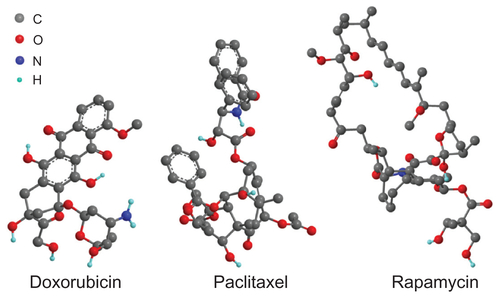
Figure S2 The cellular uptake and distribution of MMSNs on A549 cancer cells. MMSNs were labeled by FITC, and cultured with cells for 1 hour. Confocal microscopy image showed MMSNs were located in cytoplasm, and no green fluorescence was observed in nuclei which were stained by DAPI.
Note: Scale bar: 100 μm.
Abbreviations: DAPI, 4′,6-diamidino-2-phenylindole; FITC, fluorescein isothiocyanate; MMSNs, magnetic mesoporous silica nanoparticles.
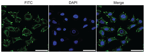
Figure S3 The cellular uptake of DOX-MMSNs and the release of DOX from MMSNs. A549 cells were treated with 50 μg/mL of DOX-MMSNs for 10, 30, 60, and 180 minutes, and observed by confocal microscopy. The internalization of MMSNs could be detected at the time point of 10 minutes as weak green fluorescence was observed. The fluorescence intensity increased accordingly with the prolonging of the treatment time. It was observed that the red fluorescence of DOX was almost undetectable until the incubating time increased to 30 min, which indicated that the nanoparticles were internalized but DOX was still kept in MMSNs until the time point of 30 minutes.
Note: Scale bar: 100 μm.
Abbreviations: DOX, doxorubicin; FITC, fluorescein isothiocyanate; MMSNs, magnetic mesoporous silica nanoparticles.
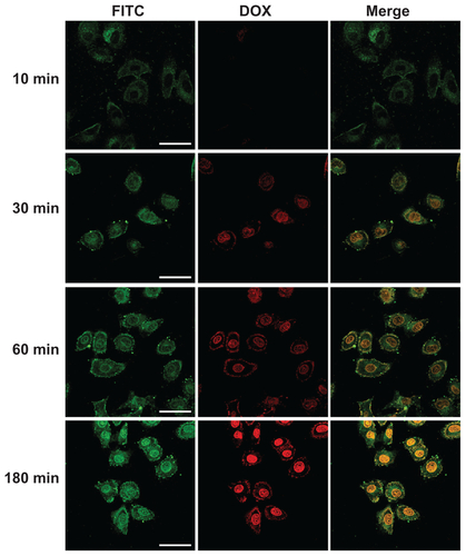
Figure S4 The fluorescence of DOX was detectable in FITC channel. (A) A549 cells were treated with free DOX in PBS at 5 μg/mL for 1 hour, fixed, and prepared for confocal microscopy. The fluorescence of DOX was mostly located in nuclei, and detectable in both 510–540 nm (FITC channel) and 560–590 nm emission range. (B) A549 cells were treated with empty MMSNs or DOX-MMSNs for 1 hour. Live cells were observed under confocal microscope. DOX-MMSNs-treated cells shows nucleic red fluorescent, whereas empty MMSNs-treated cells did not, which implies that it was the loaded DOX rather than MMSNs that entered nuclei.
Note: Scale bar: 50 μm.
Abbreviations: DOX, doxorubicin; FITC, fluorescein isothiocyanate; MMSNs, magnetic mesoporous silica nanoparticles.
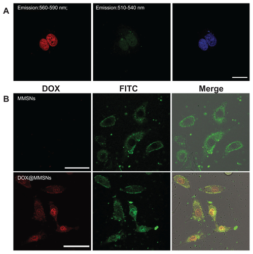
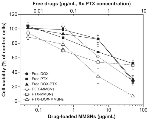
Figure S5 The growth inhibition of A549 cells induced by free or drug-loaded MMSNs with the approximate drug-loading amounts. The DOX-MMSNs loading content was 90 μg/mg, while the PTX-MMSNs loading content was 10 μg/mg. The cell growth inhibition induced by drugs loaded in MMSNs is more significant compared to that induced by free drugs.
Abbreviations: DOX, doxorubicin; MMSNs, magnetic mesoporous silica nanoparticles; PTX, paclitaxel.