Abstract
Background
CuO is one of the most important transition metal oxides due to its captivating properties. It is used in various technological applications such as high critical temperature superconductors, gas sensors, in photoconductive applications, and so on. Recently, it has been used as an antimicrobial agent against various bacterial species. Here we synthesized different sized CuO nanoparticles and explored the size-dependent antibacterial activity of each CuO nanoparticles preparation.
Methods
CuO nanoparticles were synthesized using a gel combustion method. In this approach, cupric nitrate trihydrate and citric acid were dissolved in distilled water with a molar ratio of 1:1. The resulting solution was stirred at 100°C, until gel was formed. The gel was allowed to burn at 200°C to obtain amorphous powder, which was further annealed at different temperatures to obtain different size CuO nanoparticles. We then tested the antibacterial properties using well diffusion, minimum inhibitory concentration, and minimum bactericidal concentration methods.
Results
XRD spectra confirmed the formation of single phase CuO nanoparticles. Crystallite size was found to increase with an increase in annealing temperature due to atomic diffusion. A minimum crystallite size of 20 nm was observed in the case of CuO nanoparticles annealed at 400°C. Transmission electron microscopy results corroborate well with XRD results. All CuO nanoparticles exhibited inhibitory effects against both Gram-positive and -negative bacteria. The size of the particles was correlated with its antibacterial activity.
Conclusion
The antibacterial activity of CuO nanoparticles was found to be size-dependent. In addition, the highly stable minimum-sized monodispersed copper oxide nanoparticles synthesized during this study demonstrated a significant increase in antibacterial activities against both Gram-positive and -negative bacterial strains.
Introduction
The unique, unusual and interesting physical, chemical, and biological properties of nanometer-sized materials have recently attracted a great deal of interest in the scientific community. As the size of materials is reduced to the nanometer regime the resulting properties change noticeably. Considerable efforts have been made to characterize and describe the physical and chemical properties of metal oxide nanomaterials because of their significant applications in numerous technological fields.Citation1–Citation4 The oxides of transition metals are an important class of semiconductors that have wider applications in magnetic storage media, solar energy transformation, electronics, and catalysis.Citation5–Citation12 Among various transition metal oxides, copper oxide (CuO) has attracted greater attention due to its fascinating properties such as the basis of high critical temperature (Tc) superconductors. CuO is a semiconducting compound with a narrow band gap and is used for photoconductive and photothermal applications.Citation13 Reports on the preparation and characterization of nanocrystalline CuO are few compared to those on some other transition metal oxides, such as zinc oxide, titanium dioxide, tin dioxide, and iron oxide. Some methods for the preparation of nanocrystalline CuO have been reported such as the sonochemical method,Citation14 sol-gel technique,Citation15 one-step solid state reaction method at room temperature,Citation16 electrochemical method,Citation17 and thermal decomposition of precursors.Citation18 Copper can also be used as an antimicrobial agent, and CuO nanoparticles have been investigated previously for enhancing antibacterial properties.Citation19–Citation22 Here we report a novel gel-combustion method, controlling the size of the synthesized nanoparticles and its effect on antimicrobial characteristics. The bactericidal property of such nanoparticles depends on their size, stability, and concentration added to the growth medium, since this provides greater retention time for bacterium nanoparticles interaction. In general, bacterial cells are in the micron-sized range. Most bacterial cells have cellular membranes that contain pores in the nanometer range. A unique property of crossing the cell membrane can potentially be attributed to synthesized nanoparticles through such bacterial pores. However, to make this possible, it is important to overcome challenges and prepare/design nanoparticles which are stable enough to significantly restrict bacterial growth while crossing the cell membrane.
To realize the potential of CuO nanoparticles to act as antimicrobial agents, we synthesized different sized CuO nanoparticles by controlling the annealing temperature during the gel-combustion synthesis. Furthermore, the antibacterial activities of CuO nanoparticles against two Gram-positive bacteria (Staphylococcus aureus and Bacillus subtilis) and two Gram-negative bacteria (Pseudomonas aeruginosa and Escherichia coli) were investigated.
Materials and methods
Synthesis of CuO nanoparticles
In a typical synthesis procedure, Cu(NO3)2 · 3H2O and citric acid were dissolved in distilled water with a molar ratio of 1:1. The solution was stirred with a magnetic stirrer at 100°C. Stirring continued until gel formation (approximately 1 hour). Afterwards, the gel was allowed to burn at 200°C. A light fluffy mass was obtained as a result of combustion, which was further annealed for 2 hours at varying temperatures, 400°C, 500°C, 600°C, and 700°C, to obtain the highly crystalline CuO nanoparticles.
Characterization of CuO nanoparticles
Synthesized CuO nanoparticles were characterized by X-ray diffraction (XRD), Fourier-transform infrared spectroscopy (FTIR), Raman spectroscopy, and transmission electron microscopy (TEM). Crystallinity, structure, and crystallite size of CuO nanoparticles were determined by XRD technique using a Rigaku-Miniflex X-ray diffractometer (Rigaku Corporation, Tokyo, Japan) with Cu-Kα radiations (λ = 0.15406 nm) in 2θ range from 20° to 80°. TEM analysis was carried out using a 200 kV JEOL transmission electron microscope (JEOL Ltd, Tokyo, Japan). FTIR spectra of the samples were obtained using a PerkinElmer FTIR spectrophotometer (PerkinElmer Inc, Waltham, MA).
Determination of antimicrobial activity by the well-diffusion method
Antimicrobial activities of the synthesized CuO nanoparticles of different sizes were determined using Gram-negative bacteria (E. coli and P. aeruginosa) and Gram-positive bacteria (B. subtilis and S. aureus) following a modified Kirby Bauer disc diffusion method.Citation23 In brief, the bacteria were cultured in Müller–Hinton broth at 35°C ± 2°C on an orbital shaking incubator (Remi, India) at 160 rpm. A lawn of bacterial culture was prepared by spreading 100 μL culture broth, having 106 CFU/mL of each test organism on solid nutrient agar plates. The plates were allowed to stand for 10–15 minutes. to allow for culture absorption. The 8 mm size wells were punched into the agar with the head of sterile micropipette tips. Wells were sealed with one drop of molten agar (0.8% nutrient agar) to prevent leakage from the bottom of the plate. Using a micropipette, 100 μL (50 μg) of the nanoparticles solution sample was poured into each of five wells on all plates. After incubation at 35°C ± 2°C for 24 hours, the size of the zone of inhibition was measured. A solvent blank was run as a negative control whereas the antibiotic (tetracycline) was used as a positive control.
Determination of minimal inhibitory/bactericidal concentrations (MIC/MBC)
Minimum inhibitory concentrations (MIC) and minimum bactericidal concentrations (MBC) of CuO nanoparticles were determined by the broth dilution method which conformed to the recommended standards of the National Committee for Clinical Laboratory Standards (NCCLS; now renamed the Clinical and Laboratory Standards Institute, CLSI, 2000). Tetracycline was used as a positive control. A dilution series with 10 mL nutrient broth medium containing 10–100 μg/mL copper oxide nanoparticles was prepared. Each set was inoculated aseptically with 50 μL of respective bacterial suspension (approximately 106 CFU/mL). The bacteria were plated onto solid nutrient agar plates. The lowest concentration inhibiting bacterial growth was defined as the MIC. In contrast the minimal concentration which completely inhibited the bacterial growth was defined as the minimum bactericidal concentration (MBC).Citation24 Each experiment was repeated three times, and the resulting bacterial growth on three plates corresponding to a particular sample were averaged and reported.
Results
Structural analysis
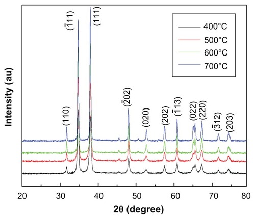
The typical XRD pattern of the CuO nanoparticles annealed at 400°C is shown in . The peak positions of the sample exhibited the monoclinic structure of CuO which was confirmed from the International Centre for Diffraction Data (ICDD) card No 801916. Further, no other impurity peak was observed in the XRD pattern, showing the single phase sample formation. The crystalline size was calculated using the Scherrer formula, D = 0.9 λ/β cosθ, where λ is the wavelength of X-ray radiation, β is the full width at half maximum (FWHM) of the peaks at the diffracting angle θ. Crystallite size calculated by the Scherrer formula was found to be 20 nm. Lattice parameters calculated by powder X software (Cheng Dong, Institute of Physics, Chinese Academy of Sciences, Beijing, China) were found to be a = 4.688 Å, b = 3.427 Å, c = 5.132 Å. These values are in good agreement with the standard values reported by the ICDD Card No 801916. In order to investigate the effect of temperature on CuO nanoparticles, samples were further annealed at 500°C, 600°C, and 700°C. exhibits the XRD spectra of CuO nanoparticles annealed at different temperatures. It is clear from that the intensity of crystalline peaks increases with temperature, indicating an improvement in the samples crystallinity. Simultaneously, the peaks become narrower as the temperature increases resulting in the increase of crystallite size. The variation of crystallite size and lattice parameters with temperature was calculated and the results are presented in . It can be seen from that crystallite size and lattice parameters increase with the increase in annealing temperature. The increase in crystallite size with temperature can be attributed to atomic diffusion. From an atomic perspective, diffusion is the stepwise migration of atoms from lattice site to lattice site. In fact, the atoms in solid materials are in constant motion, rapidly changing positions. For an atom to make such a move, the atom must have sufficient energy to break bonds with its neighbor atoms and then cause some lattice distortion during the displacement. As the temperature increases, the atoms gain sufficient energy for diffusive motion and hence increase in size takes place.
Table 1 Variation of crystallite size and lattice parameters with annealing temperature
Figure 1 XRD spectra of CuO nanoparticles annealed at 400°C.
Abbreviations: XRD, X-ray diffraction; CuO, copper oxide; AU, units of intensity.
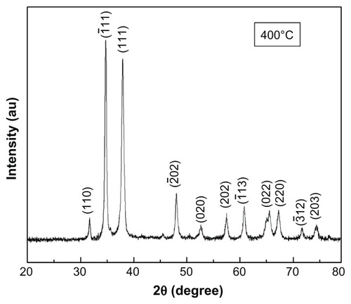
Figure 2 XRD spectra of CuO nanoparticles annealed at different temperatures.
Abbreviations: XRD, X-ray diffraction; CuO, copper oxide; AU, units of intensity.
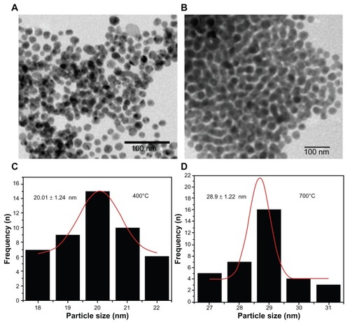
show the TEM micrographs of CuO nanoparticles sintered at 400°C and 700°C, respectively, while exhibit the size distribution of CuO nanoparticles sintered at 400°C and 700°C, respectively. Average particle sizes obtained from TEM images were found to be 20 ± 1.24 nm and 28.9 ± 1.22 nm for the samples sintered at 400°C and 700°C, respectively. The average particle sizes determined by TEM are closely matched to the crystallite size calculated from XRD results. TEM results also confirm the increase in the particle size with sintering temperature which corroborates well with the XRD results.
Figure 3 TEM image of CuO nanoparticles.
Notes: TEM image of CuO nanoparticles annealed at (a) 400°C (b) 700°C.
Abbreviations: TEM, transmission electron microscopy; CuO, copper oxide; n, number.
FTIR spectra were recorded in solid phase using the KBr pellets technique in the range of 3500–400 cm−1. FTIR spectra of CuO nanoparticles treated at 400°C, 500°C, 600°C, and 700°C are shown in . FTIR spectra exhibit only three vibrations: occurring at approximately 480 cm−1, 530 cm−1, and 580 cm−1 for all the samples, which can be attributed to the vibrations of Cu-O, confirming the formation of highly pure CuO nanoparticles. A weak band at around 2300 cm−1 may be attributed to the vibrations of atmospheric CO2. These assignments are in agreement with the values available in literature.Citation25–Citation27
Figure 4 FTIR spectra of CuO nanoparticles annealed at different temperatures.
Note: Inset shows Raman spectrum of CuO nanoparticles annealed at 400°C.
Abbreviations: FTIR, Fourier-transform infrared spectroscopy; CuO, copper oxide.
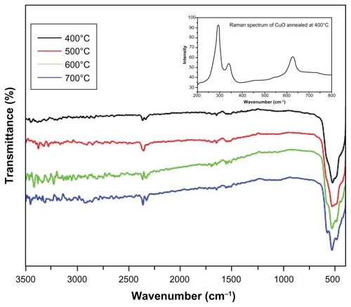
The Raman spectrum of CuO nanocrystals sintered at 400°C is shown in , which exhibits three one-phonon Raman scattering bands at approximately 277.5 cm−1 (Ag), 329.9 cm−1 (Bg), and 610.8 cm−1 (Bg), respectively.Citation28 The Raman spectra showed the presence of all three Raman active phonons (Ag + 2Bg) of CuO confirming the formation of CuO nanoparticles. The high crystalline nature of CuO nanoparticles is reflected by the significant intensity of Raman bands.
Antimicrobial properties
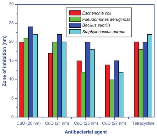
In this study, the copper oxide nanoparticles showed remarkable antibacterial activity against both Gram-positive (B. subtilis and S. aureus) and Gram-negative (E. coli and P. aeruginosa) bacteria (). The extent of inhibition of bacterial growth observed in this study was found to be variable and size-dependant. The smallest CuO nanoparticles (particle size 20 ± 1.24 nm) synthesized at the lowest temperature of 400°C, showed a significant inhibitory effect against both Gram-negative and -positive bacteria as compared to the CuO samples sintered at higher temperatures ( and ). One unique observation was that CuO nanoparticles synthesized at 400°C with the smallest particle size demonstrated the maximum zone of inhibition in the case of B. subtilis, which was 20% more than the zone of inhibition observed for tetracycline (). While comparing the effect of nanoparticles synthesized at varying temperature ranges, the greatest inhibitory effect was recorded for those CuO particles, which were synthesized at 400°C against P. aeruginosa strain. Furthermore, the smaller particles synthesized at 400°C had a zone of inhibition radii twice that of particles produced at 700°C. While comparing the effect of nanoparticles on bacterial strains, CuO nanoparticles were more toxic to E. coli regardless of the particle size except in one case ( and ). In all cases, the smaller the CuO nanoparticles, the lower the MIC and MBC values. In the case of the MIC, the most pronounced difference amongst all strains except S. aureus was between 400°C and 500°C nanoparticles. In case of S. aureus, the largest increase in MIC was observed for nanoparticles synthesized at temperatures between 500°C and 600°C (). For E. coli and S. aureus, the MIC values for particles synthesized at 700°C were threefold higher than those recorded for the nanoparticles synthesized at a comparatively smaller temperature (400°C). In general, the CuO nanoparticles had a less pronounced effect on other bacterial strains, but these effects were still twofold higher than those determined for 700°C synthesized particles. As can be seen in , the MBC values were even more drastically dependent on the particle size. Again we observed that E. coli and S. aureus had threefold lower values for the 400°C MBC than those synthesized at 700°C. As mentioned above, unlike the MIC values, each temperature dependent synthesis had more variable MBC values (); however, for the S. aureus, the largest MBC impact was from the 400°C to 500°C transition, unlike the MIC.
Table 2 Antimicrobial activity of copper oxide (CuO) nanoparticles against two Gram-positive and two Gram-negative bacteria
Table 3 MIC of copper oxide nanoparticles (annealed at different temperatures) against different laboratory bacterial strains
Table 4 MBC of copper oxide nanoparticles (annealed at different temperatures) against different laboratory bacterial strains
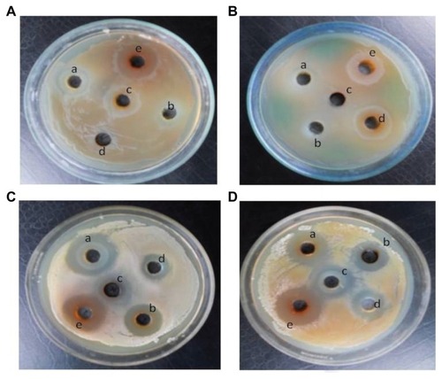
Figure 5 Zone of inhibition of copper oxide nanoparticles.
Notes: Zone of inhibition of copper oxide nanoparticles synthesized at different temperatures (a–d) and positive control, a known antibiotic tetracycline (e) against two Gram-negative bacteria (A) Escherichia coli and (B) Pseudomonas aeruginosa, and two Gram-positive bacteria (C) Bacillus subtilis and (D) Staphylococcus aureus.
Discussion
A few studies have been performed to elucidate the mechanism of bactericidal action of nanoparticles. For example, Tsao et al suggested that the exposure of Gram-positive bacteria to carboxyfullerene nanoparticles resulted in the puncturing of the bacteria leading to cell death.Citation29 Another proposed way in which the membrane can be compromised is the alteration of membrane lipid components.Citation30 It is, however, difficult to distinguish between the bactericidal activity of nanoparticles from the ions released by the nanoparticles themselves.Citation31 Previously, Ruparelia et al estimated the concentration of released ions for 10 mg of copper nanoparticles suspended in 100 mL nutrient media and distilled water.Citation32 They found that the concentration of Cu2+ ions released in nutrient media was 17 mgL−1 after 24 hours of incubation in a rotary shaker, while in distilled water under the same conditions over a period of 24 hours, the concentration of ions released was 0.5 mgL−1. These results indicate that the nutrient media can facilitate the release of Cu2+ ions. The considerably greater release of Cu2+ ions in the nutrient media is possibly due to the interaction of the media chloride ions with the oxide layer of the nanoparticles.Citation32 For oxidized copper particles embedded in an inert, Teflon-like matrix, Cioffi et al demonstrated significant antimicrobial activity due to the release of ions.Citation33 Consequently, the bactericidal effects observed in this study might have been influenced by the release of Cu2+ ions in solution. The presence of nanoparticles in suspension would ensure continuous release of ions into the nutrient media.Citation33 However, the preceding studies did not correlate their findings with the size of the nanoparticles and hence, the precise mechanisms of how nanoparticles act as biocidal agent needs to be explained. There are, however, a few mechanisms of nanoparticle toxicity suggested by other works. For example, copper ions released by the nanoparticles may attach to the negatively charged bacterial cell wall and rupture it, thereby leading to protein denaturation and cell death.Citation34 Copper ions inside the bacterial cells may bind to deoxyribonucleic acid molecules and become involved in cross-linking within and between the nucleic acid strands, resulting in the disorganized helical structure. In addition, copper ion uptake by the bacterial cells has also been found to damage important biochemical processes.Citation35,Citation36
It is clear from (maximum zone of inhibition against B. subtilis and S. aureus) that CuO nanoparticles have shown greater antimicrobial activity against B. subtilis and S. aureus. The variation in the sensitivity or resistance to both Gram-positive and -negative bacteria populations could be due to the differences in the cell structure, physiology, metabolism, or degree of contact of organisms with nanoparticles. For example, greater sensitivity among Gram-positive bacteria such as B. subtilis and S. aureus to the CuO nanoparticles has been attributed to the greater abundance of amines and carboxyl groups on their cell surface and greater affinity of copper towards these groups.Citation37 Alternatively, Gram-negative bacteria like E. coli have a special cell membrane structure which possesses an important ability to resist antimicrobial agents.Citation38 Furthermore, other factors such as nanoparticle diffusion rates may also affect bacterial strain differently. Nevertheless, further studies are required to confirm this and it is beyond the scope of this manuscript.
and exhibit the zone of inhibition of CuO nanoparticles synthesized at different temperatures (a–d) and positive control, a known antibiotic tetracycline (e), against two Gram-negative bacteria [(A) E. coli (B) P. aeruginosa], and two Gram-positive bacteria [(C) B. subtilis (D) S. aureus]. clearly indicates that the copper oxide nanoparticles inhibit the growth of both Gram- negative and -positive bacteria and the zone of inhibition decreases with the increase in annealing temperature from 400°C–700°C. shows that the zone of inhibition is maximum when the particle size is minimum (20 ± 1.24 nm). These results demonstrate the excellent antimicrobial behavior of CuO nanoparticles synthesized at low temperature. Broadly, interactions between the negative charges of microorganisms and the positive charge of nanoparticles produces an electromagnetic attraction between the microbe and effective levels of active nanoparticles. Such interactions lead to oxidation of surface molecules of microbes resulting in their death. Biodestructive effects such as degradation of deoxyribonucleic acid was observed following exposure of Gram-positive bacteria to silver and copper nanoparticles by other works,Citation39,Citation40 and are in agreement with present findings.
Figure 6 Bar graph representing the zone of inhibition for CuO nanoparticles and tetracycline against Gram-positive and -negative bacteria.
Abbreviation: CuO, copper oxide.
Conclusion
We have successfully synthesized CuO nanoparticles using a gel combustion route. XRD spectra confirmed the formation of single phase CuO nanoparticles. Crystallite size was found to increase with the increase in annealing temperature. Minimum crystallite size of 20 ± 1.24 nm was observed in the case of CuO nanoparticles annealed at 400°C. TEM results corroborate well with XRD results. FTIR and Raman spectra also validated the purity of CuO nanoparticles. Antibacterial activity experiments performed on various microorganisms clearly demonstrated the higher effectiveness of CuO nanoparticles annealed at 400°C against bacterial growth due to smaller particle size of this sample compared to other samples. Zone of inhibition for all the microorganisms reached a maximum point using CuO nanoparticles annealed at 400°C. Moreover, minimum inhibitory concentration and minimum bactericidal concentration of CuO nanoparticles annealed at 400°C was lowest for all the bacterial strains.
Acknowledgments
Mr Arham S Ahmed and Mr M Oves are thankful to CSIR, New Delhi for providing financial support in the form of SRF. Dr Adnan Memic would like to thank the Strategic Technologies Program by King Abdulaziz City for Science and Technology, grant number 10-NAN1081-3 for their partial support and funding of this project.
Disclosure
The authors report no conflicts of interest in this work.
References
- UedaNMaedaHHosonoHKawazoeHBand-gap widening of CdO thin filmsJ Appl Phys1998841161746177
- LiuHZhangXLiLRole of point defects in room-temperature ferromagnetism of Cr-doped ZnOAppl Phys Lett2007917072511072513
- ZhuHZhaoFPanLStructural and magnetic properties of Mn-doped CuO thin filmsJ Appl Phys2007101909H11109H111-113
- FerreiraFFTabacniksMHFantiniaMCAFariabICGorensteinbAElectrochromic nickel oxide thin films deposited under different sputtering conditionsSolid State Ionics199686882971976
- BradleyFNChapter 2BradleyFNMaterials for Magnetic FunctionsNew York, NYHayden1976
- MitsuyuTYamazakiOOhjiKWasaKPiezoelectric thin films of zinc oxide for saw devicesFerroelectrics1982421233240
- O’ReganBGratzelMA low-cost, high-efficiency solar cell based on dye- sensitized colloidal TiO2 filmsNature19913536346737740
- WangYChengHZhangLThe preparation, characterization, photoelectrochemical and photocatalytic properties of lanthanide metal-ion-doped TiO2 nanoparticlesJ Mol Catal A Chem20001511–2205216
- BjoerkstenUMoserJGraetzelMPhotoelectrochemical studies on nanocrystalline hematite filmsChem Mater199466858863
- DowWPHuangTJYttria-stabilized zirconia supported copper oxide catalyst: II. Effect of oxygen vacancy of support on catalytic activity for CO oxidationJournal of Catalysts19961602171182
- LarssonPOAnderssonAWallenbergRLSvenssonBCombustion of CO and Toluene; Characterisation of copper oxide supported on Titania and activity comparisons with supported cobalt, iron, and manganese oxideJournal of Catalysts19961632279293
- JiangYDeckerSMohsCKlabundeKJCatalytic solid state reactions on the surface of nanoscale metal oxide particlesJournal of Catalysts199818012435
- RakhshaniAEPreparation, characteristics and photovoltaic properties of cuprous oxide – a reviewSolid State Electron1986291717
- KumarRVDiamantYGedankenASonochemical synthesis and characterization of nanometer-size transition metal oxides from metal acetatesChem Mater200012823012305
- EliseevAALukashinAVVertegelAAHeifetsLIZhirovAITretyakovYDComplexes of Cu(II) with polyvinyl alcohol as precursors for the preparation of CuO/SiO2 nanocompositesMaterials Research Innovations200035308312
- XuJFJiWShenZXPreparation and characterization of CuO nanocrystalsJ Solid State Chem20001472516519
- BorgohainKSinghJBRama RaoMVShripathiTMahamuniSQuantum size effects in CuO nanoparticlesPhys Rev B200061161109311096
- Salavati-NiasariMDavarFSynthesis of copper and copper (I) oxide nanoparticles by thermal decomposition of a new precursorMater Lett2009633–4441443
- HoriguchiHChemistry of Antimicrobial AgentsTokyo, JapanSankyo Press198046
- OjasMBhagatMGopalakrishnanCArunachalamKDUltrafine dispersed CuO nanoparticles and their antibacterial activityJournal of Experimental Nanoscience200833185193
- LiBYuSHwangJYShiSAntibacterial vermiculite nano-materialJournal of Minerals and Materials Characterization and Engineering2002116168
- CondorelliGGCostanzoILFragalaILGiuffridaSVentimigliaGA single photochemical route for the formation of both copper nanoparticles and patterned nanostructured filmsJ Mater Chem2003131024092411
- BauerAWKirbyWMSherrisJCTurckMAntibiotic susceptibility testing by a standardized single disk methodAm J Clin Pathol19664544934965325707
- TarpayMMWelchDFMarksMIAntimicrobial susceptibility testing of Streptococcus pneumonia by micro-broth dilutionAntimicrob Agents Chemother19801845795816905715
- JagminasAKuzmarskytJNiauraGElectrochemical formation and characterization of copper oxygenous compounds in alumina template from ethanolamine solutionsAppl Surf Sci20022011–4129137
- JagminasANiauraGKuzmarskytJButkieneRSurface-enhanced Raman scattering effect for copper oxygenous compounds array within the alumina template pores synthesized by ac deposition from Cu(II) acetate solutionAppl Surf Sci20042251–4302308
- ZhangYCTangJYWangGLZhangMHuXYFacile synthesis of submicron Cu2O and CuO crystallites from a solid metallorganic molecular precursorJ Crys Growth20062942278282
- IrwinJCChrzanowskiJWeiTLockwoodDJWoldARaman scattering from single crystals of cupric oxidePhysica C Supercond19901665–6456464
- TsaoNLuhTYChouCKIn vitro action of carboxyfullereneJ Antimicrob Chemother200249464164911909838
- KochAMReynoldsFMerkleHPWeisslederRJosephsonLTransport of surface- modified nanoparticles through cellmonolayersChem bio chem200562337345
- YoonKHoon ByeonJParkJHHwangJSusceptibility constants of Escherichia coli and Bacillus subtilis to silver and copper nanoparticlesSci Total Environ20073732–357257517173953
- RupareliaJPChatterjeeAKDuttaguptaSPMukherjiSStrain specificity in antimicrobial activity of silver and copper nanoparticlesActa Biomater20084370771618248860
- CioffiNDitarantoNTorsiLAnalytical characterization of bioactive fluoropolymer ultra-thin coatings modified by copper nanoparticlesAnal Bioanal Chem2005381360761615349710
- LinYEVidicRDStoutJEMcCartneyCAYuVLInactivation of Mycobacterium avium by copper and silver ionsWater Res199832719972000
- KimJHChoHRyuSEChoiMUEffects of metal ions on the activity of protein tyrosine phosphatase VHR: highly potent and reversible oxidative inactivation by Cu2+ ionArch Biochem Biophys20003821728011051099
- StohsSJBagchiDOxidative mechanisms in the toxicity of metal ionsFree Radic Biol Med19951823213367744317
- BeveridgeTJMurrayRGSites of metal deposition in the cell wall of Bacillus subtilisJ Bacteriol198014128768876767692
- LiangXSunMLiLPreparation and antibacterial activities of polyaniline/Cu0.05Zn0.95O nanocompositesDalton Trans20124192804281122249414
- Rezaei-ZarchiSJavedAGhaniMJComparative study of antimicrobial activities of TiO2 and CdO nanoparticles against the pathogenic strain of Escherichia coliIran J Pathol2010528389
- HosseinkhaniAMZandAMImaniSRezayiMRezaei-ZarchiSDetermining the antibacterial effect of ZnO nanoparticle against the pathogenic bacterium, Shigella dysenteriae (type 1)International Journal of Nano Dimension201014279285