Abstract
Protein nanocages are self-organized complexes of oligomers whose three-dimensional architecture can been determined in detail. These structures possess nanoscale inner cavities into which a variety of molecules, including therapeutic or diagnostic agents, can be encapsulated. These properties yield these particles suitable for a new class of drug delivery carrier, or as a bioimaging reagent that might respond to biochemical signals in many different cellular processes. We report here the design, synthesis, and biological characterization of a hepatocyte-specific nanocage carrying small heat-shock protein. These nanoscale protein cages, with a targeting peptide composed of a preS1 derivative from the hepatitis B virus on their surfaces, were prepared by genetic engineering techniques. PreS1-carrying nanocages showed lower cytotoxicity and significantly higher specificity for human hepatocyte cell lines than other cell lines in vitro. These results suggested that small heat-shock protein-based nanocages present great potential for the development of effective targeted delivery of various agents to specific cells.
Acknowledgements
This work was supported by a Health Labor Sciences Research Grant (Research on Publicly Essential Drugs and Medical Devices) from the Ministry of Health Labor and the Special Coordination Funds for Promoting Science and Technology (SCF funding program “Innovation Center for Medical Redox Navigation”), Japan.
Disclosure
The authors report no conflict of interest in this work.
Supplementary figures
Figure S1 Gel permeation chromatography purification of HSPG41C nanocages and HSPG41C-preS1 nanocages. GHSPG41C nanocages (A); HSPG41C-preS1 nanocages (B). The peaks observed at 13.31 minutes in (A) and 13.26 minutes in (B) were collected individually. The collected fractions were then analyzed by SDS-PAGE using 12% gel according to the standard protocol (C).
Note: Lane 1, molecular weight standards; lane 2, HSPG41C nanocages; lane 3, HSPG41C-preS1 nanocages.
Abbreviation: SDS-PAGE, sodium dodecyl sulfate polyacrylamide gel electrophoresis.
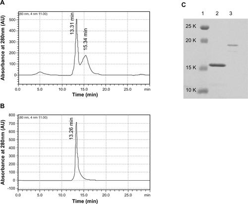
Figure S2 MALDI-TOF mass spectrum of HSPG41C nanocages and HSPG41C-preS1 nanocages. GHSPG41C nanocages (A); HSPG41C-preS1 nanocages (B).
Abbreviation: MALDI-TOF, matrix-assisted laser desorption/ionization time of flight.
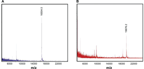
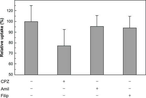
Figure S3 Inhibition of cellular uptake of HSPG41C-preS1 nanocages by endocytosis inhibitors.
Notes: HepG2 cells were harvested on poly-L-lysine-coated 48-well plates at an initial density of 50,000 cells/well and grown overnight. Cells were treated with DMEM containing chlorpromazine (10 μg/mL), amiloride (500 μM), or filipin III (10 μg/mL) for 1 hour (all from Sigma-Aldrich, St Louis, MO). Then 84 nM of fluorescent-labeled HSPG41C-preS1 nanocages solution containing the above inhibitors was added to cells. Two hours after transfection, cells were rinsed three times with PBS and then lysed in lysis buffer (pH 7.5, 20 mM Tris-HCl, 2 mM EDTA, and 0.05% Triton-X 100). Lysate solutions were replaced on black-bottomed 96-well plates and the fluorescence intensity of each sample measured using a Microplate Reader (ARVO MX 1420; Perkin Elmer Inc, Waltham, MA). Data are means ± SEM of three independent experiments.
Abbreviations: CPZ, chlorpromazine; Amil, amiloride; Filip, filipin; DMEM, Dulbecco’s Modified Eagle’s Medium; PBS, phosphate buffered saline; HCl, hydrochloride; EDTA, ethylenediaminetetraacetic acid; SEM, standard error of the mean.