Abstract
Background
Hepatocellular carcinoma (HCC) is a common and aggressive form of cancer. Due to a high rate of postoperative recurrence, the prognosis for HCC is poor. Subclinical metastasis is the major cause of tumor recurrence and patient mortality. Currently, there is no reliable prognostic method of invasion.
Aim
To investigate the feasibility of fingerprints of volatile organic compounds (VOCs) for the in-vitro prediction of metastasis.
Methods
Headspace gases were collected from 36 cell cultures (HCC with high and low metastatic potential and normal cells) and analyzed using nanomaterial-based sensors. Predictive models were built by employing discriminant factor analysis pattern recognition, and the classification success was determined using leave-one-out cross-validation. The chemical composition of each headspace sample was studied using gas chromatography coupled with mass spectrometry (GC-MS).
Results
Excellent discrimination was achieved using the nanomaterial-based sensors between (i) all HCC and normal controls; (ii) low metastatic HCC and normal controls; (iii) high metastatic HCC and normal controls; and (iv) high and low HCC. Several HCC-related VOCs that could be associated with biochemical cellular processes were identified through GC-MS analysis.
Conclusion
The presented results constitute a proof-of-concept for the in-vitro prediction of the metastatic potential of HCC from VOC fingerprints using nanotechnology. Further studies on a larger number of more diverse cell cultures are needed to evaluate the robustness of the VOC patterns. These findings could benefit the development of a fast and potentially inexpensive laboratory test for subclinical HCC metastasis.
Acknowledgements
The authors acknowledge Dr Yoav Broza, Ms Orna Barash, and Ms Meggie Hakim for their assistance and support.
Conflict of interest
None of the authors declare any conflict of interest.
Funding source
The research leading to these results was funded by the FP7’s ERC grant under DIAG-CANCER (grant agreement no 256639; HH).
Supplementary data
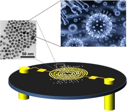
Figure S1 Schematic representation of the GNP sensors used in this study (not drawn to scale).
Note: The left inset in the sensor’s schematics shows a tunneling electron micrograph (TEM) of the GNPs in solution.
Abbreviations: GNP, gold nanoparticle; TEM, tunneling electron micrograph.