
Abstract
Antioxidant (quercetin) and hypoglycemic (voglibose) drug-loaded poly-D,L-lactideco-glycolide nanoparticles were successfully synthesized using the solvent evaporation method. The dual drug-loaded nanoparticles were incorporated into a scaffold film using a solvent casting method, creating a controlled transdermal drug-delivery system. Key features of the film formulation were achieved utilizing several ratios of excipients, including polyvinyl alcohol, polyethylene glycol, hyaluronic acid, xylitol, and alginate. The scaffold film showed superior encapsulation capability and swelling properties, with various potential applications, eg, the treatment of diabetes-associated complications. Structural and light scattering characterization confirmed a spherical shape and a mean particle size distribution of 41.3 nm for nanoparticles in the scaffold film. Spectroscopy revealed a stable polymer structure before and after encapsulation. The thermoresponsive swelling properties of the film were evaluated according to temperature and pH. Scaffold films incorporating dual drug-loaded nanoparticles showed remarkably high thermoresponsivity, cell compatibility, and ex vivo drug-release behavior. In addition, the hybrid film formulation showed enhanced cell adhesion and proliferation. These dual drug-loaded nanoparticles incorporated into a scaffold film may be promising for development into a transdermal drug-delivery system.
Acknowledgment
This work was supported by the Gachon University Research Fund of 2012 (KWU 2011-R372).
Disclosure
The authors report no conflicts of interest in this work.
Supplementary information
Figure S1 Size analysis of permeation-enhanced drug-loaded nanoparticle. Representative field emission scanning electron microscopic image of permeation-enhanced drugloaded nanoparticles. (A) 4 wt%, (B) 4.5 wt%, (C) 5 wt%, (D) 5.5 wt%, and (E) 6 wt% oleic acid (chemical permeation-enhanced) preparations.
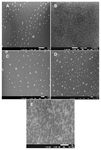
Figure S2 Calibration curve of drugs validated by UV-visible spectroscopic methods. This was found by plotting various concentrations of drugs versus absorbance with linear correlation co-efficient. (A) Voglibose 100–500 μg/mL, (B) quercetin 200–1000 μg/mL.
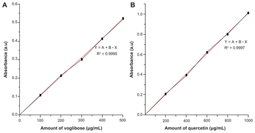
Figure S3 X-ray diffraction patterns of permeation-enhanced dual drug-loaded nanoparticle-incorporated cross-linked films (S1, S2, S3, and S4), noncross-linked film (blank), and drug-loaded nanoparticles.
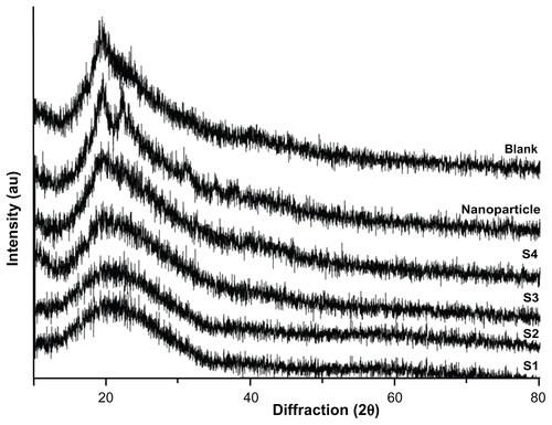
Figure S4 Optical microscopic images showing the morphology of blank film and nanoparticle-incorporated scaffold films (S1, S2, S3, and S4), before (A) and after (B) the release study. Scale bar, 20 μm at 50× magnification. (C) Field emission scanning electron microscopic images of blank film and nanoparticle-incorporated scaffold films after release study.
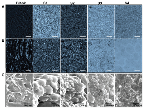
Figure S5 (A) Two-dimensional atomic force microscopic images showing the morphology of the swelling structure and surface properties of the blank, S1, S2, S3 and S4 films. (B) Corresponding three-dimensional atomic force microscopic images.
Notes: The blank film ensures the swelling structure has a relatively smooth surface and the nanoparticle-incorporated films have some projections, indicating the location of nanoparticles on the surface of the scaffold film. Scan size 1 × 1 μm.
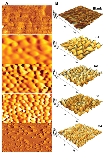
Table S1 Physicochemical characterization of permeation-enhanced dual drug-loaded nanoparticle preparations in different ratios of oleic acid concentration