Abstract
Background
Curcumin, the principal curcuminoid of the popular Indian spice turmeric, has wide spectrum of pharmaceutical properties such as antitumor, antioxidant, , and anti-inflammatory activity. However, poor aqueous and low bioavailability of curcumin is a major challenge in its as a useful drug. To enhance the aqueous solubility and of curcumin, attempts have been made to encapsulate it in , polymeric nanoparticles (NPs), lipid-based NPs, biodegradable , cyclodextrin, and hydrogels.
Methods
In this work, we attempted to entrap curcumin in novel self-assembled NPs containing a nonprotein amino acid, α, β-dehydrophenylalanine, and investigated the biological activity of -curcumin NPs in cancer models both in vitro and in vivo.
Results
Of the several dehydrodipeptides tested, methionine-dehydrophenylalanine was most suitable one for loading and release of curcumin. Loading of in the dipeptide NPs increased its solubility, improved cellular , enhanced its toxicity towards different cancerous cell lines, enhanced curcumin’s efficacy towards inhibiting tumor growth in /c mice bearing a B6F10 melanoma tumor.
Conclusion
These novel, highly biocompatible, and easy to construct dipeptide NPs with a to load and release curcumin in a sustained manner significantly curcumin’s cellular uptake without altering its anticancer other therapeutic properties. Curcumin-dipeptide NPs also showed improved vitro and in vivo chemotherapeutic efficacy compared to curcumin alone. dipeptide-NPs may also improve the delivery of other potent hydrophobic molecules that show poor cellular uptake, bioavailability, and .
Acknowledgements
SA thanks Indian Council of Medical Research for fellowship and JJP thanks -Lo’real for Young Women in Science Program for providing a . The authors thank the core funding at the International Centre for Engineering and Biotechnology, New Delhi, Department of Biotechnology and of Science and Technology, India, for financial assistance. We also thank Varshaney, ICGEB, New Delhi, for help with mass spectrometric analysis.
Disclosure
The authors report no conflicts of interest in this work.
Supplementary information
Synthesis of methionine-dehydrophenylalnine (MΔF), -dehydrophenylalanine (LΔF), and isoleucine-dehydrophenylalanine (IΔF)
Briefly, Boc-Met-OH (5 mM; 1.24 g) was dissolved in dry THF, chilled to −20μC and stirred in an ice-salt bath. Equivalent moles of -methylmorpholine (5 mM; 0.655 mL) and isobutyl chloroformate (5 mM; 0.69 mL) added to the solution. After 10 min, a precooled aqueous solution of -threo-β-phenylserine (5.5 mM; 0.99 g) and sodium hydroxide (5.5 mM; 0.22 g) were added. The reaction mixture was stirred overnight and concentrated vacuo. The residue was acidified with citric acid and extracted using ethyl . The ethyl acetate layer was washed three times with water and saturated chloride, dried over anhydrous sodium sulfate, and evaporated to obtain -Met-DL-threo-β-phenylserine, which was then mixed with anhydrous acetate (6.5 mM; 0.58 g) in freshly distilled acetic anhydride (50 mL) stirred for 36 hours. The thick slurry was poured over crushed ice and the was filtered, washed with 5% NaHCO3 followed by water, and dried in vacuo. The resulting compound (Boc-Met-ΔPhe-azalactone) was dissolved in methanol, treated with 1.5 of 1 N NaOH solution for 3–4 hours, and concentrated in . Acidification with solid citric acid liberated the peptide, which was in ethyl acetate to yield Boc-Met-ΔPhe-COOH. The Boc group was using anhydrous tetrahydrofuran (TFA). The peptide was purified on a reverse-phase C18 column (Delta-Pak, Waters Co, Milford, MA; 15 μm, internal diameter, 300 × 19 mm) using an acetonitrile-water gradient 5%–95% acetonitrile (0.1% )/water (0.1% TFA) at a flow rate of 4 mL/minute over 55 minute on a -performance liquid chromatography (HPLC) system [LC-6 AD, liquid , Shimadzu, Kyoto, Japan)]. The purified peptide was into an analytical reverse phase C18 column (Phenomenex, Hyderabad, , C18, 5 μm, internal diameter, 250 × 4.6 mm) using an -water linear gradient of 5%–95% (0.1% TFA)/water (0.1% TFA) at a flow rate of 1 /min over 45 min on a HPLC system (LC-10 AD; Shimadzu) and was found to be 98% pure with a retention time of 23 min. The purified peptide was using mass spectrometry [AppliedBiosystems QStar (Q-TOF)]. Observed mass: 295.139 Da; expected mass: 294.407 Da.
Table S1 Yield of synthesis, retention time, and observed and expected masses MΔF, LΔF, IΔF, and MF
Table S2 Morphology and average size of nanoparticles determined using dynamic scattering and transmission electron microscopy analysis
Other dipeptides such as leucine-dehydrophenylalanine (LΔF) and -dehydrophenylalanine (IΔF) were synthesized and purified the same procedure described above, starting from Boc-Leu-OH and -Ile-OH respectively (Supplementary Table 1).
Synthesis of methionine-phenylalanine (MF)
Briefly, Boc-Met-OH (5 mM; 1.24 g) was dissolved in dry THF, chilled to −20μC, and stirred in an ice-salt bath. Equivalent moles of -methylmorpholine and isobutyl chloroformate were added to the solution. 10 min, a precooled aqueous solution of NH2-Phe-OH (5.5 mM; 0.90 g) and sodium hydroxide (5.5 mM; 0.22 g) were added. The reaction was stirred overnight and concentrated in vacuo. The residue was with citric acid and extracted with ethyl acetate. The ethyl layer was washed three times with water and saturated sodium , dried over anhydrous sodium sulfate, and evaporated to obtain -Met-Phe. The Boc group was deprotected using anhydrous tetrahydrofuran. peptide was purified on a preparative reverse-phase C18 column as above. The purified peptide was analyzed using mass spectrometry [AppliedBiosystems QStar (Q-TOF)]. Observed mass: 296 Da; mass: 296.3 Da.
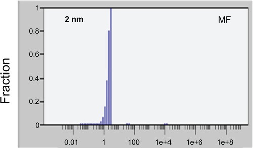
Figure S1 Mean particle size of methionine–phenylalanine (MF) in :water solution (1:1) measured using dynamic -scattering (Rh = 2 nm).