Abstract
Ovarian cancer is one of the leading causes of cancer death for women throughout the Western world. Kaempferol, a natural flavonoid, has shown promise in the chemoprevention of ovarian cancer. A common concern about using dietary supplements for chemoprevention is their bioavailability. Nanoparticles have shown promise in increasing the bioavailability of some chemicals. Here we developed five different types of nanoparticles incorporating kaempferol and tested their efficacy in the inhibition of viability of cancerous and normal ovarian cells. We found that positively charged nanoparticle formulations did not lead to a significant reduction in cancer cell viability, whereas nonionic polymeric nanoparticles resulted in enhanced reduction of cancer cell viability. Among the nonionic polymeric nanoparticles, poly(ethylene oxide)-poly(propylene oxide)-poly(ethylene oxide) (PEO-PPO-PEO) nanoparticles incorporating kaempferol led to significant reduction in cell viability of both cancerous and normal cells. Poly(DL-lactic acid-co-glycolic acid) (PLGA) nanoparticles incorporating kaempferol resulted in enhanced reduction of cancer cell viability together with no significant reduction in cell viability of normal cells compared with kaempferol alone. Therefore, both PEO-PPO-PEO and PLGA nanoparticle formulations were effective in reducing cancer cell viability, while PLGA nanoparticles incorporating kaempferol had selective toxicity against cancer cells and normal cells. A PLGA nanoparticle formulation could be advantageous in the prevention and treatment of ovarian cancers. On the other hand, PEO-PPO-PEO nanoparticles incorporating kaempferol were more effective inhibitors of cancer cells, but they also significantly reduced the viability of normal cells. PEO-PPO-PEO nanoparticles incorporating kaempferol may be suitable as a cancer-targeting strategy, which could limit the effects of the nanoparticles on normal cells while retaining their potency against cancer cells. We have identified two nanoparticle formulations incorporating kaempferol that may lead to breakthroughs in cancer treatment. Both PEO-PPO-PEO and PLGA nanoparticle formulations had superior effects compared with kaempferol alone in reducing cancer cell viability.
Introduction
Natural compounds with antioxidant properties that function to protect the human body against development of cancerCitation1,Citation2 are present in a variety of fruit and vegetables.Citation3 Natural dietary compounds have been reported to reduce the risk of development of diabetes,Citation4 cardiovascular disease,Citation5 prostate cancer,Citation6 colorectal cancer,Citation7 and ovarian cancer.Citation8 Kaempferol (3,5,7-trihydroxy-2-(4-hydroxyphenyl)-4H-1-benzopyran-4-one) is a relatively common nontoxic, natural dietary compound which has been reported to reduce the risk of ovarian cancer.Citation9 Kaempferol was found to inhibit estrogen receptor alpha expression in breast cancer cellsCitation10 and to induce apoptosis in glioblastoma cellsCitation11 and lung cancer cellsCitation12 by activation of MEK-MAPK. Studies have shown that kaempferol also has anti-inflammatory effects via inhibition of interleukin-4Citation13 and cyclo-oxygenase 2 expression by suppressing Src kinaseCitation14 and downregulating the NFκB pathway.Citation15 Kaempferol is also effective in inhibiting angiogenesis and inducing apoptosis in ovarian cancer cells.Citation16–Citation19 In human studies, a significant 40% decrease in incidence of ovarian cancer was found for individuals with the highest quintile of kaempferol intake as compared with those in the lowest quintile.Citation20 Despite promising preclinical results, the utility of such compounds for chemoprevention in humans has met with only limited success, largely due to inefficient systemic delivery and limited bioavailability of promising agents. Therefore, to achieve the maximum response to a chemopreventive agent, novel strategies are required to enhance the bioavailability of potentially useful agents and to reduce toxicity.
Nanotechnology is an emerging interdisciplinary field that encompasses biology, engineering, chemistry, and medicine. Citation21 Using nanotechnology for the development of efficient anticancer drug delivery systems is a recent advance in medical science.Citation22–Citation24 The ability of nanoparticles to incorporate entities renders them ideal carriers for various anticancer drugs.Citation25,Citation26 Because most anticancer drugs have poor solubility in water and low bioavailability, the use of nanocarriers enables cancer medications with low solubility in water to be prepared as solid or liquid formulations. Nanoparticles comprised of biodegradable polymers have been studied for delivery of drugs.Citation27,Citation28 Significant advantages of using biodegradable polymers are their safety and the ability to control the time and rate of polymer degradation as well as timely release of the drug. Nanoparticles comprised of biodegradable polymers such as poly(lactic acid), poly(DL-lactic acid-co-glycolic acid) (PLGA), poly(caprolactone), palmitic acid, and chitosan have been utilized for the delivery of anticancer drugs.Citation27,Citation28 In recent years, nanotechnology has been implemented and assessed in different areas of cancer therapeutics and management.Citation29 Siddiqui et al reported that nanoencapsulated epigallocatechin-3-gallate retains its biological effectiveness, with over a 10-fold dose reduction advantage compared with nonencapsulated epigallocatechin-3-gallate when inhibiting cell growth, and proapoptotic and angiogenic effects.Citation30
Ovarian cancer is one of the leading causes of cancer death in women throughout the Western world.Citation31 There has been limited progress in the prevention, early diagnosis, and treatment of ovarian cancer to date,Citation32,Citation33 leaving this malignancy with an unchanged death rate over decades.Citation31 Chemoprevention of ovarian cancer using natural products has received more attention recently, and our earlier studies have indicated that kaempferol, a dietary flavonoid, is effective in inhibiting angiogenesis and inducing apoptosis in ovarian cancer cells.Citation16–Citation19 However, effective concentrations are often above 20–40 μM, which are not always physiologically attainable. In this study, five nanoparticle formulations of kaempferol were developed and their efficacy in inhibiting the viability of malignant and normal ovarian cells was determined.
Materials and methods
Chemicals
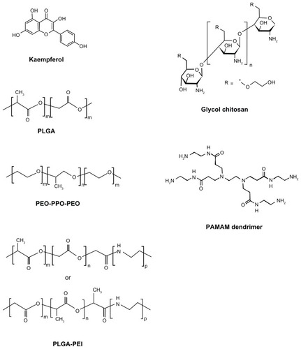
Kaempferol (soluble, 50 mg/mL in dimethyl sulfoxide), polyethylenimine (PEI, molecular weight 800 Da), PLGA (lactide to glycolide ratio 50:50, molecular weight 30–60 kDa), glycol chitosan, poly(amidoamine) (PAMAM) dendrimer (molecular weight 516), and polyvinyl alcohol (molecular weight 31–50 kDa), tetrahydrofuran, ethanol, acetone, dimethyl sulfoxide, and Dulbecco’s phosphate-buffered saline solution were purchased from Sigma-Aldrich (St Louis, MO). Poly(ethylene oxide)-poly(propylene oxide)-poly(ethylene oxide) (PEO-PPO-PEO, Pluronic P123 surfactant) was obtained from BASF Corporation (Mount Olive, NJ). The chemical structures of kaempferol, PLGA, PEO-PPO-PEO, glycol chitosan or chitosan, PLGA-PEI, and PAMAM den-drimer are shown in . The PLGA-PEI polymer was synthesized using an aminolysis approach. In brief, PLGA was dissolved in tetrahydrofuran and PEI was dissolved in ethanol. The two solutions were then mixed at a PLGA to PEI mass ratio of 10:1 and stirred at 50°C for 30 minutes to form PLGA-PEI. The polymer solution was then dialyzed using a 2 kDa dialysis membrane for 2 days and lyophilized.
Figure 1 Chemical structures of kaempferol, PLGA, PEO-PPO-PEO, glycol chitosan, PLGA-PEI, and PAMAM dendrimer.
Abbreviations: PEO-PPO-PEO, poly(ethylene oxide)-poly(propylene oxide)-poly(ethylene oxide); PLGA, poly(DL-lactic acid-co-glycolic acid); PEI, polyethyleneimine; PAMAM, poly(amidoamine).
Synthesis and characterization of nanoparticles incorporating kaempferol
The PEO-PPO-PEO, PLGA, PLGA-PEI, chitosan, and PAMAM nanoparticles were studied, and found to be biodegradable and to have good biocompatibility with normal cells and tissues. These five nanoparticles incorporated with and without kaempferol were synthesized using a nanoprecipitation method.Citation34 In brief, kaempferol was dissolved in dimethyl sulfoxide at a concentration of 0.2 M. PLGA, PAMAM, PEO-PPO-PEO, and PLGA-PEI were dissolved in acetone at a concentration of 20 g/L. Chitosan was dissolved in deionized water at a concentration of 200 g/L and further mixed with acetone to 20 g/L before synthesis of the nanoparticles. To synthesize the nanoparticles incorporating kaempferol, 250 μL of kaempferol solution was mixed with 1 mL of polymer solution. Five mL of Dulbecco’s phosphate-buffered saline solution was added dropwise and gently stirred with a magnetic bar for 30 minutes. The solution was kept under ventilation for 6 hours and then gradually vacuumed until no vapor was observed. The resulting solution was concentrated at 2.5 mM under a speed vacuum concentrator (Thermo Fisher Scientific, Waltham, MA) or lyophilized as a powder; for lyophilized samples, Dulbecco’s phosphate-buffered saline solution was replaced with deionized water. To prepare the PLGA nanoparticles, polyvinyl alcohol was used as a surfactant and added in Dulbecco’s phosphate-buffered saline solution at a concentration of 0.5 wt%. Nanoparticles incorporating kaempferol were diluted and their size and surface potential were characterized using a 2000 Zetasizer (Malvern Instruments, Worcestershire, UK).
Cell culture
IOSE397 cells are normal ovarian surface epithelial cells immortalized with SV40 T/t, and were gifted for this research by Dr Nelly Auersperg, University of British Columbia, Canada. A2780/CP70 and OVCAR-3 ovarian cancer cell lines were from Dr Bing-Hua Jiang at Thomas Jefferson University, Philadelphia, PA. All cells were maintained in RPMI 1640 medium (Sigma-Aldrich) supplemented with 10% fetal bovine serum (Life Technologies, Carlsbad, CA) at 37°C with 5% CO2.
Cell viability assay
Kaempferol alone, nanoparticles without kaempferol, kaempferol mixed with but not incorporated into nanoparticles, and nanoparticles incorporating kaempferol were tested for their effects on the viability of malignant and/or normal ovarian cells. To test the effects of kaempferol alone and nanoparticles incorporating kaempferol, the cells were seeded in microplates at 8000 cells per well, incubated at 37°C overnight, and treated with kaempferol or nanoparticles for 24 hours. Cell viability was analyzed using a CellTiter 96 Aqueous one solution cell proliferation assay kit from Promega Corporation (Madison, WI). Optical density (OD) values were recorded at 490 nm.
Statistical analysis
The results were expressed as the mean ± standard error of the mean. During the screening process, optical density values were compared by t-test. For indepth analysis of selected nanoparticles, independent experiments were normalized and combined for statistical analysis. Statistical significance was set at P < 0.05.
Results and discussion
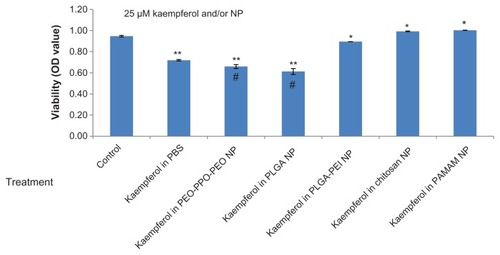
The synthesized PEO-PPO-PEO, PLGA, PLGA-PEI, chitosan, and PAMAM nanoparticles were approximately 200 nm in size (). The PEO-PPO-PEO and PLGA nanoparticles had almost no surface charge, while chitosan, PLGA-PEI, and PAMAM nanoparticles had a positive surface charge, with PAMAM having the highest charge ().
Table 1 Particle size and zeta potential of nanoparticles incorporating kaempferol (data are an average of three samples)
We screened the five different types of kaempferol nanoparticles for their ability to inhibit viability of A2780/CP70 cancer cells. As shown in , kaempferol in 25 μM phosphate-buffered saline solution did not achieve any significant reduction in cell viability compared with unexposed controls. Neither nanoparticles plus kaempferol nor nanoparticles alone resulted in any significant change in A2780/CP70 cell viability compared with kaempferol in phosphate-buffered saline solution or the control. In contrast, PEO-PPO-PEO nanoparticles incorporating kaempferol achieved significant inhibition of A2780/CP70 cells and resulted in significant reduction in cell viability compared with kaempferol in phosphate-buffered saline solution (). PLGA nanoparticles incorporating kaempferol also showed marginally significant inhibitory effects compared with kaempferol in phosphate-buffered saline solution (P = 0.07, ). The other three types of nanoparticle (ie, PLGA-PEI, chitosan, and PAMAM) did not achieve a significant reduction in A2780/CP70 cell viability compared with kaempferol in phosphate-buffered saline solution or the control, and no significant differences in ability to reduce cell viability were observed between these three nanoparticle types ().
Figure 2 Effects of nanoparticles incorporating kaempferol on A2780/CP70 ovarian cancer cells.
Notes: Ovarian cancer cells were seeded in a microplate, incubated overnight, and treated with 25 μM kaempferol for 24 hours. Cell viability was analyzed using an MTS-based method. *P < 0.05 as compared with kaempferol in phosphate-buffered saline solution and control.
Abbreviations: MTS, 3-(4,5-dimethylthiazol-2-yl)-5-(3-carboxymethoxyphenyl)-2-(4-sulfophenyl)-2H-tetrazolium; NP, nanoparticle; OD, optical density; PEO-PPO-PEO, poly(ethylene oxide)-poly(propylene oxide)-poly(ethylene oxide); PAMAM, poly(amidoamine); PBS, phosphate-buffered saline solution; PLGA, poly(DL-lactic acid-co-glycolic acid).
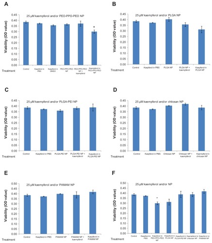
These data suggest that nanoparticle chemistry plays an important role in the treatment of cancer if nanoparticles are used. Appropriate nanoparticle formulation or chemistry (ie, PEO-PPO-PEO) can lead to significant reduction of cancer cell viability (see ). Positively charged nanoparticles did not result in reduction of A2780/CP70 cell viability, while nonionic polymeric (eg, PEO-PPO-PEO) nanoparticles led to significant reduction in A2780/CP70 cell viability.
We also examined these chemicals in another ovarian cancer cell line (ie, OVCAR-3). Consistent with the screening results for A2780/CP70 cells, PEO-PPO-PEO and PLGA nanoparticles incorporating kaempferol resulted in significantly lower OVCAR-3 cell viability compared with kaempferol in phosphate-buffered saline solution and the control (). PLGA-PEI, chitosan, and PAMAM nanoparticles resulted in higher cell viability compared with kaempferol in phosphate-buffered saline solution (). Comparing and , PEO-PPO-PEO and PLGA nanoparticles incorporating kaempferol led to lower viability of both A2780/CP70 and OVCAR-3 cancer cells than did kaempferol in phosphate-buffered saline solution, but the degree of reduction was significantly different. Moreover, PEO- PPO-PEO and PLGA nanoparticles incorporating kaempferol showed greater activity against OVCAR-3 cells than A270/CP70 cells. This is consistent with the effect of kaempferol alone because kaempferol 25 μM in phosphate-buffered saline solution significantly reduced the viability of OVCAR-3 cells () but not A2780/CP70 cells (). These findings suggest that the treatment outcome depends on the type of cancer cell, and PEO-PPO-PEO and PLGA nanoparticles incorporating kaempferol could have potential application in different forms of ovarian cancer (even when kaempferol alone does not have a significant impact).
Figure 3 Effects of nanoparticles incorporating kaempferol on OVCAR-3 ovarian cancer cells.
Notes: OVCAR-3 cells were seeded in a microplate, incubated overnight, and treated with 25 μM kaempferol for 24 hours. Cell viability was analyzed using an MTS-based method. **P < 0.01, *P < 0.05 versus control. #P < 0.01 versus kaempferol in phosphate-buffered saline solution.
Abbreviations: NP, nanoparticle; OD, optical density; PEO-PPO-PEO, poly(ethylene oxide)-poly(propylene oxide)-poly(ethylene oxide); PAMAM, poly(amidoamine); PBS, phosphate-buffered saline solution; PLGA, poly(DL-lactic acid-co-glycolic acid).
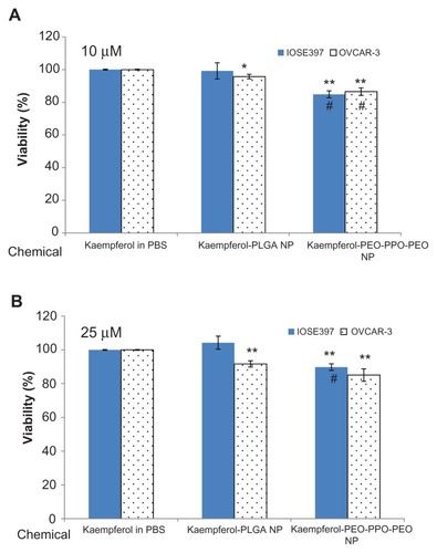
The effects of PEO-PPO-PEO and PLGA nanoparticles incorporating kaempferol were further compared with kaempferol in phosphate-buffered saline solution for their inhibitory effects on cancerous and normal ovarian cells at two different concentrations (10 μM and 25 μM). As shown in , at 10 μM, PLGA nanoparticles incorporating kaempferol significantly reduced the viability of OVCAR-3 cells but had no significant influence on the viability of IOSE397 cells. This result was also found at the 25 μM concentration. However, PEO-PPO-PEO nanoparticles incorporating kaempferol significantly reduced the viability of both OVCAR-3 and IOSE397 cells compared with kaempferol in phosphate-buffered saline solution at concentrations of 10 μM and 25 μM. Overall, the higher the concentration (from 10 μM to 25 μM), the greater the reduction in OVCAR-3 cell viability. Moreover, it seemed that, at the same concentration, PEO-PPO-PEO nanoparticles incorporating kaempferol were more effective in reducing OVCAR-3 cell viability compared with PLGA nanoparticles incorporating kaempferol, with a significant reduction seen at 10 μM (). PEO-PPO-PEO nanoparticles incorporating kaempferol also resulted in significant reduction of IOSE397 cell viability compared with PLGA nanoparticles incorporating kaempferol (). These results suggest that incorporation of kaempferol into PEO-PPO-PEO and PLGA nanoparticles enhanced the effectiveness of kaempferol in reducing the viability of cancer cells, and PEO-PPO-PEO nanoparticles incorporating kaempferol were more effective than PLGA nanoparticles incorporating kaempferol. PLGA nanoparticles incorporating kaempferol could discriminate between cancerous and normal cells, whereby the viability of cancer cells but not the normal cells was significantly affected by PLGA nanoparticles incorporating kaempferol compared with kaempferol alone.
Figure 4 Effects of PEO-PPO-PEO and PLGA nanoparticles incorporating kaempferol on OVCAR-3 ovarian cancer cells and immortalized IOSE397 epithelial ovarian cells at (A) 10 μM and (B) 25 μM. Ovarian cells were seeded in microplates, incubated overnight, and treated with kaempferol or its nanoparticles for 24 hours.
Notes: Viability of cells was measured using an MTS-based method. Data represent the mean ± standard error of the mean from a minimum of three independent experiments. *P < 0.05, **P < 0.01 versus kaempferol in phosphate-buffered saline solution. #P < 0.01 versus PLGA nanoparticles incorporating kaempferol in treating the same cell type.
Abbreviations: MTS, 3-(4,5-dimethylthiazol-2-yl)-5-(3-carboxymethoxyphenyl)-2-(4-sulfophenyl)-2H-tetrazolium; PBS, phosphate-buffered saline solution; NP, nanoparticle; PEO-PPO-PEO, poly(ethylene oxide)-poly(propylene oxide)-poly(ethylene oxide); PLGA, poly(DL-lactic acid-co-glycolic acid).
It is worth mentioning that developing effective strategies for targeting anticancer drugs or nanoparticles has also attracted attention.Citation35–Citation39 Sunoqrot et al recently reported a hybrid nanoparticle platform that may allow targeting kinetics to be effectively controlled through hybridization of targeted dendrimers using polymeric nanoparticles.Citation39 In their study, folate-targeted PAMAM dendrimers were incorporated into poly(ethylene glycol)-b-poly(D,L-lactide) nanoparticles, and their combined passive and active targeting enabled precise control over the targeting kinetics of dendrimers to folate-overexpressing cells.Citation39 Similarly, folate-targeted kaempferol complexes could be incorporated into our PEO-PPO-PEO or PLGA nanoparticles to achieve high targeting efficacy against folate-overexpressing cancerous cells while limiting potential effects on normal cells.
Conclusion
Five nanoparticle formulations incorporating kaempferol were investigated for their potential in the treatment of ovarian cancer, and their efficacy was tested in vitro. summarizes the effects of PEO-PPO-PEO and PLGA nanoparticles incorporating kaempferol on cancerous and normal ovarian cells. In A2780/CP70 ovarian cancer cells, we found that nanoparticles incorporating kaempferol with positive charges (ie, PLGA-PEI, glycol chitosan, and PAMAM dendrimer) did not significantly reduce cell viability. PEO-PPO-PEO nanoparticles incorporating kaempferol significantly reduced cell viability compared with kaempferol alone, and PLGA nanoparticles incorporating kaempferol also showed notably enhanced reduction of the viability of A2780/CP70 cells. PEO-PPO-PEO and PLGA nanoparticles incorporating kaempferol also significantly reduced the viability of OVCAR-3 cancer cells, compared with kaempferol alone. These two kaempferol nanoparticles were further compared with kaempferol alone in immortalized IOSE397 ovarian cells and OVCAR-3 ovarian cancer cells. We found that PEO-PPO- PEO nanoparticles incorporating kaempferol not only significantly reduced the viability of OVCAR-3 cancer cells but also that of normal IOSE397 cells compared with kaempferol alone. Interestingly, PLGA nanoparticles incorporating kaempferol significantly reduced the viability of OVCAR-3 cancer cells but not normal IOSE397 cells compared with kaempferol alone. Therefore, the PLGA nanoparticle formulation could be a promising candidate for cancer treatment due to its improved ability to reduce cancer cell viability along with no significant reduction in the viability of normal ovarian cells. PEO-PPO-PEO nanoparticles incorporating kaempferol were more effective in inhibiting the viability of cancer cells compared with PLGA nanoparticles incorporating kaempferol. If appropriately targeted, PEO-PPO-PEO nanoparticles incorporating kaempferol could be more effective in treating cancer. The mechanisms related to the effects of PEO-PPO-PEO and PLGA nanoparticles incorporating kaempferol on cancerous and normal ovarian cells are still unclear, so further investigation of these nanoparticles for better nanochemoprevention of cancer is warranted.
Table 2 Summary of the effects of PEO-PPO-PEO and PLGA nanoparticles incorporating kaempferol on cancerous and normal ovarian cells studied
Acknowledgments
This research was supported by a West Virginia Experimental Program to Stimulate Competitive Research grant and an NIH grant (P20 RR016477) from the National Center for Research Resources awarded to the West Virginia IDeA Network of Biomedical Research Excellence. BL also acknowledges financial support from the National Science Foundation (1003907), the West Virginia Higher Education Policy Commission/Division of Science Research, and WVNano. The authors thank Suzanne Smith for proofreading.
Disclosure
The authors report no conflicts of interest in this work.
References
- GonzalezCARiboliEDiet and cancer prevention: where we are, where we are goingNutr Cancer20065622523117474869
- HolickCNGiovannucciELRosnerBStampferMJMichaudDSProspective study of intake of fruit, vegetables, and carotenoids and the risk of adult gliomaAm J Clin Nutr20078587788617344512
- BosettiCRossiMMcLaughlinJKFlavonoids and the risk of renal cell carcinomaCancer Epidemiol Biomarkers Prev2007169810117220336
- HanhinevaKTörrönenRBondia-PonsIImpact of dietary polyphenols on carbohydrate metabolismInt J Mol Sci2010111365140220480025
- RudkowskaIJonesPJFunctional foods for the prevention and treatment of cardiovascular diseases: cholesterol and beyondExpert Rev Cardiovasc Ther2007547749017489672
- BosettiCBraviFTalaminiRFlavonoids and prostate cancer risk: a study in ItalyNutr Cancer20065612312717474856
- TheodoratouEKyleJCetnarskyjRDietary flavonoids and the risk of colorectal cancerCancer Epidemiol Biomarkers Prev20071668469317416758
- RossiMNegriELagiouPFlavonoids and ovarian cancer risk: a case-control study in ItalyInt J Cancer200812389589818491402
- DuthieGCrozierAPlant-derived phenolic antioxidantsCurr Opin Clin Nutr Metab Care2000344745111085830
- HungHInhibition of estrogen receptor alpha expression and function in MCF-7 cells by kaempferolJ Cell Physiol200419819720814603522
- SharmaVJosephCGhoshSAgarwalAMishraMKSenEKaempferol induces apoptosis in glioblastoma cells through oxidative stressMol Cancer Ther200762544255317876051
- LeungHWLinCJHourMJYangWHWangMYLeeHZKaempferol induces apoptosis in human lung non-small carcinoma cells accompanied by an induction of antioxidant enzymesFood Chem Toxicol2007452005201317583406
- CortesJRPerezGMRivasMDZamoranoJKaempferol inhibits IL-4-induced STAT6 activation by specifically targeting JAK3J Immunol20071793881388717785825
- LeeKMLeeKWJungSKKaempferol inhibits UVB-induced COX-2 expression by suppressing Src kinase activityBiochem Pharmacol2010802042204920599768
- García-MediavillaVCrespoIColladoPSThe anti-inflammatory flavones quercetin and kaempferol cause inhibition of inducible nitric oxide synthase, cyclooxygenase-2 and reactive C-protein, and down-regulation of the nuclear factor kappaB pathway in Chang Liver cellsEur J Pharmacol200755722122917184768
- LuoHRankinGOJulianoNJiangBHChenYCKaempferol inhibits VEGF expression and in vitro angiogenesis through a novel ERK-NFκB-cMyc-p21 pathwayFood Chem201213032132821927533
- LuoHRankinGOLiZDePriestLChenYCKaempferol induces apoptosis in ovarian cancer cells through activating p53 in the intrinsic pathwayFood Chem201112851351921625365
- LuoHRankinGOLiuLDaddysmanMKJiangBHChenYCKaempferol inhibits angiogenesis and VEGF expression through both HIF dependent and independent pathways in human ovarian cancer cellsNutr Cancer20096155456319838928
- LuoHJiangBHKingSMChenYCInhibition of cell growth and VEGF expression in ovarian cancer cells by flavonoidsNutr Cancer20086080080919005980
- GatesMATworogerSSHechtJLDe VivoIRosnerBHankinsonSEA prospective study of dietary flavonoid intake and incidence of epithelial ovarian cancerInt J Cancer20071212225223217471564
- FerrariMCancer nanotechnology: opportunities and challengesNat Rev Cancer2005516117115738981
- SiddiquiIAAdhamiVMAhmadNMukhtarHNanochemoprevention: sustained release of bioactive food components for cancer preventionNutr Cancer20106288389020924964
- SiddiquiIAAdhamiVMChristopherJChamcheu MukhtarHImpact of nanotechnology in cancer: emphasis on nanochemopreventionInt J Nanomedicine2012759160522346353
- MaXWangHJinSWuYLiangXJConstruction of paclitaxel-loaded poly (2-hydroxyethyl methacrylate)-g-poly (lactide)-1,2-dipalmitoylsn- glycero-3-phosphoethanolamine copolymer nanoparticle delivery system and evaluation of its anticancer activityInt J Nanomedicine201271313132822419875
- RanganathanRMadanmohanSKesavanANanomedicine: towards development of patient-friendly drug-delivery systems for oncological applicationsInt J Nanomedicine201271043106022403487
- SabnisNNairMIsraelMMcConathyWJLackoAGEnhanced solubility and functionality of valrubicin (AD-32) against cancer cells upon encapsulation into biocompatible nanoparticlesInt J Nanomedicine2012797598322393294
- GrefRMinamitakeYPeracchiaMTTrubetskoyVTorchilinVLangerRBiodegradable long-circulating polymeric nanospheresScience1994263160016038128245
- PeerDKarpJMHongSFarokhzadOCMargalitRLangerRNanocarriers as an emerging platform for cancer therapyNat Nanotechnol2007275176018654426
- NishiyamaNNanomedicine: nanocarriers shape up for long lifeNat Nanotechnol2007220320418654260
- SiddiquiIAAdhamiVMBharaliDJIntroducing nanochemoprevention as a novel approach for cancer control: proof of principle with green tea polyphenol epigallocatechin-3-gallateCancer Res2009691712171619223530
- JemalASiegelRXuJWardECancer statistics, 2010CA Cancer J Clin20106027730020610543
- BanksEThe epidemiology of ovarian cancerBartlettJMSOvarian Cancer: Methods and ProtocolsTotawa, NJHumana Press Inc2000
- AntoniouAPharoahPDPNarodSAverage risks of breast and ovarian cancer associated with BRCA1 or BRCA2 mutations detected in case series unselected for family history: a combined analysis of 22 studiesAm J Hum Genet2003721117113012677558
- GuterresSSFessiHBarratGPuisieuxFDevissaguetJPPoly (D,L-lactide) nanocapsules containing non-steroidal anti-inflammatory drugs: gastrointestinal tolerance following intravenous and oral administrationPharm Res199512154515478584497
- DuncanRPolymer conjugates as anticancer nanomedicinesNat Rev Cancer2006668870116900224
- MycAKukowska-LatalloJCaoPTargeting the efficacy of a dendrimer-based nanotherapeutic in heterogeneous xenograft tumors in vivoAnticancer Drugs20102118619220010426
- MaedaHTumor-selective delivery of macromolecular drugs via the EPR effect: background and future prospectsBioconjug Chem20102179780220397686
- TorchilinVTumor delivery of macromolecular drugs based on the EPR effectAdv Drug Deliv Rev20116313113520304019
- SunoqrotSBaeJWPearsonRMTemporal control over cellular targeting through hybridization of folate-targeted dendrimers and PEG-PLA nanoparticlesBiomacromolecules2012131223123022439905