Wang B, Liu H, Sun L, et al. In J Nanomedicine. 2017;12:111–125.
The authors have informed the journal that the scales bars in , and were missed and the images in and were mistakenly duplicated. It was also found the images selected to represent , , were selected from the wrong samples. New representative images have been selected from the original data to replace the original images used and scale bars have been added to , and . This correction does not change any description, results or conclusions of the original paper.
The authors apologize for any confusion this may have caused. The corrected versions are shown below.
Figure 6 Fluorescent microscopy images of live/dead staining of S. aureus.
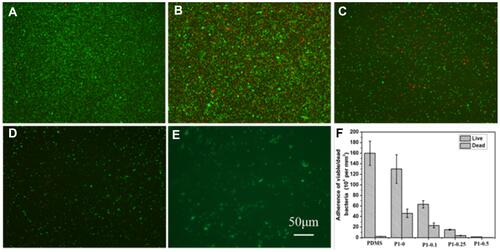
Figure 7 Fluorescent microscopy images of live/dead staining of S. aureus.
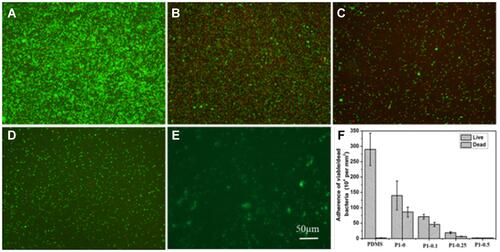
Figure 8 SEM images.
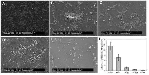
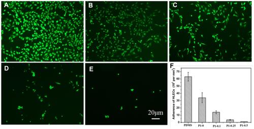
Figure 11 Growth and morphology of HLECs stained with FDA after 24 hours of incubation on various surfaces.