
Abstract
Background
Conventional nanoparticle synthesis methods involve harsh conditions, high costs, and environmental pollution. In this context, researchers are actively searching for sustainable, eco-friendly alternatives to conventional chemical synthesis methods. This has led to the development of green synthesis procedures among which the exploration of the plant-mediated synthesis of nanoparticles experienced a great development. Especially, because plant extracts can work as reducing and stabilizing agents. This opens up new possibilities for cost-effective, environmentally-friendly nanoparticle synthesis with enhanced size uniformity and stability. Moreover, bio-inspired nanoparticles derived from plants exhibit intriguing pharmacological properties, making them highly promising for use in medical applications due to their biocompatibility and nano-dimension.
Objective
This study investigates the role of specific phytochemicals, such as phenolic compounds, terpenoids, and proteins, in plant-mediated nanoparticle synthesis together with their influence on particle size, stability, and properties. Additionally, we highlight the potential applications of these bio-derived nanoparticles, particularly with regard to drug delivery, disease management, agriculture, bioremediation, and application in other industries.
Methodology
Extensive research on scientific databases identified green synthesis methods, specifically plant-mediated synthesis, with a focus on understanding the contributions of phytochemicals like phenolic compounds, terpenoids, and proteins. The database search covered the field’s development over the past 15 years.
Results
Insights gained from this exploration highlight plant-mediated green synthesis for cost-effective nanoparticle production with significant pharmacological properties. Utilizing renewable biological resources and controlling nanoparticle characteristics through biomolecule interactions offer promising avenues for future research and applications.
Conclusion
This review delves into the scientific intricacies of plant-mediated synthesis of nanoparticles, highlighting the advantages of this approach over the traditional chemical synthesis methods. The study showcases the immense potential of green synthesis for medical and other applications, aiming to inspire further research in this exciting area and promote a more sustainable future.
Introduction
The recent era emerged as a period of development for nanomaterials in diverse areas of research, since they possess exceptional physical, chemical and electronic properties.Citation1 These properties are essential in various scientific disciplines, such as catalysis, electronics, targeted drug delivery, water sensing and treatment, corrosion inhibition, oil recovery, and many more.Citation2–4 Generally, the term ‘nanoparticles’ is used to describe particles that range in size from 1–100 nanometers although, in the field of biotechnology, this is usually extended to include particles up to 500 nanometers in size.Citation5 “Size” is the parameter which makes ‘nanoparticles’ unique from other materials, usually called “bulk materials”, and able to generate certain profound physicochemical properties.Citation5,Citation6 Usually, fabricated nanoparticles are metallic in nature and display an effect commonly known as “Surface Plasmon Resonance”, which plays an important role in the quantum mechanical effects of light in UV-Visible regions, leading to unique optronics/optoelectronic properties.Citation7 Prominently, any type of change in the size or shape of a nanoparticle is reflected on its inter-particle interactions as well as absorption properties.Citation8 Due to their exceptional properties, nanoparticles are extensively utilized in various biomedical applications.Citation9–11
The numerous developments associated with nanoscale science have produced plentiful nano-dimensioned materials to enhance the related research and hence, a variety of valuable nano-sized materials are being produced on a commercial scale.Citation12 It has been assumed that, in the future, nano-sized materials and their related products will become an aid in day-to-day life. It is equally important that these nano-sized materials possess a strong ability to establish an interaction with different biological molecules, both inside as well as on the surface of cells. Their ability to reach inside cells enables these molecules to guide different cellular physicochemical and biochemical processes.Citation13 Nanoparticles can be quickly and easily absorbed into cellular compartments/organelles, due to their small dimensions. Furthermore, nano-sized particles can easily cross the placenta and the barriers of the blood-brain.Citation12,Citation14 This explains the availability of nano-sized materials in various drug formulations (approximately 45), which Weissig et al discussed in detail.Citation15 For instance, TiO2 and ZnO nanoparticles are able to resist UV-radiation, and thus have been incorporated into various cosmetic products (specifically sunscreen). On application to the skin surface, these nanoparticles remain transparent to visible light and provide better protection against UV-rays compared to ordinary sunscreen.Citation16 Furthermore, a collection of NPs based on the single-walled carbon-nanotubes (SWCNT) was introduced to the Russian market recently.Citation17 In the food market, nano-sized materials are already being used to increase the storage time of edible products and so control the spoilage rate.Citation18
Vance et al, extensively evaluated the nanotechnology-based-products in the market and reported that approximately 1814 products containing nano-sized materials, introduced by around 622 companies, are being used by about 32 countries globally. It was claimed that about 435 silver-based, nano-sized materials are readily available in the market, including veils, toothpaste, detergent, humidifiers and shampoo. Currently, there exists a high demand for nanomaterials, which was calculated to be from 300,000 to 1.6 million tonnes worldwide. The market share of the Asia-Pacific area is the greatest, (34%), followed by North America and Europe (at 31% and 30%, respectively).Citation19
The method of nano-sized materials synthesis is an important chemical process. At present, both chemical and physical methods are applied when preparing nano-sized materials, but these may not be the optimum choice, due to their high cost and potential pollution of the environment. Consequently, alternatives to these existing methods, which are environmentally-friendly (green synthesis) during the whole production procedure, must be developed for the synthesis of nano-sized materials, which has attracted the interest of researchers worldwide.Citation19,Citation20 The traditional synthesis procedures (both physical and chemical) are usually carried out under extremely harsh conditions. In contrast, the biological procedures are generally conducted under an ambient temperature and pressure, which indicates simplicity, energy saving and reduced toxicity or harm to both humans and the environment.
Bearing these advantages in mind, various biological resources, including bacteria, fungi, yeast, plants and algae, are being exploited to synthesize both intracellular and extracellular materials possessing nano-size.Citation21 Even though nanoparticles are synthesized following biological methods (biosynthesis), the mechanism of their synthesis remains challenging.Citation22 However, these methods are well established and commonly used to synthesize nano-sized materials, as they are more cost-effective and environmental-friendly compared to conventional methods. Biological methods harness the power of renewable sources such as plants and microorganisms, acting as reducing agents to stabilize and cap nanomaterials, eliminating the need for chemical additives.Citation23,Citation24 A standardized method for the synthesis of nanoparticles utilizing plant extracts involves a systematic approach, where a specific plant material is carefully selected and taxonomically identified, and the desired plant extract obtained. Subsequently to the selection of plant parts, an extraction process using an appropriate solvent/s, followed by filtration/chromatography to eliminate any impurities, is carried out. Simultaneously, a metal salt solution of choice is prepared as the nanoparticle precursor and the previously prepared plant extract is added to the metal salt solution, maintaining the appropriate temperature and pH for the reaction, and initiating a reaction that leads to the formation of nanoparticles.Citation25 In the several methods reported in earlier studies, the continuous stirring of the reaction mixture offers better results in the form of uniform-sized nanoparticles, as is visually indicated by a noticeable change in colour. Furthermore, to ensure uniform dispersion, ultrasonic treatment may be applied. Subsequently, the nanoparticles are separated from the solution by employing centrifugation before being washed to remove any remaining impurities. Optionally, the nanoparticle precipitate can be dried to eliminate any additional impurities.Citation26 Finally, the synthesized nanoparticles are thoroughly characterized using diverse analytical techniques to confirm their composition and physicochemical properties. Following synthesis, different spectroscopic and microscopic methods play a crucial role in characterizing synthesized nanoparticles. For instance, UV-Vis spectrophotometry is used to assess their optical characteristics, while FTIR spectroscopy assists the identification of the functional groups present on the nanoparticle surface. For a detailed analysis of their size, shape, and structure, Scanning Electron Microscopy (SEM) and Transmission Electron Microscopy (TEM) are employed. Additionally, Dynamic Light Scattering (DLS) and Zeta potential measurements are utilized to determine the size and surface charge of the nanoparticles. These methods provide valuable insights into the properties and behaviour of the synthesized nanoparticles.Citation27–29
Nanotechnology and Green Synthesis
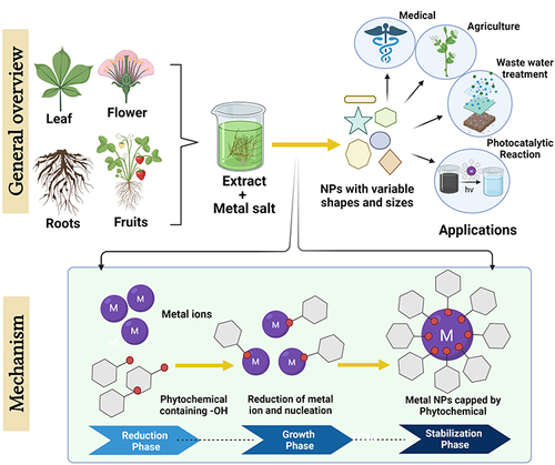
As discussed in Introduction, the synthesis of nano-sized materials employing “green” processes is relatively cost-effective and does not harm the surrounding environment, as non-toxic chemicals are employed throughout the entire process. Therefore, the usage of stabilizers and reducing agents that possess a biological origin, such as microbial entities, fauna and various other resources, is a sustainable way to produce nano-sized materials.Citation30,Citation31 Although being cost-effective and environment-friendly are key factors motivating the green synthesis of nano-sized materials, the “stability” of the produced material has attracted the attention of researchers across the globe.Citation14 Although the methods involved in green synthesis are relatively diverse, the living entities that are involved in the synthesis usually simply react with different salts (metallic) and reduce them to nano-sized materials, which can be utilized for different purposes only following appropriate characterization.Citation32,Citation33 Both microbe- and plant-mediated approaches are employed to synthesize nano-sized materials. Microbe-mediated construction products involve their inherently sophisticated biochemical machinery, which leads to well-defined nanoparticles of different chemical compositions, shapes and sizes.Citation34 Scaling-up may, however, sometimes prove challenging with regard to microbial preparations. This drawback can be easily overcome by using plant-based extracts, and the production rates can be amplified as a consequence. Plant extracts are more efficient than microbes with regard to the production rate. They reduce metal-ions faster than microbial entities and produce nano-sized materials, which are also very stable.Citation18,Citation19 Plants contain various compounds (ie, alkaloids, flavonoids, phenol, tannin, alcohol) with the capability to reduce metallic ions to nanoparticles with decent stability.Citation17,Citation21 Currently, there is evidence that the benefits of plant-based synthesis arise from the synergy of those compounds. A very-general scheme of “green-synthesis of nano-sized material” is shown in .
Figure 1 Schematic illustration of the method and mechanism involved in green synthesis of nanoparticles using plants as reducing agents.
Medicinal Plants Exploited for Nanomaterial Synthesis
Plants are rich sources of medicinal compounds that possess the capacity to produce simple ions by reducing complex metallic ions. The accumulation of metallic-ions in plant cells and tissues inspired the idea of applying metal-reduction to nano-sized materials.Citation35 For instance, Alfalfa (Medicago sativa) and Brown Mustard (Brassica juncea), developed in the presence of silver nitrate (AgNO3), accrue 12.4 and 13.6 wt% of Ag, respectively, with Ag-NPs being around 50 nm in size.Citation21 Similarly, 4 nm sized gold icosahedra were observed in Alfalfa (Medicago sativa),Citation36 and around 2 nm sized semi-spherical Cu-NPs were found in Iris pseudacorus, when grown in the salts. Various in-vitro methods, including the utilization of herbal extracts as reducers for synthesizing nano-sized materials, have recently been developed.Citation35 Extracts of many plant-species, several acids and the salts of different metals (eg, Cu, Au, Ag, Pt, Se and Fe) have been employed in the green synthesis of nano-sized materials.Citation37,Citation38 Because no bacteria or toxic chemical contaminants are present, plant materials are more favourable when used for NP fabrication compared to methods involving microbes or deleterious chemicals. Furthermore, this approach consumes less energy and has both simple and broad applications.Citation39,Citation40 For the bio-friendly synthesis of NPs, diverse plant extracts obtained from various components, including the plant stems, bark, roots, floral parts, leaves, seeds and fruit have been employed.Citation41–43 In recent years, a range of green synthesized nano-sized materials, obtained from various medicinal plant species, have been described (). Different features of these synthesized, nano-sized materials, such as their shape, size and quality, are highly dependent on various aspects, such as the concentration of the extract used, and specifically its composition and pH, as well as the temperature at which the reaction occurs.Citation44–46
Table 1 Metallic NPs synthesized using plant-extracts
Mechanistic Investigation of Green Synthesis
Both microbe- and plant-mediated approaches are being employed to synthesize nano-sized materials. Microbes are less efficient than plant extracts in terms of their production rate. In contrast to microorganisms, which contain important enzymes that can serve as a reductant as well as a stabilizer for nano synthesis, plants often possess a phytochemical composition, including “alcohols, phenols, terpenes, alkaloids and proteins”.Citation74 The mechanistic investigation of the green synthesis of nanoparticles is described in detail in the following subsections.
Plant-Mediated Green Synthesis
The utilization of plant extracts has become one of the main biosynthesis techniques being addressed, since the majority of plants are often affordable, accessible and safe to use. Additionally, a variety of phytochemicals, mentioned above, are found in abundance in plant extracts, which function as a reductant as well as a stabilizer in the synthesis of plant derived NPs.Citation74 The precise mechanisms of phytosynthesis for biogenic NPs are not yet fully known, and several plausible pathways have been postulated for its synthesis.Citation75–77 Since numerous phytochemicals are present, it is challenging to identify any specific bio-reductant and stabilizer mediators that are responsible for the production and stabilization of metallic nanoparticles (MNPs).Citation78 The potential influence of various phytochemicals on NP synthesis will be discussed in the following sub-sections.
Influence of Phenolic Compounds in Nano-Synthesis
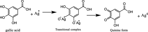
In the polyphenol family, phenolic acid plays a significant role in nano-synthesis. Its structure consists of a “phenolic ring and a functional carboxylic acid group”. The nucleophilic aromatic rings of such compounds offer potent support for antioxidant activity and metal chelation.Citation74,Citation79 Several known phenolic acids function as reductants in the biosynthesis of MNPs, including “caffeic acid, gallic acid and ellagic acid”.Citation80,Citation81 According to Raja et al, the development of a transitional complex between silver ions (Ag+) and gallic acid’s phenolic hydroxyl groups may enable the synthesis of silver nanoparticles (AgNPs). Thereafter, it transforms into quinine via the oxidation process, producing silver nanoparticles ().Citation82–84
Scheme 1 A plausible mechanism for AgNP synthesis using plant extract containing gallic acid.
Wang et al, used Melaleuca nesophila, Rosmarinus officinalis and Eucalyptus tereticornis leaf extracts to create “iron-polyphenol” NPs, which were then FTIR-characterized. They demonstrated that charged iron combines with polyphenol to create complex nanoparticles, ranging in size from 50–90 nm, with an organic-looking surface.Citation85 The glycosidic water-soluble tannin in Gloriosa superba leaf extract aid the reduction of gold and silver ions into spherical nanoscale particles of gold and silver.Citation86 Jha et al, reported that the occurrence of “protocatechuic acid, catechol and ascorbic acid” accounts for silver bio-reduction, caused by the production of “H+” by Hydrilla species of the hydrophyte genera. In a similar manner, in Cyperus sp., the ability of tautomerization of benzoquinone derivatives has been reported, leading to reduction of silver metal along with formation of coalescent cluster.Citation87
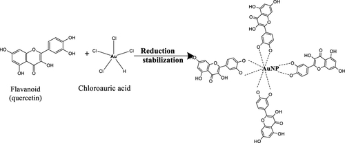
A family of secondary metabolites, known as flavonoids, are indeed the components of plant pigments. There exist numerous types of flavonoids that may be discovered in plants. The capacity of flavonoids to donate “H” atoms or electrons is associated with their reducing capability, which is regarded as the primary bio-reducing component of plant extracts.Citation88,Citation89 Ahmad et al, suggested the H+, created in the course of the keto-enol transformation of the flavonoids “luteolin and rosmarinic acid”, could be employed to reduce silver metal ions and create silver nanoparticles.Citation90,Citation91 Sahu et al, employed the poly-hydroxylated secondary metabolites of plants to bio-reduce Ag+ ions to silver nanoparticles. Moreover, these flavonoids serve as capping agents, which impact on the antibacterial properties of silver nanoparticles.Citation92,Citation93 Zhou et al, found that bio-compounds interacted with “tetrachloroaurate ion” via an ionic bond or electrostatic force. It was discovered that flavonoids and reducing sugars were the bio-reductants responsible for converting gold ion to gold nanoparticles (AuNP). Proteins were not found to be reductants in this study but were shown to be capping mediators in Au nanoparticles synthesis.Citation89 Raki et al, explained the mechanism for the formation and stabilization of AuNPs by the flavonoids present in plant extract. The neighboring hydroxyl groups of the polyphenolic molecules first bind with gold to create a chelate ring containing five members. The chelated “ortho- dihydroxyl” moieties are converted into quinones while simultaneously reducing the Au, due to the extremely high oxidation-reduction potential of Au. In order to create AuNPs, neighboring Au atoms collide, and the quinones and polyphenolic molecules work together to stabilize the resulting AuNPs ().Citation94 In a surfactant-free condition, Nasrollahzadeh et al, synthesized copper nanoparticles through the bio-reduction of copper ions using “quercetin” from the leaf extract of G. biloba.Citation95,Citation96 A redox process caused the -OH in the reduced form of polyphenolics to change into a -C=O, which then caused the metal ions to undergo reduction. The metal nanoparticles are electrostatically stabilized by these carbonyl groups on the oxidized polyphenol.Citation95
Scheme 2 Mechanism underlying the formation and stabilization of gold nanoparticles (AuNPs) using flavonoids present in plant extracts.
Influence of Terpenoids During Nano-Synthesis
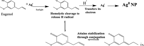
Terpenoids are obtained from the essential oils of plants, which are intricate blends of volatile organic molecules that are produced by plants as secondary metabolites. Terpenoids are the oxygenated version of terpenes. They may operate as surface active chemicals that can stabilize and decrease nanoparticles, but the mechanism through which they participate in nanoparticle formation is not yet thoroughly understood.Citation97 Shankar et al, utilized C. zeylanicum extracts to reduce chloroauric acid and silver nitrate to gold and silver nanoparticles, respectively, due to the significant concentration of the fundamental terpenoid “eugenol”.Citation98 According to Singh et al, the “eugenol” in S. aromaticum extract acts as a bio-reductant to yield gold and silver nanoparticles.Citation90,Citation99 A clear, concise, effective and useful approach to the fabrication of silver nanoparticles was devised by Brahmachari et al, which entailed employing an O. sanctum leaf extract in water under the impact of direct sunlight.Citation100 The researchers postulated that, upon exposure to sunlight, the “phenolic O-H bond” of eugenol experiences a homolytic breakage to produce a H+ radical, which then donates its electron to Ag+ and converts to Ag0 NP. With prolonged conjugation, the oxygen radical component is stabilized in the solution ().Citation95,Citation100
Scheme 3 Photo-induced bio reduction of Ag+ to Ag0 nanoparticles using biological agents.
Influence of Proteins During Nano-Synthesis
Proteins may contribute to the biosynthesis and stability of metallic nanoparticles. The -C=O of amino acid sequences often serve as capping ligands for NPs, and FTIR spectra amply illustrate the involvement of diverse C=O groups on the surface of NPs, thus avoiding their aggregation and maintaining their stability during the aqueous phase.Citation95,Citation101 Proteins and amino acids bearing exposed disulfide bonds and thiols function throughout biosynthesis as non-enzymatic reducers and stabilisers.Citation95,Citation102 A tripeptide sequence that contains a “C-terminus with tyrosine remnants” that acts as a reductant was used by Bhattacharjee et al, to create gold nanoparticles. This sequence contains an open “N terminal” that may be linked to the developing nanoparticles and generate permanent gold nanoparticles.Citation103 Li et al, postulated that, during the biosynthesis of AgNPs using Capsicum annuum extract, the electrostatic interactions cause the silver ions to become trapped on the surface of the peptides. These silver ions are reduced by the protein, which leads to changes in the 2° structure and the formation of silver nuclei. These nuclei grow further through the accumulation of ions. The versatile protein-biomolecule interaction that was found in the reaction conditions may have contributed to the formation of the sturdy silver nanoparticles ().Citation104 Stabilizer proteins offer advantages over polymers and surfactants, such as being lower cost, more environmentally friendly and not needing complicated processes. They can also serve as an anchorage layer for medications or genes that need to be introduced into living cells.Citation95,Citation105
Scheme 4 The green synthesis of AgNPs utilizing plant extract-derived proteins as efficient reducing agents.

Physicochemical factors affecting Green Synthesis
A number of factors are associated with the synthesis of NPs utilizing different plant extracts, that influence the nanoparticle’s shape and dimension. Even these factors have a straight relation with the characterization procedures of nanoparticles as well as their application. The “adsorbate” type and catalyst activity have a huge impact on the synthesized nanoparticles, as reported by various researchers.Citation106,Citation107 Also, the time and environment of the reaction have a huge impact on the synthesized nano-material.Citation107 In addition, the temperature, pH, extract concentrations, raw materials size and methodology employed to synthesize the nanomaterials also influence the resulting products.Citation108 Indeed, as the temperature of the reaction rises, the particle size also increases, suggesting that the Ostwald ripening, from which the larger sized particles grow, is due to the dissolution of the smaller ones.Citation109 The phytochemicals present in plant extracts are of chief importance in boosting dispersion and eventually decreasing agglomeration.Citation110 Moreover, it was believed and later confirmed that these phytochemicals are the deciding factors regarding the origin of novel properties of synthesized materials and play a major role in green synthesis.Citation111,Citation112 Furthermore, the effect of salt’s concentration on the morphology of different metallic particles has been reported by several researchers.Citation113,Citation114 Some of the major factors that influence the preparation of nanomaterials are summarized below in and .
Figure 2 The impact of diverse factors on the morphology and yield of nanoparticles.
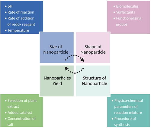
Table 2 Major factors influencing the synthesis of nano-material
Synthesized Nano-Sized Materials and Their Characterization
When synthesizing nanoparticles, a number of challenges emerge with regard to characterizing and describing them, so it becomes important to choose an appropriate method for characterizing nanoparticles from among the existing techniques for determining their size, shape, aggregation, chemistry, crystallinity, orientation, fractal dimensions, dispersion and other parameters.Citation107 In this sense, various analytical methods have been employed to characterize in detail the properties (both chemical as well as physical) of green synthesized nanoparticles. Recently, Catalano et al, highlighted the necessity to develop validated procedures for the characterization of nanomaterials that have been synthesized following green practices to achieve their sustainable, safe application.Citation33 In this sense, techniques that are frequently employed in this field are “Scanning Electron Microscopy”, “Transmission Electron Microscopy”, “Fourier Transform Infrared Spectroscopy”, and “X-ray Diffraction”, among others. A list of techniques is presented in below, providing some insight into the methods.
Table 3 An assortment of techniques involved in the characterisation nanoparticles with associated rationale(s)
Plant vs Chemical Synthesis: Advantages for Biomedical Applications
Recent studies that have compared the plant-based green synthesis of nanoparticles with chemical methods have consistently demonstrated the advantages of the former. Green synthesis offers a sustainable, environmentally friendly approach to synthesizing nanomaterials, including metal and metal oxide nanoparticles, with wide-ranging applications.Citation123 Plant-based green synthesis ensures non-toxic, biocompatible nanoparticles, making it a safe, eco-friendly alternative to chemical methods, particularly with regard to biomedical applications.Citation124,Citation125 In relation to this, Sabeena et al, conducted a comparative study of the green and chemical synthesis of CuO-NPs, evaluating their in vitro and in vivo bioactivity and toxicity in zebrafish (Danio rerio) embryos. The green synthesis method utilized a leaf extract from Salacia reticulata, which acted as both a reducing and capping agent, enabling the conversion of copper ions to CuO-NPs and ensuring their stability. In contrast, the chemical synthesis method employed sodium hydroxide. The in vitro assessments revealed that the green CuO-NPs exhibited higher antibacterial activity against both Gram-negative and Gram-positive bacteria, increased cytotoxicity in human breast cancer cells (MCF-7), and greater antidiabetic as well as anti-inflammatory effects compared to the chemically synthesized CuO nanoparticles. Notably, the green CuO-NPs demonstrated reduced toxicity in zebrafish embryos. These findings highlight the significance of environmentally friendly green synthesis for CuO nanoparticles in diverse biomedical applications.Citation126 In another study, Sudhasree et al, performed a comparative analysis of nickel nanoparticle synthesis via the chemical and green routes to evaluate the biological and toxicological effects of nickel nanoparticles. Chemical synthesis involved the use of polyethylene glycol and hydrazine hydrate as stabilizing and reducing agents, while green synthesis utilized the aqueous root extract of Desmodium gangeticum without any additional agents. The characterization techniques revealed that both methods produced similar nanoparticles, but that green-synthesized Ni-NPs exhibited a smaller size and better uniformity. The green-synthesized Ni-NPs demonstrated significant antioxidant and antibacterial activity. Additionally, the toxicity assessments of the animals and cell lines confirmed the non-toxic nature of the Ni-NPs that had been synthesized through the green route. This study highlighted the comparable biological activity and lower toxicity of green-synthesized nickel nanoparticles compared to those that had been synthesized chemically.Citation127
Additionally, plant extracts facilitate the controlled synthesis of nanoparticles, allowing a precise control of their size, shape and composition, along with the presence of natural stabilizers and reducing agents for enhanced biomedical compatibility.Citation128 A comparative analysis of green synthesis and chemical synthesis of SiO2 NPs was carried out by Rahimzadeh et al, using Rhus coriaria L. extract and sodium metasilicate. Characterization techniques, such as FTIR, UV-Vis, XRD, FESEM, EDX, zeta potential, DLS, TGA and DSC, were employed to analyze the structure, thermal properties, and morphology of both types of nanoparticles. The results demonstrated that the green-synthesized SiO2 nanoparticles outperformed the chemically synthesized ones. The researchers concluded that the presence of phytochemicals in Rhus coriaria L. extract enhanced the stability, improved the thermal properties, and increased the surface area of the nanoparticles.Citation129
Abdelmigid et al, further suggested the equivalent antimicrobial efficacy of synthesized silver nanoparticles (Ag-NPs) using both chemical and biological methods. In this study, trisodium citrate, pomegranate fruit peel extract and coffee ground waste extract were used as the reductant agents. Ag-NPs that had been synthesized chemically (AgNPs_Chem) exhibited higher stability and negativity of the zeta potential compared to Ag-NPs synthesized using coffee ground waste extract (Ag-NPs_CE) and pomegranate peel extract (Ag-NPs_PPE). All the synthesized Ag-NPs showed antimicrobial activity against Enterobacter aerogenes, Klebsiella pneumoniae, Pseudomonas aeruginosa and methicillin resistant Staphylococcus aureus. The most effective concentration was found to be 8 mg/mL, with the Ag-NPs demonstrating higher efficacy against K. pneumoniae. This study supports the eco-friendly synthesis of Ag-NPs using agro-waste as a viable alternative to chemical methods. The biologically synthesized Ag-NPs exhibited similar antimicrobial properties to their chemically synthesized counterparts, suggesting the potential for sustainable nanoparticle production.Citation130,Citation131 Correspondingly, Aravind et al, synthesized titanium dioxide nanoparticles (TiO2 NPs) using both chemical and green synthesis methods. The green synthesis involved using jasmine flower extract as a reducing and stabilizing agent. The TiO2 NPs exhibited a rutile phase, with a crystalline size of 31–42 nm. UV-Visible spectroscopy confirmed their presence in the visible spectrum. SEM images showed spherical-shaped NPs, arranged randomly. The green-synthesized TiO2 NPs demonstrated higher photocatalytic degradation efficiency for methylene blue dye compared to chemically synthesized NPs. They also exhibited enhanced antibacterial activity against both gram-positive and gram-negative strains. These findings suggest that the green-synthesized TiO2 NPs possess the potential for application in environmental and biomedical fields.Citation132
In addition to the points noted above, the plant-based, green synthesis of nanoparticles is cost-effective, utilizing readily available plants instead of expensive chemicals or metal precursors. It allows diverse nanoparticles’ compositions and surface functionalities, offering a high level of tolerability, reproducibility and biocompatibility. Plant extracts enable precise control over the size, shape, and composition while at the same time acting as natural stabilizers and reducing agents. Notably, combined plant extracts can exhibit synergistic effects, thus enhancing nanoparticle properties. Therefore, plant-based green synthesis provides a cost-effective, versatile and controlled approach for synthesizing nanoparticles with diverse biomedical applications.Citation128,Citation133,Citation134
Bio-Derived Nanomaterials and Future Prospects
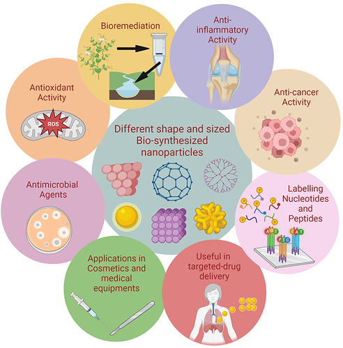
The ability of living things to produce nanoparticles and nanodevices with a variety of applications is vast (). In the reaction mixture, it is possible to produce nanoparticles and nanodevices with a specified form and size, ranging from simple microbes to highly complex organisms. Although nano-biotechnology remains in its infancy, the numerous examples used to demonstrate this science and its implications in this article will draw readers’ attention to its potential uses. Various researchers have suggested that diverse reductases from these species fulfil a pivotal function in the construction of nanoparticles of various shapes and sizes, although far more research is needed to determine the optimum method for creating nanoparticles using living things.Citation108
Figure 3 Illustrative representation of different sized and shaped nanoparticles with their potential application.
Currently, it is vital to create and develop unique drugs due to the numerous limitations imposed on the conventional procedures, such as their escalating cost and perniciousness. The opportunity to use a variety of nanosized materials in a more environmentally responsible manner, that is also economical, readily accessible, and secure without any involvement of harsh substances (chemicals), thus opens up a fresh avenue. Nanotechnology has advanced rapidly over the past decade, fostering healthcare and industrial uses, such as drug administration, imaging, and detection. According to the literature, it appears that in vitro research on bio-derived nanomaterials has taken place, but there have been no reports of in vivo uses. The potential toxicological processes of metallic NPs are also poorly understood. Therefore, before embarking on preclinical research, extensive in vivo experiments should be conducted to produce figures that represent nano-structured medicine’s behavior.
The number of applications for biologically driven nano-sized materials will increase, and are anticipated to reveal more about their long-term effects on people, animals and the ecosystem. To fully comprehend the true mechanism of nanomaterials at the molecular level and the associated dynamics contained by bodily tissue, more research is needed. Metallic nanoparticles produced through biosynthesis will significantly impact the nanodrug business and provide other commercial applications in the coming years.
Potential Applications of Synthesized Nanomaterials
Therapeutic Application of Nanoparticles
Anti-Bacterial Activities of Metallic Nanoparticles
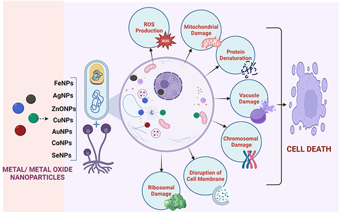
Numerous studies are presently being conducted worldwide to explore how metal and metal oxide nanoparticles interact with bacteria. Researchers have demonstrated that unbound metal nanoparticles cause the bacterial surface’s outer membrane to dissolve in a hazardous manner, while the main mechanism related to metal oxide nanoparticles is oxidative stress brought on by reactive oxygen species (ROS).Citation135 According to the literature, Ag NPs cause perforations and openings to appear in the bacterial membranes by releasing ions following interactions with the enzyme’s disulphide or sulfhydryl groups, and ultimately break off the metabolically important pathways, causing bacterial cell death.Citation136 Several researchers have found that ZnO NPs increased the production of ROS on the membrane’s surface, which led to membrane malfunction and bacterial cell death.Citation137 In the case of TiO2 NPs, a comparable means of oxidative stress, mediated by ROS production, has already been recognized. It has been demonstrated that TiO2 NPs can generate ROS, which can in turn alter the fluidity and stability of the bacterial cell wall by causing lipid peroxidation. Gold nanoparticles have been found to be effective against several bacterial strains, including Pseudomonas aeruginosa and Staphylococcus aureus. They can disrupt the bacterial cell membrane and inhibit bacterial growth. In a recent study, gold nanoparticles were found to be effective against multidrug-resistant strains of Acinetobacter baumannii.Citation138 Copper nanoparticles have been shown to exhibit strong antimicrobial activity against a wide range of bacterial strains, including E. coli and Salmonella. They can disrupt the bacterial cell membrane and interfere with the bacterial DNA replication. In a recent work, copper nanoparticles were found to be effective against drug-resistant strains of Pseudomonas aeruginosa ().Citation139
Figure 4 Diverse mechanisms involved in the antimicrobial activities exhibited by metal and metal-oxide nanoparticles.
Anti-Fungal Activities of Metallic Nanoparticles
The most extensively researched nanoparticles for preventing the growth of fungi that are pathogenic to various plant species are Ag and Cu. Other metal nanoparticles, such as Se,Citation140–142 Ni,Citation143,Citation144 Mg,Citation144 PdCitation145 and Fe,Citation146 have been tested as antifungal agents, and the findings have been encouraging. Se nanoparticles were recently tested in vivo at dosages ranging from 0–1000 ppm against S. graminicola. Approximately six strains of species belonging to Trichoderma (T. longibrachiatum, T. atroviride, T. asperellum, T. harzianum, T. brevicompactum and T. virens) were employed to create these nanoparticles, using filtrates of culture and the lysates of the cells as well as the cell wall in a crude form. T. asperellum in culture filtrate produced superlative results, revealing the ability of Se-based nano-sized-particles to prevent the germination of fungi.Citation142 Another study used T. viride biologically to create Se nanoparticles, which were then tested in vitro against A. solani at various concentrations (50, 100, 200, 300, 400, 500, 600, 700 and 800 ppm). The growth of the fungus was shown to be inhibited by Se nanoparticles at 800 ppm.Citation140 Finally, the effectiveness of chemically produced Se nanoparticles against certain strains of fungi (D. longicolla, M. Phaseolina and S. sclerotiorum) was assessed at variable doses (100, 50, 10, 5, 1, 0.5 and 0.1 ppm). The Se NPs restrained the growth of D. longicolla at concentrations up to 10 ppm and of M. phaseolina at concentrations of 50–100 ppm. For S. sclerotiorum, however, the various doses failed to exhibit any kind of inhibition, instead permitting the pathogen to proliferate and develop.Citation141
The Ability of Nanoparticles to Reduce Inflammation
An essential component of the wound healing process is “anti-inflammation”. This is a cyclic process that results in the production of inter leukines and cytokinins like immuno-responsive substances, usually produced by B and T lymphocytes, and macrophages, among other keratinocytes.Citation147 The endocrine system secretes a variety of inflammatory mediators, including enzymes and antibodies. The key immune organs also release other cytokines, interleukin-1 and 2, which have a capacity to reduce inflammation. These agents that reduce inflammation promote healing.Citation148 Inflammatory mediators also regulate the biochemical processes and eventually control the spread of illnesses. Biosynthetic Au nanoparticles improve tissue regeneration and wound healing processes in inflammatory function.Citation149
The following are some of the ways in which nanoparticles can reduce inflammation:
Targeted drug delivery: Nanoparticles can be engineered to deliver drugs directly to inflamed tissue, thereby reducing inflammation without affecting healthy tissue. For example, in one study, researchers developed chitosan nanoparticles that were able to deliver curcumin, a potent anti-inflammatory agent, to the colon, where it reduced inflammation in a rat model of inflammatory bowel disease.Citation150,Citation151
Inhibition of inflammatory cytokines: Nanoparticles can also be used to inhibit the production of pro-inflammatory cytokines, which play a key role in the inflammatory response. For example, a study showed that gold nanoparticles had the ability to inhibit the production of tumour necrosis factor-alpha (TNF-alpha), a pro-inflammatory cytokine, in macrophages.Citation152
Scavenging of reactive oxygen species (ROS): Nanoparticles can also scavenge ROS, which are highly reactive molecules that contribute to inflammation.Citation153 For example, a work reported that cerium oxide nanoparticles were able to scavenge ROS and reduce inflammation in a rat model of acute lung injury.Citation154
While nanoparticles show promise as anti-inflammatory agents, it is important to note that further research is needed in order fully to understand their potential benefits and risks. It is important to ensure that any nanoparticles used in medical applications are safe and do not cause unintended harm to patients.
Investigations into the Role of Plant-Mediated Nanoparticles in Cancer Prevention
Raghunandan et al, investigated and reported the in vitro anti-cancer effectiveness of biofunctionalized nano-sized-particles of Au and Ag nanoparticles against four distinct cell lines of cancer, including human cancer cells related to the large intestine (colorectal adenocarcinoma), human glomerular cells, leukaemia cells related to human bone marrow, and cells associated with the human cervix.Citation155 They claimed that clove bud extract in aqueous form, combined with AuNPs functionalized with flavonoids, offered greater anti-cancer potential than guava leaf extract. The irregular shaped, functionalized Au nanoparticles prepared using clove bud extract (aqueous) demonstrated acceptable anti-tumour activity on the tested cell lines, according to the MTT assay and microscopic investigations. The same extracts were used to create silver nanoparticles, but these lacked any anti-cancer properties. The MTT experiment showed that the cell lines of cancer were cytotoxic in a “dose-dependent” manner. Also, the anticancer impact of gold nanoparticles is caused by free radicals.Citation155
Several studies have shown that plant-mediated nanoparticles can inhibit cancer cell growth and induce cell death, thereby preventing cancer development and progression. For example, nanoparticles synthesized from green tea leaves have been shown to inhibit the growth of breast cancer cells and induce cell death in colon cancer cells.Citation156 Similarly, nanoparticles synthesized from turmeric have been shown to inhibit the growth of prostate cancer cells and induce cell death in lung cancer cells. These nanoparticles are thought to work by targeting and damaging cancer cells while leaving healthy cells unharmed. One of the advantages of plant-mediated nanoparticles as a potential cancer prevention strategy is their biocompatibility and low toxicity compared to synthetic nanoparticles.Citation26 Plant-mediated nanoparticles are also readily available and easy to synthesize, making them a cost-effective, sustainable alternative to synthetic nanoparticles.
Despite the promising results of preclinical studies, more research is needed to understand fully the mechanisms of plant-mediated nanoparticles and their potential applications regarding cancer prevention. Further studies should focus on the safety and efficacy of these nanoparticles in both animal and human studies, as well as their potential interactions with other drugs and therapies.
Green Nanoparticles: Targeting Cancer Cells and Mitochondria for Effective Cancer Treatment
Green nanoparticles have displayed great potential for use as targeted cancer treatments by specifically targeting cells and mitochondria, and offer several advantages regarding cancer therapy. Mitochondria, once considered solely responsible for energy production, have emerged as important drug targets in diseases like cancer. Their dysfunction plays a role in various human conditions. Targeting mitochondria with nanoparticles provides new therapeutic approaches, thereby overcoming the limitations of conventional drugs. In a mini-review, Tabish and Hamblin introduced the concept of “mitoNANO”, which refers to the use of nanoparticles for targeting mitochondria. They explore the design and application of mitoNANO as a promising approach to advanced cancer therapies. MitoNANO has the potential to overcome drug resistance and minimize the side effects, making it an exciting avenue for future cancer treatments.Citation157
George et al, established the cytotoxic effects of Rubus-conjugated silver nanoparticles (RAgNPs) on MCF-7 cells, with a focus on mitochondrial-mediated intrinsic apoptosis. The RAgNPs exhibit dose-dependent cytotoxicity, decreased proliferation and increased cell death. They induce nuclear damage, intracellular ROS production and apoptotic protein upregulation (caspase 3, Bax, and P53). These findings highlight the potential of RAgNPs to target mitochondria and trigger cell death through the intrinsic apoptosis pathway, making them promising candidates for anticancer drug development.Citation158
In another study, the mitochondria-targeted delivery of chemotherapeutic drugs using TPP-Pluronic F127-hyaluronic acid (TPH) nanomicelles showed promise in overcoming multidrug resistance in cancer. PTX-loaded (a natural plant product derived from the bark of Taxus brevifolia) TPH (TPH/PTX) nanomicelles efficiently entered acidic lysosomes and underwent lysosomal escape, ultimately localizing to mitochondria in drug-resistant cancer cells. This leads to mitochondrial outer membrane permeabilization, cytochrome C release and caspase enzyme activation, resulting in significant antitumor efficacy in xenograft tumour models. This study highlights the potential of mitochondria-targeted nano-micelles as a nontoxic nanoparticle platform for combating drug-resistant cancers.Citation159
Anti-Diabetic Management Employing Metallic Nanoparticles
The majority of medicines are obtained from nature and are herbal in nature. The care of diabetes and diabetic complications has a long history of the successful use of herbal medications.Citation160,Citation161 It has been reported that a number of medicinal plants, including Allium sativum, Asparagus racemosus, Azadirachta indica, Emblica officinalis, Eugenia jambolana, Gymnema sylvestre, Inula racemosa, Momordica charantia, Pterocarpus marsupium, Syzygium cumini, Tinospora cordifolia and Trigonella foenum gracecum exhibited some effectiveness regarding the treatment of diabetes. Diabetes management has been reported to benefit more from the green synthesis of polymeric or metallic nanoparticles made from herbal products than from native crude materials.Citation162,Citation163 Numerous commercially available, anti-diabetic medications are clinically equal to phyto-nanotherapy, which has greater biopharmaceutical properties. Additionally, a synergistic effect can be used by plant-metal nanoparticles to achieve special medicinal qualities. Green preparations of Ag, Au and zinc oxide (ZnO) nano-formulations have attracted considerable attention due to their ability to enhance the stability, pharmacokinetics and biopharmaceutical outcomes of plant-based chemicals in herbal drug formulations.Citation164 These advances aim to harness their augmented therapeutic potential regarding diabetes prevention and management.Citation165
There has been a growing interest in the use of metallic nanoparticles for antidiabetic management. Metallic nanoparticles, such as gold, silver and platinum, possess unique physicochemical properties that make them promising candidates for drug delivery and therapeutic treatment. One approach is to use AuNPs to deliver insulin to the pancreas. In a study, an AuNPs-based insulin delivery system was able effectively to lower blood glucose levels in diabetic mice.Citation166 The AuNPs were coated with a polymer that protected the insulin from degradation and allowed it to be released in a controlled manner.
Another approach is to use silver nanoparticles to improve insulin sensitivity. A recent published study reported that AgNPs were able to improve insulin sensitivity in diabetic mice by reducing inflammation and oxidative stress in liver.Citation167 Platinum nanoparticles (Pt NPs) have also been investigated for their potential regarding antidiabetic management.Citation168 Alternatively, researchers developed a Pt NP-based glucose biosensor that could detect glucose levels in diabetic rat models. The biosensor was found to be highly sensitive and selective, with potential use as a diagnostic tool for diabetes.Citation169
It should be noted, however, that the use of metallic nanoparticles in antidiabetic management remains in its infancy, and further research is needed to understand fully their potential benefits and risks. It is important to ensure that any nanoparticles used in medical applications are safe and do not cause any unintended harm to patients.
Other Applications of Bio-Derived Nanoparticles
Agriculture
The antibacterial action described above may also be useful for crop protection, when agricultural diseases are the focus. In particular, ZnO NPs have shown their potential for broad agricultural application by demonstrating activity against “plant pathogens” (both bacteria and fungi), obtained from Citrus limon (L.) Burm (against soft-rot bacteria). It is notable that TiO2 NPs prepared utilizing lemon fruit exhibited an anti-bacterial action against D. dadantii that was comparable to ZnO NPs.Citation170
AgNPs created from wheat extract helped to mitigate the detrimental effects of salinity stress considerably on wheat-crop by altering the concentration of “abscisic acid”, ion-homeostasis and defense mechanisms embodying both enzymatic and non-enzymatic antioxidants.Citation171 Strikingly, ZnO NPs displayed less toxicity and an ability to strengthen flax seedlings’ antioxidant defense mechanisms.Citation172
Antioxidant Action
A range of cell biomolecules, such as DNA, polypeptides and membrane lipids, might experience oxidative injury resulting from high oxidative stress brought on by the deed of “mitochondria” and other internal or external causes. This deterioration can result in neurodegenerative disorders and senescence.Citation173 Antioxidants have the potential to stop these harmful processes and be utilised to manage ailments associated with ageing. Ag NPs made from leaf extract prepared using C. carandas,Citation174 nanoparticles of silver and gold attained from leaf extract of C. inermeCitation175 or nickel oxide-based nano-sized particles prepared using leaf extract of Stevia rebaudianaCitation42 have been documented for their antioxidant potential. The phytochemicals deposited on the surface of the NPs undoubtedly play a significant role in the reported antioxidant effect. For assaying antioxidant activity, usually, only one in vitro test is performed, such as a “DPPH-assay.” However, “antioxidant activity” should not be validated by a solitary technique. The assessment of antioxidant activity, therefore, relies mainly on the associated reaction system, due to the intricate nature of phytochemicals.Citation176 The reliability of the findings from in vitro cell-free antioxidant testing must be restricted to the evaluation in the context of chemical reactivity, since in vivo substantiation is strongly advised.
Bioremediation
There are also descriptions of additional possible applications, including (i) “photo-catalytic” applications and (ii) “absorption” applications. Silver based nano-sized particles, prepared by the employment of chamomilla, exhibits strong activity against “Rhodamine B” when exposed to ultra-violet radiation, making them a potential wastewater treatment material.Citation177 The capacity to remove diverse both organic and inorganic pollutants has been demonstrated by magnesium oxide-based nano-sized particles generated from leaf extract of “Indian mallow” which also showed excellent photocatalytic activity and effective absorption related properties against the high-density metallic element viz. Cr(VI).Citation175 Finally, it should be mentioned that reduced forms of graphene oxide, synthesised employing leaf extract of Stevia rebaudiana, were coated on “Palladium-silver” bimetallic nano-sized structures to facilitate photocatalytic H2 synthesis.Citation178
Challenges
The synthesis of plant-derived nanoparticles for various applications presents several challenges. Standardizing the extraction methods is crucial to ensure consistent, reproducible synthesis. The contamination and impurities in plant extracts must be effectively removed to maintain the purity and functionality of the nanoparticles. Scaling-up production while maintaining quality and stability is a significant consideration. Advanced characterization techniques specific to plant-derived nanoparticles are needed to understand their complex structures. Their long-term stability and storage, as well as regulatory considerations, must be addressed for their successful translation into clinical use. Batch-to-batch variability in composition also poses a challenge that needs to be minimized. Overall, interdisciplinary collaboration, method optimization and protocol standardization are essential if we are to overcome these challenges and unlock the full potential of plant-derived nanoparticles.
Conclusion
The synthesis of nanoparticles through plant-mediated green synthesis represents a significant advance in the field of nanoscience and green chemistry. This review paper provides an up to date comprehensive overview of the research conducted in this field, highlighting the advantages and potential applications of this eco-friendly approach. The utilization of plant extracts as reducing and stabilizing agents for nanoparticle synthesis offers numerous advantages over the traditional chemical methods, including cost-effectiveness, scalability and the absence of toxic contaminants, thus making plant-mediated synthesis a sustainable alternative to its counterparts. The use of renewable plant-based resources facilitates controlled synthesis processes, resulting in nanoparticles with enhanced size uniformity and stability making them highly suitable for various applications. Furthermore, the bio-inspired nanoparticles derived from plants exhibit intriguing pharmacological properties, such as biocompatibility and nano-dimensions, which make them highly promising for various biomedical applications and the targeting of specific cells in a controlled manner. These applications include drug delivery, disease management, agriculture, bioremediation, and other industrial applications.
While the plant-mediated green synthesis of nanoparticles has demonstrated tremendous potential, several challenges and areas for future research have been identified. The relation between metal salt concentration and nanoparticle yield, as well as the optimization of the parameters to overcome polydispersity, require further investigation. Understanding the chemical components and underlying mechanisms involved in the synthesis, action and stabilization of biological nanoparticles is crucial for their effective utilization. Moreover, addressing issues related to the distribution profile, excretion, clearance, biocompatibility and bioavailability of biological nanoparticles through conducting extensive in vivo trials and research is essential if we are to exploit their biomedical applications to the full. The convergence of green chemistry and nanotechnology has paved the way for the development of environmentally friendly nanomaterial synthesis methods, and plant-mediated nanoparticle production has emerged as a promising field, with further applications related to catalysis, agriculture, water treatment, biotechnology, electronics and other industries. Green plant-based nanoparticles offer potential benefits, related to areas such as phytopathogen treatment in the field of agriculture, and water disinfection for environmental clean-up. It is important to consider the long-term impacts of these nanoparticles on animals, humans, and the environment, however, and further research is required to address concerns regarding the accumulation and influence of green nanoparticles, ensuring their safe, sustainable utilization.
In conclusion, this review paper provides valuable insights into the scientific intricacies of the plant-mediated synthesis of nanoparticles. The adoption of green synthesis approaches using plant extracts as reducing agents has immense potential regarding cost-effective, environmentally friendly nanoparticle production, with significant pharmacological properties. It is hoped that this review will serve as a comprehensive resource for researchers and scientists working in the field, and inspire further exploration and innovation in this rapidly-growing area of nanoscience and green chemistry.
Disclosure
The authors report no conflicts of interest in this work.
Acknowledgments
The authors extend their appreciation to the Deputyship for Research & Innovation, Ministry of Education in Saudi Arabia for funding this research work through the project number: IFP22UQU4420118DSR061.
References
- El-Shafai N, El-Khouly ME, El-Kemary M, Ramadan M, Eldesoukey I, Masoud M. Graphene oxide decorated with zinc oxide nanoflower, silver and titanium dioxide nanoparticles: fabrication, characterization, DNA interaction, and antibacterial activity. RSC Adv. 2019;9(7):3704–3714. doi:10.1039/c8ra09788g
- EL-Sheshtawy HS, El-Hosainy HM, Shoueir KR, El-Mehasseb IM, El-Kemary M. Facile immobilization of Ag nanoparticles on g-C3N4/V2O5 surface for enhancement of post-illumination, catalytic, and photocatalytic activity removal of organic and inorganic pollutants. Appl Surf Sci. 2019;467:268–276. doi:10.1016/j.apsusc.2018.10.109
- Kaviya S. Synthesis, self-assembly, sensing methods and mechanism of bio-source facilitated nanomaterials: a review with future outlook. Nano Struct Nano Objects. 2020;23:100498. doi:10.1016/j.nanoso.2020.100498
- Al-Anssari S, Ali M, Alajmi M, et al. Synergistic Effect of Nanoparticles and Polymers on the Rheological Properties of Injection Fluids: Implications for Enhanced Oil Recovery. Energy Fuels. 2021;35(7):6125–6135. doi:10.1021/acs.energyfuels.1c00105/asset/images/medium/ef1c00105_0011.gif
- Khan I, Saeed K, Khan I. Nanoparticles: properties, applications and toxicities. Arab J Chem. 2019;12(7):908–931. doi:10.1016/j.arabjc.2017.05.011
- Yokoyama T, Masuda H, Suzuki M, et al. Basic properties and measuring methods of nanoparticles. Nanoparticle Technol Handb. 2008:3–48. doi:10.1016/B978-044453122-3.50004-0
- Dessie Y, Tadesse S, Eswaramoorthy R, Abdisa E. Bimetallic Mn–Ni oxide nanoparticles: green synthesis, optimization and its low-cost anode modifier catalyst in microbial fuel cell. Nano Struct Nano Objects. 2021;25:100663. doi:10.1016/j.nanoso.2020.100663
- Sharma VK, Yngard RA, Lin Y. Silver nanoparticles: green synthesis and their antimicrobial activities. Adv Colloid Interface Sci. 2009;145(1–2):83–96. doi:10.1016/J.CIS.2008.09.002
- Wagner AM, Knipe JM, Orive G, Peppas NA. Quantum dots in biomedical applications. Acta Biomater. 2019;94:44–63. doi:10.1016/j.actbio.2019.05.022
- Lenders V, Koutsoumpou X, Sargsian A, Manshian BB. Biomedical nanomaterials for immunological applications: ongoing research and clinical trials. Nanoscale Adv. 2020;2(11):5046–5089. doi:10.1039/d0na00478b
- Mitarotonda R, Giorgi E, Eufrasio-da-Silva T, et al. Immunotherapeutic nanoparticles: from autoimmune disease control to the development of vaccines. Biomater Adv. 2022;135:212726. doi:10.1016/j.bioadv.2022.212726
- Kessler R. Engineered Nanoparticles in Consumer Products: understanding a New Ingredient. Environ Health Perspect. 2011;119(3):a120–5. doi:10.1289/ehp.119-a120
- Mody V, Siwale R, Singh A, Mody H. Introduction to metallic nanoparticles. J Pharm Bioallied Sci. 2010;2(4):282. doi:10.4103/0975-7406.72127
- Trickler WJ, Lantz SM, Murdock RC, et al. Silver nanoparticle induced blood-brain barrier inflammation and increased permeability in primary rat brain microvessel endothelial cells. Toxicol Sci. 2010;118(1):160–170. doi:10.1093/TOXSCI/KFQ244
- Weissig V, Pettinger TK, Murdock N. Nanopharmaceuticals (part 1): products on the market. Int J Nanomedicine. 2014;9:4357–4373. doi:10.2147/IJN.S46900
- Gulson B, Mccall M, Korsch M, et al. Small amounts of zinc from zinc oxide particles in sunscreens applied outdoors are absorbed through human skin. Toxicol Sci. 2010;118(1):140–149. doi:10.1093/TOXSCI/KFQ243
- Krestinin AV, Dremova NN, Knerel’Man EI, Blinova LN, Zhigalina VG, Kiselev NA. Characterization of SWCNT products manufactured in Russia and the prospects for their industrial application. Nanotechnol Russ. 2015;10(7–8):537–548. doi:10.1134/S1995078015040096
- Ravichandran R. Nanotechnology Applications in Food and Food Processing: Innovative Green Approaches, Opportunities and Uncertainties for Global Market. Int J Green Nanotechnol. 2010;1(2):P72–P96. doi:10.1080/19430871003684440
- Vance ME, Kuiken T, Vejerano EP, McGinnis SP, Hochella MF, Hull DR. Nanotechnology in the real world: redeveloping the nanomaterial consumer products inventory. Beilstein J Nanotechnol. 2015;6(1):1769–1780. doi:10.3762/BJNANO.6.181
- Santo-Orihuela PL, Desimone MF, Catalano PN. Green Synthesis: A Land of Complex Nanostructures. Curr Pharm Biotechnol. 2022;24(1):3–22. doi:10.2174/1389201023666220512094533
- Makarov VV, Мв В, Love AJ, et al. “Green” nanotechnologies: synthesis of metal nanoparticles using plants. Acta Naturae. 2014;6(1):35–44. doi:10.32607/20758251-2014-6-1-35-44
- Velusamy P, Kumar GV, Jeyanthi V, Das J, Pachaiappan R. Bio-Inspired Green Nanoparticles: Synthesis, Mechanism, and Antibacterial Application. Toxicol Res. 2016;32(2):95. doi:10.5487/TR.2016.32.2.095
- Galdopórpora JM, Ibar A, Tuttolomondo MV, Desimone MF. Dual-effect core–shell polyphenol coated silver nanoparticles for tissue engineering. Nano Struct Nano Objects. 2021;26:100716. doi:10.1016/J.NANOSO.2021.100716
- Bandeira M, Possan AL, Pavin SS, et al. Mechanism of formation, characterization and cytotoxicity of green synthesized zinc oxide nanoparticles obtained from Ilex paraguariensis leaves extract. Nano Struct Nano Objects. 2020;24:100532. doi:10.1016/J.NANOSO.2020.100532
- Safat S, Buazar F, Albukhaty S, Matroodi S. Enhanced sunlight photocatalytic activity and biosafety of marine-driven synthesized cerium oxide nanoparticles. Sci Rep. 2021;11(1):1–11. doi:10.1038/s41598-021-94327-w
- Alhujaily M, Albukhaty S, Yusuf M, et al. Recent Advances in Plant-Mediated Zinc Oxide Nanoparticles with Their Significant Biomedical Properties. Bioengineering. 2022;9(10):541. doi:10.3390/bioengineering9100541
- Khane Y, Benouis K, Albukhaty S, et al. Green synthesis of silver nanoparticles using aqueous citrus limon zest extract: characterization and evaluation of their antioxidant and antimicrobial properties. Nanomaterials. 2022;12(12):2013. doi:10.3390/nano12122013
- Alzubaidi AK, Al-Kaabi WJ, Al AA, et al. Green synthesis and characterization of silver nanoparticles using flaxseed extract and evaluation of their antibacterial and antioxidant activities. Appl Sci. 2023;13(4):2182. doi:10.3390/app13042182
- Mahmood RI, Kadhim AA, Ibraheem S, et al. Biosynthesis of copper oxide nanoparticles mediated Annona muricata as cytotoxic and apoptosis inducer factor in breast cancer cell lines. Sci Rep. 2022;12(1):1–10. doi:10.1038/s41598-022-20360-y
- Potbhare AK, Chaudhary RG, Chouke PB, et al. Phytosynthesis of nearly monodisperse CuO nanospheres using Phyllanthus reticulatus/Conyza bonariensis and its antioxidant/antibacterial assays. Mater Sci Eng C Mater Biol Appl. 2019;99:783–793. doi:10.1016/J.MSEC.2019.02.010
- Zikalala N, Matshetshe K, Parani S, Oluwafemi OS. Biosynthesis protocols for colloidal metal oxide nanoparticles. Nano Struct Nano Objects. 2018;16:288–299. doi:10.1016/J.NANOSO.2018.07.010
- Kagdi AR, Pullar RC, Meena SS, et al. Green synthesis based X-type Ba–Zn hexaferrites: their structural, hysteresis, mӧssbauer, dielectric and electrical properties. Mater Chem Phys. 2022:282. doi:10.1016/J.MATCHEMPHYS.2022.125914
- Catalano PN, Chaudhary RG, Desimone MF, Santo-Orihuela PL. A survey on analytical methods for the characterization of green synthesized nanomaterials. Curr Pharm Biotechnol. 2021;22(6):823–847. doi:10.2174/1389201022666210104122349
- Antezana PE, Municoy S, Desimone MF. Building nanomaterials with microbial factories. Biog Sustain Nanotechnol Trends Prog. 2022;1–39. doi:10.1016/B978-0-323-88535-5.00012-3
- Das SK, Dickinson C, Lafir F, Brougham DF, Marsili E. Synthesis, characterization and catalytic activity of gold nanoparticles biosynthesized with Rhizopus oryzae protein extract. Green Chem. 2012;14(5):1322–1334. doi:10.1039/C2GC16676C
- Gardea-Torresdey JL, Parsons JG, Gomez E, et al. Formation and Growth of Au Nanoparticles inside Live Alfalfa Plants. NanoL. 2002;2(4):397–401. doi:10.1021/NL015673
- Mondal A, Umekar MS, Bhusari GS, et al. Biogenic Synthesis of Metal/Metal Oxide Nanostructured Materials. Curr Pharm Biotechnol. 2021;22(13):1782–1793. doi:10.2174/1389201022666210111122911
- Singh NB, Jain P, De A, Tomar R. Green synthesis and applications of nanomaterials. Curr Pharm Biotechnol. 2021;22(13):1705–1747. doi:10.2174/1389201022666210412142734
- Rai M, Yadav A. Plants as potential synthesiser of precious metal nanoparticles: progress and prospects. IET Nanobiotechnol. 2013;7(3):117–124. doi:10.1049/IET-NBT.2012.0031
- Nande A, Raut S, Michalska-Domanska M, Dhoble SJ. Green synthesis of nanomaterials using plant extract: a review. Curr Pharm Biotechnol. 2020;22(13):1794–1811. doi:10.2174/1389201021666201117121452
- Masum MI, Siddiqa MM, Ali KA, et al. Biogenic synthesis of silver nanoparticles using Phyllanthus emblica fruit extract and its inhibitory action against the pathogen Acidovorax oryzae strain RS-2 of rice bacterial brown stripe. Front Microbiol. 2019;10(APR):820. doi:10.3389/FMICB.2019.00820/BIBTEX
- Yasir M, Singh J, Tripathi MK, Singh P, Shrivastava R. Green synthesis of silver nanoparticles using leaf extract of common arrowhead houseplant and its anticandidal activity. Pharmacogn Mag. 2018;13(Suppl 4):S840–S844. doi:10.4103/PM.PM_226_17
- Pilaquinga F, Morejón B, Ganchala D, et al. Green synthesis of silver nanoparticles using Solanum mammosum L. (Solanaceae) fruit extract and their larvicidal activity against Aedes aegypti L. (Diptera:Culicidae). PLoS One. 2019;14(10):e0224109. doi:10.1371/JOURNAL.PONE.0224109
- Rautela A, Rani J, Debnath (Das) M. Green synthesis of silver nanoparticles from Tectona grandis seeds extract: characterization and mechanism of antimicrobial action on different microorganisms. J Anal Sci Technol. 2019;10(1):1–10. doi:10.1186/S40543-018-0163-Z/FIGURES/14
- Mittal AK, Chisti Y, Banerjee UC. Synthesis of metallic nanoparticles using plant extracts. Biotechnol Adv. 2013;31(2):346–356. doi:10.1016/J.BIOTECHADV.2013.01.003
- Shah M, Fawcett D, Sharma S, Tripathy SK, Poinern GEJ. Green synthesis of metallic nanoparticles via biological entities. Mater. 2015;8(11):7278–7308. doi:10.3390/MA8115377
- Rajiv P, Rajeshwari S, Venckatesh R. Bio-Fabrication of zinc oxide nanoparticles using leaf extract of Parthenium hysterophorus L. and its size-dependent antifungal activity against plant fungal pathogens. Spectrochim Acta A Mol Biomol Spectrosc. 2013;112:384–387. doi:10.1016/J.SAA.2013.04.072
- Otsuka H, Nagasaki Y, Kataoka K. PEGylated nanoparticles for biological and pharmaceutical applications. Adv Drug Deliv Rev. 2003;55(3):403–419. doi:10.1016/S0169-409X(02)00226-0
- Prasad KS, Pathak D, Patel A, et al. Biogenic synthesis of silver nanoparticles using Nicotiana tobaccum leaf extract and study of their antibacterial effect. Afr J Biotechnol. 2011;10(41):8122–8130. doi:10.5897/AJB11.394
- Lal S, Jana U, Manna PK, Mohanta GP, Manavalan R, Pal SL. Nanoparticle: an overview of preparation and characterization. J Appl Pharm Sci. 2011;2011(6):228–234.
- Kowalczyk B, Lagzi I, Grzybowski BA. Nanoseparations: strategies for size and/or shape-selective purification of nanoparticles. Curr Opin Colloid Interface Sci. 2011;16(2):135–148. doi:10.1016/J.COCIS.2011.01.004
- Brice-Profeta S, Arrio MA, Tronc E, et al. Magnetic order in γ-Fe2O3 nanoparticles: a XMCD study. J Magn Magn Mater. 2005;288:354–365. doi:10.1016/J.JMMM.2004.09.120
- Faraji M, Yamini Y, Rezaee M. Magnetic nanoparticles: synthesis, stabilization, functionalization, characterization, and applications. J Iran Chem Soc. 2010;7(1):1–37. doi:10.1007/BF03245856/METRICS
- Tiwari DK, Behari J, Sen P. Time and dose-dependent antimicrobial potential of Ag nanoparticles synthesized by top-down approach. Curr Sci. 2008;95(5):647–655.
- Gupta V, Gupta AR, Kant V. Synthesis, characterization and biomedical applications of nanoparticles. Sci Int. 2013;1(5):167–174. doi:10.5567/SCIINTL.2013.167.174
- Rajeshkumar S, Bharath LV. Mechanism of plant-mediated synthesis of silver nanoparticles – A review on biomolecules involved, characterisation and antibacterial activity. Chem Biol Interact. 2017;273:219–227. doi:10.1016/J.CBI.2017.06.019
- De Jaeger N, Demeyere H, Finsy R, et al. Particle sizing by photon correlation spectroscopy part I: monodisperse latices: influence of scattering angle and concentration of dispersed material. Part Part Syst Charact. 1991;8(1–4):179–186. doi:10.1002/PPSC.19910080134
- Magdy G, Aboelkassim E, El-Domany RA, Belal F. Green synthesis, characterization, and antimicrobial applications of silver nanoparticles as fluorescent nanoprobes for the spectrofluorimetric determination of ornidazole and miconazole. Sci Rep. 2022;12(1):1–15. doi:10.1038/s41598-022-25830-x
- Amooaghaie R, Saeri MR, Azizi M. Synthesis, characterization and biocompatibility of silver nanoparticles synthesized from Nigella sativa leaf extract in comparison with chemical silver nanoparticles. Ecotoxicol Environ Saf. 2015;120:400–408. doi:10.1016/J.ECOENV.2015.06.025
- Vishveshvar K, Aravind Krishnan MV, Haribabu K, Vishnuprasad S. Green synthesis of copper oxide nanoparticles using Ixiro coccinea plant leaves and its characterization. Bionanoscience. 2018;8(2):554–558. doi:10.1007/S12668-018-0508-5/METRICS
- Sankar V, Salinraj P, Athira R, Soumya RS, Raghu KG. Cerium nanoparticles synthesized using aqueous extract of Centella asiatica: characterization, determination of free radical scavenging activity and evaluation of efficacy against cardiomyoblast hypertrophy. RSC Adv. 2015;5(27):21074–21083. doi:10.1039/C4RA16893C
- Alsammarraie FK, Wang W, Zhou P, Mustapha A, Lin M. Green synthesis of silver nanoparticles using turmeric extracts and investigation of their antibacterial activities. Colloids Surf B Biointerfaces. 2018;171:398–405. doi:10.1016/J.COLSURFB.2018.07.059
- Krishnaraj C, Muthukumaran P, Ramachandran R, Balakumaran MD, Kalaichelvan PT. Acalypha indica Linn: biogenic synthesis of silver and gold nanoparticles and their cytotoxic effects against MDA-MB-231, human breast cancer cells. Biotechnol Rep. 2014;4(1):42–49. doi:10.1016/J.BTRE.2014.08.002
- Tyavambiza C, Elbagory AM, Madiehe AM, Meyer M, Meyer S. The antimicrobial and anti-inflammatory effects of silver nanoparticles synthesised from Cotyledon orbiculata aqueous extract. Nanomaterials. 2021;11(5):1343. doi:10.3390/nano11051343
- Hemlata MPR, Singh AP, Tejavath KK. Biosynthesis of silver nanoparticles using Cucumis prophetarum aqueous leaf extract and their antibacterial and antiproliferative activity against cancer cell lines. ACS Omega. 2020;5(10):5520–5528. doi:10.1021/acsomega.0c00155
- Fadaka AO, Meyer S, Ahmed O, et al. Broad spectrum anti-bacterial activity and non-selective toxicity of Gum Arabic silver nanoparticles. Int J Mol Sci. 2022;23(3):1799. doi:10.3390/ijms23031799
- Liang T, Qiu X, Ye X, et al. Biosynthesis of selenium nanoparticles and their effect on changes in urinary nanocrystallites in calcium oxalate stone formation. Biotech. 2020;10(1):1–6. doi:10.1007/s13205-019-1999-7
- Raut RW, Haroon ASM, Malghe YS, Nikam BT, Kashid SB. Rapid Biosynthesis Of Platinum And Palladium Metal Nanoparticles Using Root Extract Of Asparagus Racemosus Linn. Adv Mater Lett. 2013;4(8):650–654. doi:10.5185/AMLETT.2012.11470
- Rabiee N, Bagherzadeh M, Kiani M, Ghadiri AM. Rosmarinus officinalis directed palladium nanoparticle synthesis: investigation of potential anti-bacterial, anti-fungal and Mizoroki-Heck catalytic activities. Adv Powder Technol. 2020;31(4):1402–1411. doi:10.1016/J.APT.2020.01.024
- Katata-Seru L, Moremedi T, Aremu OS, Bahadur I. Green synthesis of iron nanoparticles using Moringa oleifera extracts and their applications: removal of nitrate from water and antibacterial activity against Escherichia coli. J Mol Liq. 2018;256:296–304. doi:10.1016/J.MOLLIQ.2017.11.093
- Kora AJ, Rastogi L. Green synthesis of palladium nanoparticles using gum ghatti (Anogeissus latifolia) and its application as an antioxidant and catalyst. Arab J Chem. 2018;11(7):1097–1106. doi:10.1016/j.arabjc.2015.06.024
- Kanimozhi S, Durga R, Sabithasree M, et al. Biogenic synthesis of silver nanoparticle using Cissus quadrangularis extract and its invitro study. J King Saud Univ Sci. 2022;34(4):101930. doi:10.1016/j.jksus.2022.101930
- Sameem S, Neupane NP, Saleh Ansari SM, et al. Phyto-fabrication of silver nanoparticles from Ziziphus mauritiana against hepatic carcinoma via modulation of Rho family-alpha serine/threonine protein kinase. J Drug Deliv Sci Technol. 2022;70:103227. doi:10.1016/J.JDDST.2022.103227
- Ovais M, Khalil AT, Islam NU, et al. Role of plant phytochemicals and microbial enzymes in biosynthesis of metallic nanoparticles. Appl Microbiol Biotechnol. 2018;102(16):6799–6814. doi:10.1007/S00253-018-9146-7
- Ayinde WB, Gitari WM, Munkombwe M, Amidou S. Green synthesis of Ag/MgO nanoparticle modified nanohydroxyapatite and its potential for defluoridation and pathogen removal in groundwater. Phys Chem Earth. 2018;107:25–37. doi:10.1016/j.pce.2018.08.007
- Dauthal P, Mukhopadhyay M. Phyto-synthesis and structural characterization of catalytically active gold nanoparticles biosynthesized using Delonix regia leaf extract. Biotech. 2016;6(2). doi:10.1007/S13205-016-0432-8
- Sathiya CK, Akilandeswari S. Fabrication and characterization of silver nanoparticles using Delonix elata leaf broth. Spectrochim Acta Part A Mol Biomol Spectrosc. Spectrochim Acta A Mol Biomol Spectrosc. 2014;128:337–341. doi:10.1016/J.SAA.2014.02.172
- Ayaz M, Junaid M, Ullah F, et al. Anti-Alzheimer’s studies on ß-sitosterol isolated from Polygonum hydropiper L. Front Pharmacol. 2017;8(OCT). doi:10.3389/FPHAR.2017.00697
- Jha AK, Prasad K. Mechanistic plethora of biogenetic nanosynthesis: an evaluation. Nanotechnol Life Sci. 2018;1–24. doi:10.1007/978-3-319-99570-0_1/COVER
- Edison TJI, Sethuraman MG. Instant green synthesis of silver nanoparticles using Terminalia chebula fruit extract and evaluation of their catalytic activity on reduction of methylene blue. Process Biochem. 2012;47(9):1351–1357. doi:10.1016/J.PROCBIO.2012.04.025
- Saad AM, El-Saadony MT, El-Tahan AM, et al. Polyphenolic extracts from pomegranate and watermelon wastes as substrate to fabricate sustainable silver nanoparticles with larvicidal effect against Spodoptera littoralis. Saudi J Biol Sci. 2021;28(10):5674–5683. doi:10.1016/J.SJBS.2021.06.011
- Kanwal U, Bukhari NI, Ovais M, Abass N, Hussain K, Raza A. Advances in nano-delivery systems for doxorubicin: an updated insight. J Drug Target. 2018;26(4):296–310. doi:10.1080/1061186X.2017.1380655
- Londhe S, Haque S, Patra CR. Silver and gold nanoparticles: potential cancer theranostic applications, recent development, challenges, and future perspectives. Gold Silver Nanopart. 2023;247–290. doi:10.1016/B978-0-323-99454-5.00006-8
- Raja S, Ramesh V, Thivaharan V. Green biosynthesis of silver nanoparticles using Calliandra haematocephala leaf extract, their antibacterial activity and hydrogen peroxide sensing capability. Arab J Chem. 2017;10(2):253–261. doi:10.1016/J.ARABJC.2015.06.023
- Wang Z, Fang C, Megharaj M. Characterization of iron–polyphenol nanoparticles synthesized by three plant extracts and their Fenton oxidation of azo dye. ACS Sustain Chem Eng. 2014;2(4):1022–1025. doi:10.1021/sc500021n
- Gopinath K, Kumaraguru S, Bhakyaraj K, et al. Green synthesis of silver, gold and silver/gold bimetallic nanoparticles using the Gloriosa superba leaf extract and their antibacterial and antibiofilm activities. Microb Pathog. 2016;101:1–11. doi:10.1016/J.MICPATH.2016.10.011
- Jha AK, Prasad K, Prasad K, Kulkarni AR. Plant system: nature’s nanofactory. Colloids Surf B Biointerfaces. 2009;73(2):219–223. doi:10.1016/J.COLSURFB.2009.05.018
- Hussain M, Raja NI, Iqbal M, Aslam S. Applications of plant flavonoids in the green synthesis of colloidal silver nanoparticles and impacts on human health. Iran J Sci Technol Trans a Sci. 2019;43(3):1381–1392. doi:10.1007/S40995-017-0431-6/METRICS
- Zhou Y, Lin W, Huang J, et al. Biosynthesis of gold nanoparticles by foliar broths: roles of biocompounds and other attributes of the extracts. Nanoscale Res Lett. 2010;5(8):1351–1359. doi:10.1007/S11671-010-9652-8
- Ahmad N, Sharma S, Alam MK, et al. Rapid synthesis of silver nanoparticles using dried medicinal plant of basil. Colloids Surf B Biointerfaces. 2010;81(1):81–86. doi:10.1016/J.COLSURFB.2010.06.029
- Dubey SP, Lahtinen M, Särkkä H, Sillanpää M. Bioprospective of Sorbus aucuparia leaf extract in development of silver and gold nanocolloids. Colloids Surf B Biointerfaces. 2010;80(1):26–33. doi:10.1016/j.colsurfb.2010.05.024
- Sahu N, Soni D, Chandrashekhar B, et al. Synthesis of silver nanoparticles using flavonoids: hesperidin, naringin and diosmin, and their antibacterial effects and cytotoxicity. Int Nano Lett. 2016;6(3):173–181. doi:10.1007/S40089-016-0184-9
- Sharma V, Janmeda P. Extraction, isolation and identification of flavonoid from Euphorbia neriifolia leaves. Arab J Chem. 2017;10(4):509–514. doi:10.1016/J.ARABJC.2014.08.019
- Rakhi M, Gopal BB. Terminalia Arjuna Bark Extract Mediated Size Controlled Synthesis of Polyshaped Gold Nanoparticles and Its Application in Catalysis. Int J Res Chem Environ. 2012;2(338):338–344.
- El-Seedi HR, El-Shabasy RM, Khalifa SAM, et al. Metal nanoparticles fabricated by green chemistry using natural extracts: biosynthesis, mechanisms, and applications. RSC Adv. 2019;9(42):24539–24559. doi:10.1039/C9RA02225B
- Nasrollahzadeh M, Sajadi SM. Green synthesis of copper nanoparticles using Ginkgo biloba L. leaf extract and their catalytic activity for the Huisgen [3+2] cycloaddition of azides and alkynes at room temperature. J Colloid Interface Sci. 2015;457:141–147. doi:10.1016/J.JCIS.2015.07.004
- Rehman Mashwani UR, Khan Z, Khan T, et al. Applications of plant terpenoids in the synthesis of colloidal silver nanoparticles. Adv Colloid Interface Sci. 2016;234:132–141. doi:10.1016/J.CIS.2016.04.008
- Shankar SS, Ahmad A, Pasricha R, Sastry M. Bioreduction of chloroaurate ions by geranium leaves and its endophytic fungus yields gold nanoparticles of different shapes. J Mater Chem. 2003;13(7):1822–1826. doi:10.1039/B303808B
- Chandran SP, Chaudhary M, Pasricha R, Ahmad A, Sastry M. Synthesis of gold nanotriangles and silver nanoparticles using Aloe vera plant extract. Biotech Prog. 2008;22(2):577–583. doi:10.1021/bp0501423
- Brahmachari G, Sarkar S, Ghosh R, et al. Sunlight-induced rapid and efficient biogenic synthesis of silver nanoparticles using aqueous leaf extract of Ocimum sanctum Linn. with enhanced antibacterial activity. Org Med Chem Lett. 2014;4(1). doi:10.1186/S13588-014-0018-6
- Ahmad T, Wani IA, Manzoor N, Ahmed J, Asiri AM. Biosynthesis, structural characterization and antimicrobial activity of gold and silver nanoparticles. Colloids Surf B Biointerfaces. 2013;107:227–234. doi:10.1016/J.COLSURFB.2013.02.004
- Durán M, Silveira CP, Durán N. Catalytic role of traditional enzymes for biosynthesis of biogenic metallic nanoparticles: a mini-review. IET Nanobiotechnol. 2015;9(5):314–323. doi:10.1049/IET-NBT.2014.0054
- Bhattacharjee RR, Das AK, Haldar D, Si S, Banerjee A, Mandal TK. Peptide-assisted synthesis of gold nanoparticles and their self-assembly. J Nanosci Nanotechnol. 2005;5(7):1141–1147. doi:10.1166/JNN.2005.166
- Li S, Shen Y, Xie A, et al. Green synthesis of silver nanoparticles using Capsicum annuum L. extract. Green Chem. 2007;9(8):852–858. doi:10.1039/B615357G
- Rodriguez PL, Harada T, Christian DA, Pantano DA, Tsai RK, Discher DE. Minimal “Self” peptides that inhibit phagocytic clearance and enhance delivery of nanoparticles. Science. 2013;339(6122):971–975. doi:10.1126/SCIENCE.1229568
- Priya MM, Karunai Selvi B, Paul JAJ. green synthesis of silver nanoparticles from the leaf extracts of Euphorbia hirta and Nerium indicum. Dig J Nanomater Biostructures. 2011;6(2):869–877.
- Molpeceres J, Aberturas MR, Guzman M. Biodegradable nanoparticles as a delivery system for cyclosporine: preparation and characterization. J Microencapsul. 2000;17(5):599–614. doi:10.1080/026520400417658
- Chauhan RP, Gupta C, Prakash D. Methodological advancements in green nanotechnology and their applications in biological synthesis of herbal nanoparticles. Int J Bioassays. 2012;1(7):6–10.
- Galdopórpora JM, Municoy S, Ibarra F, et al. A green synthesis method to tune the morphology of CuO and ZnO nanostructures. Curr Nanosci. 2021;19(2):186–193. doi:10.2174/1573413717666210921152709
- Chauhan CC, Gupta T, Meena SS, et al. Tailoring magnetic and dielectric properties of SrFe12O19/NiFe2O4 ferrite nanocomposites synthesized in presence of Calotropis gigantea (crown) flower extract. J Alloys Compd. 2022;900:163415. doi:10.1021/ac0206723
- Rehana D, Mahendiran D, Kumar RS, Rahiman AK. In vitro antioxidant and antidiabetic activities of zinc oxide nanoparticles synthesized using different plant extracts. Bioprocess Biosyst Eng. 2017;40(6):943–957. doi:10.1007/S00449-017-1758-2
- Chauhan CC, Gor AA, Gupta T, Desimone MF, Patni N, Jotania RB. Investigation of structural, optical, magnetic, and dielectric properties of calcium hexaferrite synthesized in presence of Azadirachta indica and Murraya koenigii leaves extract. Ceram Int. 2022;48(14):20134–20145. doi:10.1016/J.CERAMINT.2022.03.292
- Chen Z, Balankura T, Fichthorn KA, Rioux RM. Revisiting the polyol synthesis of silver nanostructures: role of chloride in nanocube formation. ACS Nano. 2019;13(2):1849–1860. doi:10.1021/ACSNANO.8B08019
- Nalajala N, Chakraborty A, Bera B, Neergat M. Chloride (Cl−) ion-mediated shape control of palladium nanoparticles. Nanotechnology. 2016;27(6):065603. doi:10.1088/0957-4484/27/6/065603
- Rajeshkumar S. Synthesis of silver nanoparticles using fresh bark of Pongamia pinnata and characterization of its antibacterial activity against gram positive and gram-negative pathogens. Resour Technol. 2016;2(1):30–35. doi:10.1016/J.REFFIT.2016.06.003
- López-Serrano A, Olivas RM, Landaluze JS, Cámara C. Nanoparticles: a global vision. Characterization, separation, and quantification methods. Potential environmental and health impact. Anal Methods. 2013;6(1):38–56. doi:10.1039/C3AY40517F
- Bootz A, Vogel V, Schubert D, Kreuter J. Comparison of scanning electron microscopy, dynamic light scattering and analytical ultracentrifugation for the sizing of poly (butyl cyanoacrylate) nanoparticles. Eur J Pharm Biopharm. 2004;57(2):369–375. doi:10.1016/S0939-6411(03)00193-0
- Mavrocordatos D, Perret D, Leppard GG. Strategies and advances in the characterisation of environmental colloids by electron microscopy. IUPAC Ser Anal Phys Chem Environ Syst. 2007;10:345. doi:10.1002/9780470024539.ch8
- Balnois E, Papastavrou G, Wilkinson KJ. Environmental colloids and particles: current knowledge and future development. Anal Phys Chem Environ Syst. 2007;2007:1.
- Bundschuh T, Yun JI, Knopp R. Determination of size, concentration and elemental composition of colloids with laser-induced breakdown detection/spectroscopy (LIBD/S). Fresenius J Anal Chem. 2001;371(8):1063–1069. doi:10.1007/S002160101065/METRICS
- Bundschuh T, Knopp R, Kim JI. Laser-induced breakdown detection (LIBD) of aquatic colloids with different laser systems. Colloids Surf a Physicochem Eng Asp. 2001;177(1):47–55. doi:10.1016/S0927-7757(99)00497-5
- Cai Y, Peng WP, Chang HC. Ion Trap Mass Spectrometry of Fluorescently Labeled Nanoparticles. Anal Chem. 2003;75(8):1805–1811. doi:10.1021/AC0206723
- Singh J, Dutta T, Kim KH, Rawat M, Samddar P, Kumar P. “Green” synthesis of metals and their oxide nanoparticles: applications for environmental remediation. J Nanobiotechnology. 2018;16(1):1–24. doi:10.1186/s12951-018-0408-4
- Mustapha T, Misni N, Ithnin NR, Daskum AM, Unyah NZ. A review on plants and microorganisms mediated synthesis of silver nanoparticles, role of plants metabolites and applications. Int J Environ Res Public Health. 2022;19(2):674. doi:10.3390/ijerph19020674
- Ramazanli VN, Ahmadov IS. Synthesis of silver nanoparticles by using extract of Olive leaves. Adv Biol Earth Sci. 2022;7(3):238–244.
- Sabeena G, Rajaduraipandian S, Pushpalakshmi E, et al. Green and chemical synthesis of CuO nanoparticles: a comparative study for several in vitro bioactivities and in vivo toxicity in zebrafish embryos. J King Saud Univ Sci. 2022;34(5):102092. doi:10.1016/j.jksus.2022.102092
- Sudhasree S, Shakila Banu A, Brindha P, Kurian GA. Synthesis of nickel nanoparticles by chemical and green route and their comparison in respect to biological effect and toxicity. Toxicol Environ Chem. 2014;96(5):743–754. doi:10.1080/02772248.2014.923148
- Hano C, Abbasi BH. Plant-based green synthesis of nanoparticles: production, characterization and applications. Biomolecules. 2022;12(1):31. doi:10.3390/biom12010031
- Rahimzadeh CY, Barzinjy AA, Mohammed AS, Hamad SM, Mukherjee A. Green synthesis of SiO2 nanoparticles from Rhus coriaria L. extract: comparison with chemically synthesized SiO2 nanoparticles. PLoS One. 2022;17(8):e0268184. doi:10.1371/journal.pone.0268184
- Abdelmigid HM, Morsi MM, Hussien NA, Alyamani AA, Al Sufyani NM, Premkumar T. Comparative analysis of nanosilver particles synthesized by different approaches and their antimicrobial efficacy. J Nanomater. 2021;2021:1–12. doi:10.1155/2021/2204776
- Baran A, Fırat Baran M, Keskin C, et al. Investigation of Antimicrobial and Cytotoxic Properties and Specification of Silver Nanoparticles (AgNPs) Derived from Cicer arietinum L. Green Leaf Extract. Front Bioeng Biotechnol. 2022;10:855136. doi:10.3389/fbioe.2022.855136
- Aravind M, Amalanathan M, Mary MSM. Synthesis of TiO2 nanoparticles by chemical and green synthesis methods and their multifaceted properties. SN Appl Sci. 2021;3(4):1–10. doi:10.1007/s42452-021-04281-5
- Meyer RA, Sunshine JC, Green JJ. Biomimetic particles as therapeutics. Trends Biotechnol. 2015;33(9):514–524. doi:10.1016/j.tibtech.2015.07.001
- Helmy A, El-Shazly M, Seleem A, et al. The synergistic effect of biosynthesized silver nanoparticles from a combined extract of parsley, corn silk, and gum Arabic: In vivo antioxidant, anti-inflammatory and antimicrobial activities. Mater Res Express. 2020;7(2):025002. doi:10.1371/journal.pone.0268184
- Qanash H, Bazaid AS, Aldarhami A, et al. Phytochemical characterization and efficacy of Artemisia judaica extract loaded chitosan nanoparticles as inhibitors of cancer proliferation and microbial growth. Polym. 2023;15(2):391. doi:10.3390/POLYM15020391
- Dizaj SM, Lotfipour F, Barzegar-Jalali M, Zarrintan MH, Adibkia K. Antimicrobial activity of the metals and metal oxide nanoparticles. Mater Sci Eng C Mater Biol Appl. 2014;44:278–284. doi:10.1016/J.MSEC.2014.08.031
- Kailasa SK, Park TJ, Rohit JV, Koduru JR. Antimicrobial activity of silver nanoparticles. Nanopart Pharmacother. 2019;461–484. doi:10.1016/B978-0-12-816504-1.00009-0
- Pajerski W, Ochonska D, Brzychczy-Wloch M, et al. Attachment efficiency of gold nanoparticles by Gram-positive and Gram-negative bacterial strains governed by surface charges. J Nanoparticle Res. 2019;21(8). doi:10.1007/s11051-019-4617-z
- Ilbasmis-Tamer S, Turk M, Evran Ş, Boyaci IH, Ciftci H, Tamer U. Cytotoxic, apoptotic and necrotic effects of starch coated copper nanoparticles on Capan 1 pancreatic cancer cells. J Drug Deliv Sci Technol. 2023;79. doi:10.1016/j.jddst.2022.104077
- Ismail AW, Sidkey N, Arafa R, Fathy R, El-Batal A. Evaluation of in vitro antifungal activity of silver and selenium nanoparticles against Alternaria solani caused early blight disease on potato. Br Biotechnol J. 2016;12(3):1–11. doi:10.9734/BBJ/2016/24155
- Vrandečić K, Ćosić J, Ilić J, et al. Antifungal activities of silver and selenium nanoparticles stabilized with different surface coating agents. Pest Manag Sci. 2020;76(6):2021–2029. doi:10.1002/PS.5735
- Nandini B, Hariprasad P, Prakash HS, Shetty HS, Geetha N. Trichogenic-selenium nanoparticles enhance disease suppressive ability of Trichoderma against downy mildew disease caused by Sclerospora graminicola in pearl millet. Sci Reports. 2017;7(1):1–11. doi:10.1038/s41598-017-02737-6
- Il Raj Yadav AISAD. Applications of Nickel Nanoparticles for Control of Fusarium Wilt on Lettuce and Tomato. Int J Innov Res Sci Eng Technol. 2016;5(5):7378–7385. doi:10.15680/IJIRSET.2016.0505132
- Divya J, Hegde Yashoda R, Rajasekhar L. Green Nanoparticles - A Novel Approach for the Management of Banana Anthracnose Caused by Colletotrichum musae. Int J Curr Microbiol Appl Sci. 2017;6(10):1749–1756. doi:10.20546/ijcmas.2017.610.211
- Osonga FJ, Kalra S, Miller RM, Isika D, Sadik OA. Synthesis, characterization and antifungal activities of eco-friendly palladium nanoparticles. RSC Adv. 2020;10(10):5894–5904. doi:10.1039/C9RA07800B
- Asghar MA, Zahir E, Shahid SM, et al. Iron, copper and silver nanoparticles: green synthesis using green and black tea leaves extracts and evaluation of antibacterial, antifungal and aflatoxin B1 adsorption activity. LWT. 2018;90:98–107. doi:10.1016/J.LWT.2017.12.009
- Jacob SJ, Finub JS, Narayanan A. Synthesis of silver nanoparticles using Piper longum leaf extracts and its cytotoxic activity against Hep-2 cell line. Colloids Surf B Biointerfaces. 2012;91. doi:10.1016/J.COLSURFB.2011.11.001
- Satyavani K, Gurudeeban S, Ramanathan T, Balasubramanian T. Biomedical potential of silver nanoparticles synthesized from calli cells of Citrullus colocynthis (L.) Schrad. J Nanobiotechnology. 2011;9(1):1–8. doi:10.1186/1477-3155-9-43/FIGURES/7
- Gurunathan S, Lee KJ, Kalishwaralal K, Sheikpranbabu S, Vaidyanathan R, Eom SH. Antiangiogenic properties of silver nanoparticles. Biomaterials. 2009;30(31):6341–6350. doi:10.1016/J.BIOMATERIALS.2009.08.008
- Fuhrmann G. Drug delivery as a sustainable avenue to future therapies. J Control Release. 2023;354:746–754. doi:10.1016/j.jconrel.2023.01.045
- Kulkarni N, Jain P, Shindikar A, Suryawanshi P, Thorat N. Advances in the colon-targeted chitosan based multiunit drug delivery systems for the treatment of inflammatory bowel disease. Carbohydr Polym. 2022;288:119351. doi:10.1016/j.carbpol.2022.119351
- Nishanth RP, Jyotsna RG, Schlager JJ, Hussain SM, Reddanna P. Inflammatory responses of RAW 264.7 macrophages upon exposure to nanoparticles: role of ROS-NFκB signaling pathway. Nanotoxicology. 2011;5(4):502–516. doi:10.3109/17435390.2010.541604
- Zhang J, Fu Y, Yang P, Liu X, Li Y, Gu Z. ROS Scavenging Biopolymers for Anti-Inflammatory Diseases: Classification and Formulation. Adv Mater Interfaces. 2020;7(16). doi:10.1002/admi.202000632
- Zhou D, Fang T, Qing Lu L, Yi L. Neuroprotective potential of cerium oxide nanoparticles for focal cerebral ischemic stroke. J Huazhong Univ Sci Technol Med Sci. 2016;36(4):480–486. doi:10.1007/s11596-016-1612-9
- Raghunandan D, Ravishankar B, Sharanbasava G, et al. Anti-cancer studies of noble metal nanoparticles synthesized using different plant extracts. Cancer Nanotechnol. 2011;2(1):57–65. doi:10.1007/S12645-011-0014-8
- Naseer F, Ahmed M, Majid A, Kamal W, Phull AR. Green nanoparticles as multifunctional nanomedicines: insights into anti-inflammatory effects, growth signaling and apoptosis mechanism in cancer. Semin Cancer Biol. 2022;86:310–324. doi:10.1016/j.semcancer.2022.06.014
- Tabish TA, Hamblin MR. Mitochondria-targeted nanoparticles (mitoNANO): An emerging therapeutic shortcut for cancer. Biomater Biosyst. 2021;3:100023. doi:10.1016/J.BBIOSY.2021.100023
- Plackal Adimuriyil George B, Kumar N, Abrahamse H, Ray SS. Apoptotic efficacy of multifaceted biosynthesized silver nanoparticles on human adenocarcinoma cells. Sci Rep. 2018;8(1):1–14. doi:10.1038/s41598-018-32480-5
- Wang H, Zhang F, Wen H, et al. Tumor- And mitochondria-targeted nanoparticles eradicate drug resistant lung cancer through mitochondrial pathway of apoptosis. J Nanobiotechnology. 2020;18(1):1–21. doi:10.1186/S12951-019-0562-3/FIGURES/8
- Modak M, Dixit P, Londhe J, Ghaskadbi S, Devasagayam TPA. Indian herbs and herbal drugs used for the treatment of diabetes. J Clin Biochem Nutr. 2007;40(3):163. doi:10.3164/JCBN.40.163
- Khan V, Najmi AK, Akhtar M, Aqil M, Mujeeb M, Pillai KK. A pharmacological appraisal of medicinal plants with antidiabetic potential. J Pharm Bioallied Sci. 2012;4(1):27. doi:10.4103/0975-7406.92727
- Alamoudi EF, Khalil WKB, Ghaly IS, Hassan NHA. Nanoparticles from of Costus speciosus extract improves the antidiabetic and antilipidemic effects against STZ-induced diabetes mellitus in Albino rats. Int J Pharm Sci. 2014;2014:1.
- Al Rashid H. Preparation and characterization of PLGA loaded nanoparticles obtained from D. melanoxylon Roxb. leaves for their antiproliferative and antidiabetic activity. Int J Green Pharm. 2017;11(03). doi:10.22377/IJGP.V11I03.1154
- Qanash H, Bazaid AS, Alharazi T, et al. Bioenvironmental applications of myco-created bioactive zinc oxide nanoparticle-doped selenium oxide nanoparticles. Biomass Convers Biorefin. 2023:1–12. doi:10.1007/S13399-023-03809-6
- Deng W, Wang H, Wu B, Zhang X. Selenium-layered nanoparticles serving for oral delivery of phytomedicines with hypoglycemic activity to synergistically potentiate the antidiabetic effect. Acta Pharm Sin B. 2019;9(1):74–86. doi:10.1016/J.APSB.2018.09.009
- Imran M, Hameed A, Hafizur RM, et al. Fabrication of Xanthan stabilized green gold nanoparticles based tolbutamide delivery system for enhanced insulin secretion in mice pancreatic islets. J Macromol Sci Part A Pure Appl Chem. 2018;55(11–12):729–735. doi:10.1080/10601325.2018.1510290
- Shaheen TI, El-Naggar ME, Hussein JS, et al. Antidiabetic assessment; in vivo study of gold and core-shell silver-gold nanoparticles on streptozotocin-induced diabetic rats. Biomed Pharmacother. 2016;83:865–875. doi:10.1016/j.biopha.2016.07.052
- Kuppusamy P, Yusoff MM, Maniam GP, Govindan N. Biosynthesis of metallic nanoparticles using plant derivatives and their new avenues in pharmacological applications – an updated report. Saudi Pharm J. 2016;24(4):473–484. doi:10.1016/j.jsps.2014.11.013
- Claussen JC, Kim SS, Haque AU, Artiles MS, Porterfield DM, Fisher TS. Electrochemical glucose biosensor of platinum nanospheres connected by carbon nanotubes. J Diabetes Sci Technol. 2010;4(2):312–319. doi:10.1177/193229681000400211
- Ahmad H, Venugopal K, Rajagopal K, et al. Green Synthesis and Characterization of Zinc Oxide Nanoparticles Using Eucalyptus globules and Their Fungicidal Ability Against Pathogenic Fungi of Apple Orchards. Biomol. 2020;10(3):425. doi:10.3390/BIOM10030425
- Wahid I, Kumari S, Ahmad R, et al. Silver nanoparticle regulates salt tolerance in wheat through changes in ABA concentration, ion homeostasis, and defense systems. Biomolecules. 2020;10(11):1–19. doi:10.3390/biom10111506
- Zaeem A, Drouet S, Anjum S, et al. Effects of biogenic zinc oxide nanoparticles on growth and oxidative stress response in flax seedlings vs. in vitro cultures: a comparative analysis. Biomolecules. 2020;10(6):1–16. doi:10.3390/BIOM10060918
- Hano C, Tungmunnithum D. Plant polyphenols, more than just simple natural antioxidants: oxidative stress, aging and age-related diseases. Medicines. 2020;7(5):26. doi:10.3390/MEDICINES7050026
- Singh R, Hano C, Nath G, Sharma B. Green Biosynthesis of Silver Nanoparticles Using Leaf Extract of Carissa carandas L. and Their Antioxidant and Antimicrobial Activity against Human Pathogenic Bacteria. Biomol. 2021;11(2):299. doi:10.3390/BIOM11020299
- Khan SA, Shahid S, Shahid B, Fatima U, Abbasi SA. Green synthesis of MnO nanoparticles using Abutilon indicum leaf extract for biological, photocatalytic, and adsorption activities. Biomol. 2020;10(5):785. doi:10.3390/BIOM10050785
- Tungmunnithum D, Drouet S, Kabra A, Hano C. enrichment in antioxidant flavonoids of stamen extracts from Nymphaea lotus l. using ultrasonic-assisted extraction and macroporous resin adsorption. Antioxidants. 2020;9(7):576. doi:10.3390/ANTIOX9070576
- Alshehri AA, Malik MA. Phytomediated photo-induced green synthesis of silver nanoparticles using Matricaria chamomilla l. and its catalytic activity against rhodamine B. Biomolecules. 2020;10(12):1–24. doi:10.3390/BIOM10121604
- Mallikarjuna K, Nasif O, Alharbi SA, et al. Phytogenic synthesis of Pd-Ag/rGO nanostructures using Stevia leaf extract for photocatalytic H2 production and antibacterial studies. Biomol. 2021;11(2):190. doi:10.3390/BIOM11020190