
Li F, Li J, Song X, et al. Int J Nanomedicine. 2022;17:6561-6578.
The authors have advised due to an error that occurred inadvertently at the time of figure assembly, Figure 4G on page 6571 is incorrect. The correct Figure 4 is as follows.
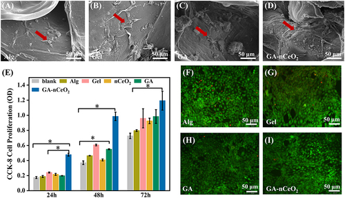
Figure 4 In vitro biocompatibility of MC3T3-E1 cells on the hydrogel scaffold: (A-D) the SEM images of MC3T3-E1 cells cultured on the Alg, Gel, GA, and GA-nCeO2 hydrogel scaffold for 3 days (red arrows showed cells stretched by adhesion on hydrogels); (E) CCK-8 assay for cells cultured for 1, 2, and 3 days on Alg, Gel, GA, and GAnCeO2 hydrogel scaffold; (F–I) Live and dead staining for cells on Alg, Gel, GA, and GA-nCeO2 hydrogel scaffold at 3 days. Scale bar: 50μm. The asterisks indicate a statistically significant difference from the groups (*p < 0.05).
The authors apologize for this error and advise it does not affect the results and conclusion of the paper.