
Huang Y, Chen Y, Cheng G, et al. Int J Nanomedicine. 2024;19:231–245.
The authors have advised due to an error that occurred inadvertently at the time of figure assembly, on page 240 is incorrect. The correct is as follows.
Figure 5 Continued.
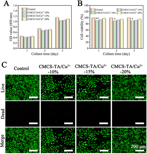
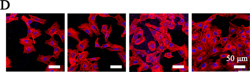
Figure 5 Results of in vitro cytocompatibility testing. (A) CCK-8 assay and (B) cell viability on hydrogels after 1, 3 and 5 days of culture. (C) Images of live/dead staining of cells following incubation on hydrogels. (D) Images taken using a confocal laser scanning microscope (CLSM) show HUVECs stained with rhodamine-coupled phalloidin (red) and DAPI (blue) after three days of growth. A one-way ANOVA was used to determine statistical significance: *p < 0.05, **p < 0.01.
The authors apologise for this error.