
Abstract
In the present study, the strain Brevibacterium frigoritolerans DC2 was explored for the efficient and extracellular synthesis of silver nanoparticles. These biosynthesized silver nanoparticles were characterized by ultraviolet-visible spectrophotometry, which detected the formation of silver nanoparticles in the reaction mixture and showed a maximum absorbance at 420 nm. In addition, field emission transmission electron microscopy revealed the spherical shape of the nanoparticles. The dynamic light scattering results indicated the average particle size of the product was 97 nm with a 0.191 polydispersity index. Furthermore, the product was analyzed by energy dispersive X-ray spectroscopy, X-ray diffraction, and elemental mapping, which displayed the presence of elemental silver in the product. Moreover, on a medical platform, the product was checked against pathogenic microorganisms including Vibrio parahaemolyticus, Salmonella enterica, Bacillus anthracis, Bacillus cereus, Escherichia coli, and Candida albicans. The nanoparticles demonstrated antimicrobial activity against all of these pathogenic microorganisms. Additionally, the silver nanoparticles were evaluated for their combined effects with the commercial antibiotics lincomycin, oleandomycin, vancomycin, novobiocin, penicillin G, and rifampicin against these pathogenic microorganisms. These results indicated that the combination of antibiotics with biosynthesized silver nanoparticles enhanced the antimicrobial effects of antibiotics. Therefore, the current study is a demonstration of an efficient biological synthesis of silver nanoparticles by B. frigoritolerans DC2 and its effect on the enhancement of the antmicrobial efficacy of well-known commercial antibiotics.
Introduction
Currently, the development of a biological methodology for the synthesis of nanoparticles as well as for their characterization and applications on medical and environmental platforms are important aspects of nanotechnology.Citation1,Citation2 There is a particularly pressing need for new and efficient methodologies for the synthesis of nanomaterials. Various physical and chemical methods have been reported along with many drawbacks that limit the use of nanoparticles in biological applications; these disadvantages include the use of toxic solvents, the generation of hazardous byproducts, and high energy consumption. The demand for a methodology that would avoid the hazardous byproducts associated with current physicochemical processes leads to the exploration of a biological species capable of synthesizing nanoparticles.Citation3 The concept of exploiting biological diversity for the synthesis of nanoparticles promises to be quite simple and economical. In addition, these species have been proved to be free of the limitations associated with chemical and physical synthesis. Furthermore, the use of these organisms represents new product development and may allow for applications of biological resources in various fields.Citation4 In the recent past, nanoparticles have been synthesized by using various plant extracts, actinomycetes, fungi, and bacteria.Citation5–Citation8 The microbial resource is a striking opportunity for nanotechnology, as a number of microorganisms are available and can be explored for this action.Citation9
From a medical perspective, the development of resistance mechanisms against antibiotics by pathogenic microorganisms has been a subject of major concern.Citation10 These resistance mechanisms, due to several enzymatic and genetic mutations in the pathogens that cause infectious diseases, has encouraged researchers to design new antimicrobial agents against pathogens to control infections.Citation10,Citation11 Therefore, there is always a need to develop new alternatives to control pathogenic organisms.
Metal nanoparticles are an effective way to control many pathogenic and antibiotic-resistant microorganisms.Citation12 Nanoparticles are applicable in diverse areas such as medicine, diagnostic agents, drug and gene delivery, electronics, cosmetics, coatings, biosensors, imaging, and environmental remediation. Among the many metal nanoparticles, silver nanoparticles have been intensely studied because of the distinct properties of their optical behavior, conductivity, chemical stability, and catalytic activity.Citation13 In addition, silver nanoparticles exhibit broad spectrum bactericidal and fungicidal activity. These properties make them suitable for use as a disinfectant in many medical devices and goods. In addition, silver nanoparticles and antibiotic conjugates can be made efficiently and could result in an increase in the efficacy of antibiotics against various pathogenic, antibiotic-resistant microorganisms.Citation14 Because of the applications of silver nanoparticles in the fields of biology, environment, and technology, there is a growing need for the development of a cost-effective method for the biosynthesis of silver nanoparticles.
The present research was carried out to explore a biological method for the synthesis of silver nanoparticles and to apply these nanoparticles in controlling pathogenic microorganisms. Silver nanoparticles were biosynthesized by Brevibacterium frigoritolerans DC2, which was isolated from a fermented food product. These nanoparticles were tested against different pathogenic microorganisms for their antibacterial efficacy both independently and in combination with various conventional antibiotics.
Materials and methods
Materials
All of the media were purchased from MB Cell (Los Angeles, CA, USA). Analytical grade silver nitrate (AgNO3) and cycloheximide were purchased from Sigma-Aldrich Co. (St Louis, MO, USA). Standard antibiotic discs with standard concentrations for their action against microorganisms were used for the antimicrobial tests. The antibiotics discs that were used are as follows: 1) vancomycin, 30 μg/disc; 2) rifampicin, 5 μg/disc; 3) oleandomycin, 15 μg/disc; 4) penicillin G, 10 μg/disc; 5) novobiocin, 30 μg/disc; and 6) lincomycin, 15 μg/disc. All of the discs were purchased from Oxoid Ltd. (Basingstoke, England).
The pathogenic bacterial strains Vibrio parahaemolyticus ATCC 33844, Bacillus anthracis NCTC 10340, Salmonella enterica ATCC 13076, Bacillus cereus ATCC 14579, Escherichia coli ATCC 10798, and Candida albicans KACC 30062 were used. The bacterial strains were cultured on nutrient agar media at 37°C and preserved at -70°C in glycerol stock vials for further study. C. albicans was cultured on Sabouraud dextrose agar at 28°C and preserved at -70°C in glucose yeast peptone broth glycerol stock vials.
Isolation and molecular identification of bacteria
Fermented skim yogurt was obtained from Indonesia. The food sample was serially diluted in sterile 0.8% NaCl and then spread onto Tryptic soy agar (TSA) media to obtain isolated colonies. To check the metal-tolerating capacity of isolated strains, the strains were further streaked on TSA medium supplemented with 1 mM filter-sterilized AgNO3. The plate was then incubated at room temperature for 48 hours and observed for bacterial growth. The isolated colonies were subcultured and obtained in pure form for further experiments.
Molecular identification of the isolated strain was carried out using a 16S ribosomal ribonucleic acid (rRNA) sequencing-based method. The genomic deoxyribonucleic acid (DNA) was extracted by using a commercial genomic DNA extraction kit (Core Bio System, Seoul, Korea). The 16S rRNA gene was amplified from the chromosomal DNA of the isolated strain by using the universal bacterial primer sets 27F, 518F, 800R, and 1512R.Citation15,Citation16 The purified polymerase chain reaction products were sequenced by Genotech (Daejeon, Korea). The nearly complete sequence (1485 base pairs) of the 16S rRNA was compiled by using SeqMan software (version 4.1). The 16S rRNA gene sequences of related taxa were obtained from the GenBank database and EzTaxon-e server.Citation17
Biosynthesis of silver nanoparticles
For biological synthesis of silver nanoparticles, the selected bacterial isolate was inoculated into a 250 mL Erlenmeyer flask containing 100 mL of sterile Tryptic soy broth. The cultured flasks were incubated in a rotating shaker set at 37°C for 120 rotations per minute (rpm) for 24 hours. After the incubation time, the culture was centrifuged at 8,000 rpm for 10 minutes to remove the bacterial pellet. The supernatant was obtained and was mixed with a filter-sterilized AgNO3 solution (1 mM final concentration) for the extracellular production of silver nanoparticlesCitation1,Citation4,Citation7,Citation8 Furthermore, the culture supernatant with 1 mM AgNO3 was incubated in an orbital shaker at 200 rpm and 25°C. The synthesis of silver nanoparticles was monitored by visual inspection for a change in the color of the culture medium. After the completion of the incubation period, the mixture was first centrifuged at 2,000 rpm for 5 minutes to remove any medium components, and then the silver nanoparticles were collected by high speed centrifugation at 16,000 rpm for 20 minutes. The obtained product was washed several times by centrifugation and was redispersed in water to remove the unconverted silver ions and any medium components. Finally, the silver nanoparticles were collected in the form of a pellet and were used for characterization.Citation18
Characterization of silver nanoparticles
To verify the reduction of silver ions, the solution was scanned in the range of 200–800 nm in a ultraviolet-visible (UV-Vis) spectrophotometer (Ultrospec 2100 Pro; GE Healthcare Bio-Sciences Corp., Piscataway, NJ, USA). The size-distribution profile of silver nanoparticles was studied by using dynamic light scattering with a particle size analyzer (Photal, Otsuka Electronics Co., Osaka, Japan). The hydrodynamic diameters and polydispersity index were analyzed at 25°C. As a reference dispersive medium, pure water with a refractive index 1.3328, viscosity 0.8878 and dielectric constant 78.3 was used. The shape, morphology, and elemental distribution of the nanoparticles were analyzed by using field emission transmission electron microscopy (FE-TEM), energy dispersive X-ray spectroscopy (EDX), and elemental mapping with a JEM-2100F (JEOL, Tokyo, Japan) operated at 200 kV. The sample was prepared by placing a drop of collected silver nanoparticles on a carbon-coated copper grid and subsequently drying the sample in an oven at 60°C before transferring it to the microscope. The X-ray diffraction (XRD) analyses were performed on an X-ray diffractometer, D8 Advance (Bruker, Billerica, MA, USA), which was set to these configurations: 1) 40 kV; 2) 40 mA; 3) CuKα radiation; 4) a scanning rate of 6°/min; and 5) step size 0.02, over the 2θ range of 20°–80°. The silver nanoparticles were collected by centrifugation and several washings with sterile water. Finally, the particles were recovered by air drying the samples and were obtained in powder form.
The stability of silver nanoparticles was observed by keeping the silver nanoparticles in solution at room temperature for different time intervals. In addition, the effect of change in pH on the stability of the silver nanoparticles was studied. The sodium hydroxide (base) was added in the pH range of 4–10, and then the solution was scanned by using the UV-Vis spectrophotometer to observe the absorbance.
Analysis of antimicrobial activity of silver nanoparticles
The antimicrobial activity of the biologically synthesized silver nanoparticles against pathogenic microorganisms B. anthracis, V. parahaemolyticus, S. enterica, E. coli, B. cereus. and C. albicans was measured on Muller-Hinton agar (MHA) plates by using the well diffusion method. An overnight log culture of each pathogenic strain (100 μL) was spread evenly on a MHA plate by using a glass spreader. Wells were made on the MHA plates by using a gel puncture. Then, 50 μL of the silver nanoparticle reaction mixture was added into each well, and the wells were incubated at 37°C for 24 hours. As a control, 50 μL of 1 mM silver nitrate solution was used, this concentration is the same as the one used to biosynthesize the nanoparticles. After incubation, the zones of inhibition were measured by measuring the diameter of the zone formed around each well. Similarly, the experiment was conducted with C. albicans on Sabouraud dextrose agar plates. The study was done in duplicate to check the reproducibility.Citation19
Simultaneously, the disc diffusion method was used to investigate the synergistic effect of antibiotics with silver nanoparticles for antimicrobial effect against test strains. In this assay, the MHA medium plates were spread evenly with 100 μL of an overnight log culture of test organisms. Each standard antibiotic disc of lincomycin, oleandomycin, vancomycin, novobiocin, penicillin G, and rifampicin was further impregnated with 30 μL (100 mg/L) of freshly prepared, partially purified silver nanoparticle solution and was placed onto the agar plates. For each sample, a corresponding control was maintained by using an antibiotic disc without the silver nanoparticle solution. In addition, a control of only silver nanoparticles and silver nitrate at the same concentrations (30 μL [100 mg/L]) was maintained. Then, the plates were incubated at 37°C for 24 hours. In case of C. albicans, cycloheximide (10 μg/disc) was used as a control. After the incubation period, the zones of inhibition around each disc were measured and compared with the corresponding control.Citation20
Results and discussion
Screening and identification of bacteria
After the incubation period, the bacterial strain DC2 showed growth on the TSA plate supplemented with 1 mM AgNO3; this suggests that the bacterial strain DC2 was capable of tolerating silver metal at a 1 mM concentration. On the basis of the molecular characterization of the bacterial isolate, DC2 showed 99.8% similarity with B. frigoritolerans. B. frigoritolerans is strictly aerobic chemo-organotroph, and it is not as well-known as the other members of the genus Brevibacterium.Citation21 Brevibacterium casei has been studied for the biosynthesis of silver and gold nanoparticles.Citation22 Biosurfactant-mediated synthesis of silver nanoparticles by marine B. casei MSA19 has been studied.Citation23 The reports suggested that the some of the species of the genus do show activity for the biosynthesis of metal nanoparticles; however, to our knowledge, this is the first report of the biosynthesis of silver nanoparticles by B. frigoritolerans DC2 isolated from a fermented food product. The 16S rRNA sequence of the strain B. frigoritolerans DC2 has been submitted to National Center for Biotechnology Information with the accession number KM583447. The strain has been deposited in Korean Collection for Type Culture (KCTC 29680).
Synthesis and characterization of silver nanoparticles
In the present study, silver nanoparticles were successfully synthesized in the culture supernatant of B. frigoritolerans DC2. The formation of silver nanoparticles by the reduction of AgNO3 was indicated by the color change of the reaction mixture (). As the biosynthesis proceeded over 48 hours, the color changed from light yellow to dark brown. This brown color could be due to the excitation of surface plasmon vibrations; if so, this would indicate the formation of silver nanoparticles in the reaction mixture.Citation24 On the other hand, no color change was observed in the control flask that contained medium with only the 1 mM AgNO3 solution and had been kept under the same conditions as the other flask (). Because the nanoparticle synthesis was extracellular, the need for downstream processing that would have been otherwise essential for intracellular synthesis was avoided and thereby made the process simpler and more cost-effective. The exact mechanism behind the extracellular synthesis of silver nanoparticles in the supernatant remains to be elucidated. However, reports have suggested that the extracellular enzyme secreted by microorganism in the culture supernatant is responsible for the reduction of silver ions to silver nanoparticles.Citation25,Citation26 The study based on extracellular synthesis of silver nanoparticles by Bacillus licheniformis showed that the nitrate reductase enzyme extracellularly secreted by the bacteria in the medium was responsible for the synthesis of silver nanoparticles.Citation8
Figure 1 Chromatic properties of culture supernatant of Brevibacterium frigoritolerans DC2.
Notes: Culture supernatant of B. frigoritolerans DC2 after incubation with AgNO3 (silver nitrate) (1 mM) (A) and control with medium and AgNO3 (1 mM) after incubation period (B). UV-Vis spectra of culture supernatant of B. frigoritolerans DC2 treated with 1 mM AgNO3 (C).
Abbreviations: OD, optical density; UV-Vis, ultraviolet-visible.
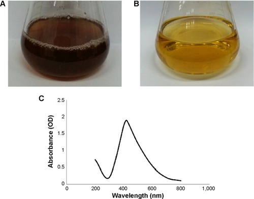
For the characterization of the silver nanoparticles, the reaction mixture was monitored by using UV-Vis spectrophotometer for spectral analysis. After the incubation period, the reaction mixture was scanned in the range of 200–800 nm. In the UV-Vis absorption spectrum, a strong peak at about 420 nm was observed; this is attributed to the surface plasmon resonance band of the silver nanoparticles (). Reports suggest that the band located in this region corresponds to the surface plasmon resonance of silver nanoparticles.Citation27,Citation28 Thus, the reaction mixture indicates the formation of silver nanoparticles.
Various studies have characterized the shape and size of silver nanoparticles by TEM.Citation22,Citation24,Citation27,Citation29 In the present study, field emission TEM images of silver nanoparticles revealed that the shape of the nanoparticles was spherical (). The results showed that particles are uniform in shape with 10 nm to 30 nm in size. The dynamic light scattering particle size analysis results indicated that the hydrodynamic diameter of the particles range was 50–150 nm with a 0.191 polydispersity index (). The average particle size was 97 nm.Citation30,Citation31 The purity and elemental composition of the biosynthesized silver nanoparticles were determined by EDX, XRD, and elemental mapping. In the EDX spectrum, silver nanocrystallites displayed an optical absorption band peak at approximately 3 keV (); this property is due to the absorption of metallic silver nanocrystallites that correspond to surface plasmon resonance.Citation8 shows the XRD pattern of silver nanoparticles, which exhibited intense peaks throughout the whole spectrum of 2θ value, which ranged from 20° to 80°; this pattern was similar to the Braggs’s reflection of silver nanocrystals. Our results are similar to those of previous reports of characterization of silver nanoparticles by XRD.Citation32–Citation35
Figure 2 Properties of silver nanoparticles synthesized by Brevibacterium frigoritolerans DC2.
Notes: Transmission electron micrograph of silver nanoparticles synthesized by B. frigoritolerans DC2 (A and B). Particles size distribution of silver nanoparticles according to volume (C), number (D), and intensity (E).
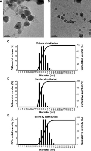
Figure 3 Silver nanoparticles examined under a variety of conditions.
Notes: Energy dispersive X-ray spectroscopy of the whole scan area showing major peak of silver nanoparticles at 3 keV (A); X-ray diffraction patterns of silver nanoparticles obtained by Brevibacterium frigoritolerans DC2 (B); transmission electron micrograph of silver nanoparticle pellet solution (C); silver nanoparticles, green (D).
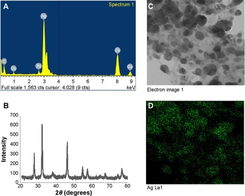
The elemental mapping results of the biosynthesized silver nanoparticles () indicate that silver was distributed maximally (50.30%); this finding suggests that silver was the predominant metal. The other elements, such as copper (26.98%), carbon (14.98%), and sulfur (1.44%) were also observed. The distribution of carbon and copper was due to the use of a TEM grid, and slight contamination by chlorine and sulfur appeared. Thus, the results indicate that the strain B. frigoritolerans DC2 was found to be capable of synthesizing spherical silver nanoparticles.
The stability of silver nanoparticles was determined by keeping the nanoparticle solution at room temperature for different time intervals over many days. There was no observable variation in the UV-Vis spectrum of the reaction mixture even after 1 month; this indicated the stable nature of the silver nanoparticles in the reaction mixture. The results proved that the nanoparticles were stable for more than 1 month. In addition, the effect of the change in pH on the stability of the silver nanoparticles in the pH range of 4–10 was studied. The silver nanoparticle solution was observed before and after the addition of sodium hydroxide (base). No major change in the wavelength was observed; this result further confirmed the stable nature of the silver nanoparticles.
Antimicrobial activity of silver nanoparticles
Metal nanoparticles have been shown to be effective against many pathogenic and multidrug-resistant bacteria. For instance, silver nanoparticles have demonstrated activity against methicillin-resistant Staphylococcus aureus (MRSA) and methicillin-resistant Staphylococcus epidermidis (MRSE).Citation36 Various studies have reported antimicrobial action of silver nanoparticles against microorganisms.Citation4,Citation37,Citation38 In our study, the biosynthesized silver nanoparticles displayed antimicrobial activity against a range of pathogenic microorganisms, such as C. albicans, V. parahaemolyticus, S. enterica, B. anthracis, B. cereus, and E. coli.
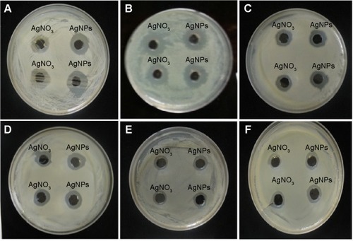
The antimicrobial activity of the reaction mixture was compared to that of silver nitrate solution after incubation period. Antimicrobial activity against tested microorganisms was measured by measuring the diameter of the zone of inhibition of each well. The results of mean diameters of zones of inhibition are interpreted in . The results demonstrate that the reaction mixture containing silver nanoparticles exhibited antimicrobial activity against the tested microorganisms in the following manner, C. albicans followed by V. parahaemolyticus, B. anthracis and B. cereus, S. enterica, and then E. coli (, respectively). Although the activity of reaction mixture exhibit slightly more antimicrobial activity than silver nitrate solution. The activity was further analyzed after the purification of nanoparticles and compared with that of the same concentration of silver nitrate. The exact mechanism of antimicrobial action is not clearly known, but studies suggest that silver nanoparticles have the ability to damage the cell membrane permeability, damage the respiration functions of the cell, and encourage the formation of free radicals. These factors cause the antimicrobial effect of silver nanoparticles.Citation9,Citation39
Table 1 Diameters of zones of inhibition (mm) for 50 μL of reaction mixture containing silver nanoparticles after incubation with various microorganisms
Figure 4 Zones of inhibition for various pathogens.
Notes: Zones of inhibition of 50 μL of reaction mixture containing silver nanoparticles and 50 μL of 1mM AgNO3 (silver nitrate) against the pathogenic strains Candida albicans (A), Vibrio parahaemolyticus (B), Bacillus anthracis (C), Bacillus cereus (D), Salmonella enterica (E), and Escherichia coli (F).
Abbreviation: AgNPs, silver nanoparticles.
Several studies have shown that the combination of silver nanoparticles with antibiotics leads to an enhanced effect of the antibiotics against microorganisms.Citation14,Citation20 This action is most probably due to an increase in cell wall penetration by these antibiotics with the nanoparticles. Combinations of antibiotics with nanoparticles have many advantages that minimize the side effects of broad-spectrum antibiotics; these benefits include increasing the concentration of the local antibiotic at the target site and facilitating the binding of antibiotics to microorganisms. Moreover, nanoparticle–antibiotic conjugates lower the amounts of both the drugs and the nanoparticles in the dosage; this property reduces the side effects of the medication while increasing its antimicrobial properties.Citation14 In our study, the synergistic effect of silver nanoparticles with different commercial antibiotics was investigated against six pathogenic microorganisms by using the disc diffusion method. represents the antimicrobial activity of partially purified silver nanoparticles and silver nitrate of the same concentration (1 mM) against S. enterica, E. coli, V. parahaemolyticus, B. anthracis, and B. cereus. The appearance of zones of inhibition indicated that the biosynthesized silver nanoparticles had stronger antimicrobial properties against the tested microorganisms than did silver nitrate when both treatments were used at the same concentration. In addition, the synergistic effect of the antibiotics (lincomycin, oleandomycin, novobiocin, vancomycin, penicillin G, and rifampicin) in conjugation with biosynthesized silver nanoparticles increased the sensitivity of the tested microorganisms. The results show that S. enterica, E. coli, and V. parahaemolyticus () were completely resistant to the antibiotics; however, the addition of the silver nanoparticle solutions to the discs rendered the bacterial strains sensitivity to those discs and thereby resulted in the formation of zones of inhibition (). The maximum increase in fold area has been calculated for antibiotics with silver nanoparticles with respect to standard antibiotics.Citation40 The average of maximum increase in fold area against the antibiotic-resistant microorganisms S. enterica, E. coli, and V. parahaemolyticus () observed in this study was highest for novobiocin (4.82 fold), then for lincomycin, oleandomycin, and rifampicin (4.23 fold), then for vancomycin (3.98 fold), and finally for penicillin G (3.66 fold). Various researchers have demonstrated the combined effect of biosynthesized silver nanoparticles with commercial antibiotics.Citation4,Citation14,Citation20 The results demonstrate that the silver nanoparticles obtained by B. frigoritolerans DC2 enhance the antimicrobial activity of commercial antibiotics against S. enterica, E. coli, and V. parahaemolyticus.
Figure 5 Effects of partially purified silver nanoparticle pellet solution on various pathogens.
Notes: Zones of inhibition of partially purified silver nanoparticle pellet solution and silver nitrate (AgNO3) (both at same concentration, 30 μL [100 mg/L]) against Salmonella enterica (A), Escherichia coli (B), Vibrio parahaemolyticus (C), Bacillus anthracis (D), and Bacillus cereus (E). Zones of inhibition of standard antibiotic discs against pathogenic bacteria Salmonella enterica (F), Escherichia coli (G), Vibrio parahaemolyticus (H), Bacillus anthracis (I), and Bacillus cereus (J). Zones of inhibition of standard antibiotic discs with silver nanoparticles against Salmonella enterica (K), Escherichia coli (L), Vibrio parahaemolyticus (M), Bacillus anthracis (N), and Bacillus cereus (O). Zones of inhibition of silver nanoparticles and silver nitrate against Candida albicans (P), cycloheximide (Q-7), Cycloheximide with AgNPs (Q-8,9). For (A–Q), the numbers, the corresponding antibiotics, and the corresponding concentrations of antibiotics are as follows: 1) lincomycin (MY15), 15 μg/disc; 2) oleandomycin (OL15), 15 μg/disc; 3) novobiocin (NV30), 30 μg/disc; 4) vancomycin (VA30), 30 μg/disc; 5) penicillin G (P10), 10 μg/disc; 6) rifampicin (RD5) 5 μg/disc; 7) cycloheximide, 10 μg/disc; 8) cycloheximide (10 μg/disc) with silver nanoparticles; and 9) cycloheximide (10 μg/disc) with silver nanoparticles.
Abbreviation: AgNPs, silver nanoparticles.
![Figure 5 Effects of partially purified silver nanoparticle pellet solution on various pathogens.Notes: Zones of inhibition of partially purified silver nanoparticle pellet solution and silver nitrate (AgNO3) (both at same concentration, 30 μL [100 mg/L]) against Salmonella enterica (A), Escherichia coli (B), Vibrio parahaemolyticus (C), Bacillus anthracis (D), and Bacillus cereus (E). Zones of inhibition of standard antibiotic discs against pathogenic bacteria Salmonella enterica (F), Escherichia coli (G), Vibrio parahaemolyticus (H), Bacillus anthracis (I), and Bacillus cereus (J). Zones of inhibition of standard antibiotic discs with silver nanoparticles against Salmonella enterica (K), Escherichia coli (L), Vibrio parahaemolyticus (M), Bacillus anthracis (N), and Bacillus cereus (O). Zones of inhibition of silver nanoparticles and silver nitrate against Candida albicans (P), cycloheximide (Q-7), Cycloheximide with AgNPs (Q-8,9). For (A–Q), the numbers, the corresponding antibiotics, and the corresponding concentrations of antibiotics are as follows: 1) lincomycin (MY15), 15 μg/disc; 2) oleandomycin (OL15), 15 μg/disc; 3) novobiocin (NV30), 30 μg/disc; 4) vancomycin (VA30), 30 μg/disc; 5) penicillin G (P10), 10 μg/disc; 6) rifampicin (RD5) 5 μg/disc; 7) cycloheximide, 10 μg/disc; 8) cycloheximide (10 μg/disc) with silver nanoparticles; and 9) cycloheximide (10 μg/disc) with silver nanoparticles.Abbreviation: AgNPs, silver nanoparticles.](/cms/asset/bb7d9c9a-7ad5-4110-9ed2-a4d6b0f835ae/dijn_a_72313_f0005_c.jpg)
Figure 6 Graphical representation of comparative study between antibiotics and antibiotics with AgNPs.
Notes: Salmonella enterica (A), Escherichia coli (B), Vibrio parahaemolyticus (C), Bacillus anthracis (D), and Bacillus cereus (E), respectively. Results interpreting the antimicrobial activity of commercial antibiotics alone with that of antibiotics combined with silver nanoparticles.
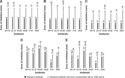
The other pathogenic strains, B. anthracis and B. cereus (), showed a sensitivity pattern to antibiotics. However, when the discs were combined with silver nanoparticles, an increase in the diameters of the zones of inhibition was observed (). The results clearly indicate that the addition of silver nanoparticles enhanced the effect of the antibiotics. Thus, an increase in fold area was observed in all of the antibiotics tested against antibiotic-sensitive and antibiotic-resistant pathogenic microorganisms. Similarly, shows the antimicrobial activity of silver nanoparticles against C. albicans. shows that C. albicans was completely resistant to cyclohexamide; however, when the silver nanoparticles were added to the cyclohexamide, antimicrobial activity was enhanced and resulted in the formation of a zone of inhibition. The results showed that the average of maximum increase in fold area against antibiotic-sensitive microorganisms B. anthracis and B. cereus () were, in descending order, as follows: 1) rifampicin (3.69 fold); 2) oleandomycin (1.91 fold); 3) penicillin G (0.36 fold); 4) novobiocin (0.26 fold); 5) vancomycin (0.22 fold); and 6) lincomycin (0.14 fold). Our results are in agreement with other studies that have shown the antimicrobial activity of silver nanoparticles against many pathogenic and multidrug-resistant microorganisms.Citation4,Citation14 In our findings, biosynthesized silver nanoparticles showed antimicrobial activity against pathogenic microorganisms. Moreover, the silver nanoparticles enhanced the antimicrobial activity of commercial antibiotics against pathogenic microorganisms. However, the exact action mechanism still must be explored.
Conclusion
In our study, the method for biological synthesis of silver nanoparticles by B. frigoritolerans DC2 was established. This biogenic process will be helpful for minimizing the need for chemical and physical methodologies. In addition to this, the extracellular synthesis of nanoparticles could be highly advantageous for facilitating downstream processing in large-scale operations. The silver nanoparticles that were obtained showed antimicrobial effects against various pathogenic microorganisms. Furthermore, they enhanced the antimicrobial activity of various commercial antibiotics against pathogenic microorganisms.
Acknowledgments
This research was supported by the program of Agriculture Science & Technology Development (grant#: PJ008813042014), Rural Development Administration and also supported by a grant from the Next-Generation BioGreen 21 Program (SSAC, grant#: PJ00952903).
Disclosure
The authors report no conflicts of interest in this work.
References
- ShankarSSRaiAAhmadASastryMRapid synthesis of Au, Ag, and bimetallic Au core–Ag shell nanoparticles using Neem (Azadirachta indica) leaf brothJ Colloid Interf Sci20042752496502
- RautRWLakkakulaJRKolekarNSMendhulkarVDKashidSBPhytosynthesis of silver nanoparticle using Gliricidia sepium (Jacq)Curr Nanosci20095117122
- NadagoudaMNVarmaRSGreen synthesis of silver and palladium nanoparticles at room temperature using coffee and tea extractGreen Chem2008108859862
- DarMAIngleARaiMEnhanced antimicrobial activity of silver nanoparticles synthesized by Cryphonectria sp. evaluated singly and in combination with antibioticsNanomedicine20139110511022633901
- KumarVYadavSKPlant-mediated synthesis of silver and gold nanoparticles and their applicationsJ Chem Technol Biotechnol2009842151157
- AhmadASenapatiSKhanMIIntracellular synthesis of gold nanoparticles by a novel alkalotolerant actinomycete, Rhodococcus speciesNanotechnology200314824828
- BasavarajaSBalajiSDLagashettyARajasabAHVenkataramanAExtracellular biosynthesis of silver nanoparticles using the fungus Fusarium semitectumMater Res Bull200843511641170
- KalimuthuKBabuRSVenkataramanDBilalMGurunathanSBiosynthesis of silver nanocrystals by Bacillus licheniformisColloids Surf B Biointerfaces200865115015318406112
- PrabhuSPouloseEKSilver nanoparticles: mechanism of antimicrobial action, synthesis, medical applications, and toxicity effectsInternational Nano Letters201221110
- SibandaTOkohAIThe challenges of overcoming antibiotic resistance: Plant extracts as potential sources of antimicrobial and resistance modifying agentsAfrican Journal of Biotechnology200762528862896
- KollefMHGolanYMicekSTShorrAFRestrepoMIAppraising contemporary strategies to combat multidrug resistant gram-negative bacterial infections–proceedings and data from the Gram-Negative Resistance SummitClin Infect Dis201153Suppl 2S33S55 quiz S56–S5821868447
- RaiMKDeshmukhSDIngleAPGadeAKSilver nanoparticles: the powerful nanoweapon against multidrug-resistant bacteriaJ Appl Microbiol2012112584185222324439
- RosarinFSMirunaliniSNobel metallic nanoparticles with novel biomedical propertiesJ Bioanal Biomed20113085091
- FayazAMBalajiKGirilalMYadavRKalaichelvanPTVenketesanRBiogenic synthesis of silver nanoparticles and their synergistic effect with antibiotics: a study against gram-positive and gram-negative bacteriaNanomedicine20106110310919447203
- WeisburgWGBarnsSMPelletierDALaneDJ16S ribosomal DNA amplification for phylogenetic studyJ Bacteriol199117326977031987160
- KimKKKimMKLimJHParkHYLeeSTTransfer of Chryseobacterium meningosepticum and Chryseobacterium miricola to Elizabethkingia gen. nov. as Elizabethkingia meningoseptica comb. nov. and Elizabethkingia miricola comb. novInt J Syst Evol Microbiol200555Pt 31287129315879269
- KimOSChoYJLeeKIntroducing EzTaxon-e: a prokaryotic 16S rRNA gene sequence database with phylotypes that represent uncultured speciesInt J Syst Evol Microbiol201262Pt 371672122140171
- SuprajaSMohammed AliSChakravarthyNGreen synthesis of silver nanoparticles from Cynodon dactylon leaf extractInt J Chem Tech201351271277
- LogeswariPSilambarasanSAbrahamJEcofriendly synthesis of silver nanoparticles from commercially available plant powders and their antibacterial propertiesScientia Iranica201320310491054
- NaqviSZHKiranUAliMICombined efficacy of biologically synthesized silver nanoparticles and different antibiotics against multidrug-resistant bacteriaInt J Nanomedicine201383187319523986635
- SelvakumarGSushilSNStanleyJBrevibacterium frigoritolerans a novel entomopathogen of Anomala dimidiata and Holotrichia longipennis (Scarabaeidae: Coleoptera)Biocontrol Science and Technology2011217821827
- KalishwaralalKDeepakVPandianSRKBiosynthesis of silver and gold nanoparticles using Brevibacterium caseiColloids Surf B Biointerfaces20107725726220197229
- KiranGSSabuASelvinJSynthesis of silver nanoparticles by glycolipid biosurfactant produced from marine Brevibacterium casei MSA19J Biotechnol2010148422122520600381
- BalajiDSBasavarajaSDeshpandeRMaheshDPrabhakarBKVenkataramanAExtracellular biosynthesis of functionalized silver nanoparticles by strains of Cladosporium cladosporioides fungusColloids Surf B Biointerfaces2009681889218995994
- DasVLThomasRVargheseRTSoniyaEVMathewJRadhakrishnanEKExtracellular synthesis of silver nanoparticles by the Bacillus strain CS 11 isolated from industrialized area3 Biotech201442121126
- DuránNMarcatoPDAlvesOLDe SouzaGEspositoEMechanistic aspects of biosynthesis of silver nanoparticles by several Fusarium oxysporum strainsJ Nanobiotechnol20053817
- VidhuVKAromalSPhilipDGreen synthesis of silver nanoparticles using Macrotyloma uniflorumSpectrochimica Acta Part A: Molecular and Biomolecular Spectroscopy2011831392397
- WaniIAKhatoonSGangulyAAhmedJGanguliAKAhmadTSilver nanoparticles: Large scale solvothermal synthesis and optical propertiesMater Res Bull201045810331038
- LitvinVAMinaevBFSpectroscopy study of silver nanoparticles fabrication using synthetic humic substances and their antimicrobial activitySpectrochim. Acta, Part A2013108115122
- VithiyaKKumarRSenSBacillus sp. mediated extracellular synthesis of silver nanoparticlesInternational Journal of Pharmcy and Pharmaceutical Sciences20146Suppl 2525527
- OtariSVPatilRMNadafNHGhoshSJPawarSHGreen biosynthesis of silver nanoparticles from an actinobacteria Rhodococcus spMater Lett2012729294
- LitvinVAGalaganRLMinaevBFKinetic and mechanism formation of silver nanoparticles coated by synthetic humic substancesColloids Surf A Physicochem Eng Asp2012414234243
- WaniIAGangulyAAhmedJAhmadTSilver nanoparticles: Ultrasonic wave assisted synthesis, optical characterization and surface area studiesMater Lett2011653520522
- NabikhanAKandasamyKRajAAlikunhiNMSynthesis of antimicrobial silver nanoparticles by callus and leaf extracts from saltmarsh plant, Sesuvium portulacastrumColloids Surf B Biointerfaces201079248849320627485
- GnanadesiganMAnandMRavikumarSAntibacterial potential of biosynthesised silver nanoparticles using Avicennia marina mangrove plantAppl Nanosci201222143147
- SaravananMNandaAExtracellular synthesis of silver bionanoparticles from Aspergillus clavatus and its antimicrobial activity against MRSA and MRSEColloids Surf B Biointerfaces201077221421820189360
- AhmadTWaniIAManzoorNAhmedJAsiriAMBiosynthesis, structural characterization and antimicrobial activity of gold and silver nanoparticlesColloids Surf B Biointerfaces201310722723423500733
- WaniIAKhatoonSGangulyAAhmedJAhmadTManzoorNStructural characterization and antimicrobial properties of silver nanoparticles prepared by inverse microemulsion methodColloids Surf B Biointerfaces201310124325023010026
- LemireJAHarrisonJJTurnerRJAntimicrobial activity of metals: mechanisms, molecular targets and applicationsNat Rev Microbiol201311637138423669886
- BirlaSSTiwariVVGadeAKIngleAPYadavAPRaiMKFabrication of silver nanoparticles by Phoma glomerata and its combined effect against Escherichia coli, Pseudomonas aeruginosa and Staphylococcus aureusLett Appl Microbiol200948217317919141039