

Abstract
The broad application of electrospun nanofibrous scaffolds in tissue engineering is limited by their small pore size, which has a negative influence on cell migration. This disadvantage could be significantly improved through the combination of nano- and microfibrous structure. To accomplish this, different nano/microfibrous scaffolds were produced by hybrid electrospinning, combining solution electrospinning with melt electrospinning, while varying the content of the nanofiber. The morphology of the silk fibroin (SF)/poly(ε-caprolactone) (PCL) nano/microfibrous composite scaffolds was investigated with field-emission scanning electron microscopy, while the mechanical and pore properties were assessed by measurement of tensile strength and mercury porosimetry. To assay cell proliferation, cell viability, and infiltration ability, human mesenchymal stem cells were seeded on the SF/PCL nano/microfibrous composite scaffolds. From in vivo tests, it was found that the bone-regenerating ability of SF/PCL nano/microfibrous composite scaffolds was closely associated with the nanofiber content in the composite scaffolds. In conclusion, this approach of controlling the nanofiber content in SF/PCL nano/microfibrous composite scaffolds could be useful in the design of novel scaffolds for tissue engineering.
Supplementary materials
Figure S1 Change of morphology and fiber distribution of the electrospun SF nanofibers with various electrospinning processing conditions.
Notes: (A) SF/PCL 2/98, (B) SF/PCL 4/96, (C) SF/PCL 6/94, and (D) SF/PCL 8/92.
Abbreviations: PCL, poly(ε-caprolactone); SF, silk fibroin.
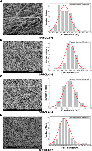
Figure S2 Changes in morphology of water vapor-treated SF/PCL nano/microfibrous composite scaffolds after water immersion for 1 hour.
Notes: (A) SF/PCL 2/98, (B) SF/PCL 4/96, (C) SF/PCL 6/94, and (D) SF/PCL 8/92.
Abbreviations: PCL, poly(ε-caprolactone); SF, silk fibroin.
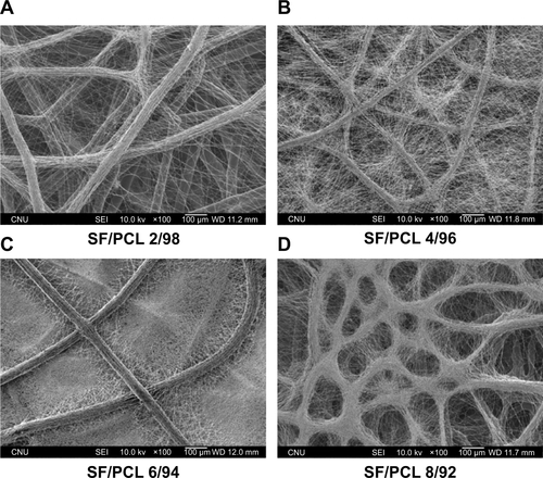
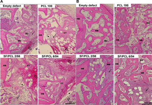
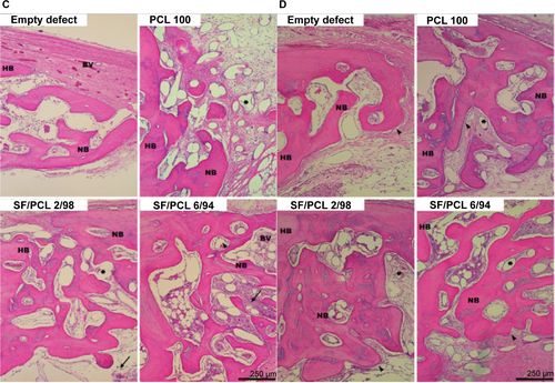
Figure S3 Hematoxylin and eosin-stained histological sections after implantation at high magnification.
Notes: Representative histological sections show cross sections of calvarial defects with native bone at the edge; (A) 1 week, (B) 2 weeks, (C) 4 weeks, and (D) 8 weeks. Note the presence of blood clots (empty arrows), inflammatory cells (thin black arrows), osteoblasts (arrowheads), and remaining scaffolds (asterisks). Scale bar 250 μm. Original magnification 100×.
Abbreviations: PCL, poly(ε-caprolactone); SF, silk fibroin; HB, host bone; NB, new bone; BV, blood vessel; CT, connective tissue.
Acknowledgments
This research was supported by the Nuclear R&D program (NRF-2012M2A2A6035747) through the National Research Foundation, funded by the Ministry of Science, ICT and Future Planning, South Korea.
Disclosure
The authors report no conflicts of interest in this work.