

Abstract
Magnetically induced heating of magnetic nanoparticles (MNP) in an alternating magnetic field (AMF) is a promising minimal invasive tool for localized tumor treatment that eradicates tumor cells by applying thermal stress. While temperatures between 42°C and 45°C induce apoptosis and sensitize the cells for chemo- and radiation therapies when applied for at least 30 minutes, temperatures above 50°C, so-called thermoablative temperatures, rapidly induce irreversible cell damage resulting in necrosis. Since only little is known concerning the protein expression of anti-apoptotic B-cell lymphoma 2 (BCL2), fibroblast growth factor receptor 1 (FGF-R1), and heat shock protein (HSP70) after short-time magnetic thermoablative tumor treatment, these relevant tumor proteins were investigated by immunohistochemistry (IHC) in a human BT474 breast cancer mouse xenograft model. In the investigated sample groups, the application of thermoablative temperatures (<2 minutes) led to a downregulation of BCL2 and FGF-R1 on the protein level while the level of HSP70 remained unchanged. Coincidently, the tumor tissue was damaged by heat, resulting in large apoptotic and necrotic areas in regions with high MNP concentration. Taken together, thermoablative heating induced via magnetic methods can reduce the expression of tumor-related proteins and locally inactivate tumor tissue, leading to a prospectively reduced tumorigenicity of cancerous tissues. The presented data allow a deeper insight into the molecular mechanisms in relation to magnetic thermoablative tumor treatments with the aim of further improvements.
Supplementary material
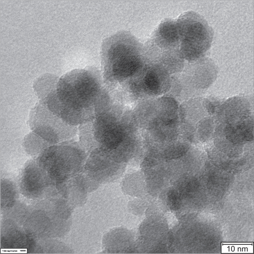
Figure S1 High-resolution transmission electron microscopy (HRTEM) micrograph of 200 nm fluidMAG-D magnetic nanoparticles (MNP).
Notes: HRTEM micrograph reveals smaller core particles of approximately 10–12 nm in size clustered to a particle core size of approximately 70 nm in total. The HRTEM micrograph was prepared as described elsewhere.Citation37
Acknowledgments
M Stapf is the main author of this manuscript. The present investigation was supported in part by the “Deutsche Forschungsgemeinschaft” (HI-689/7-2). We acknowledge valuable technical assistance provided by S Burgold, J Göring, B Grobis, Y Ozegowski, and B Ziegenhardt. We acknowledge Dr B Romeike for assisting in examining the histological slices and Dr L Leistritz for his advice in statistical calculations. We thank S Nietzsche (Center of Electron Microscopy, University Hospital Jena, Germany) for kindly providing the HRTEM micrographs of 200 nm fluidMAG-D MNP (Figure S1). This work is dedicated to WA Kaiser who passed away in 2013.
Disclosure
The authors report no conflicts of interest in this work.