

Abstract
Background
Cancer stem cells (CSCs) possess the characteristics associated with normal stem cells and are responsible for cancer initiation, recurrence, and metastasis. CD133 is regarded as a CSCs marker of osteosarcoma, which is the most common primary bone malignancy in childhood and adolescence. Salinomycin, a polyether ionophore antibiotic, has been shown to kill various CSCs, including osteosarcoma CSCs. However, salinomycin displayed poor aqueous solubility that hinders its clinical application. The objective of this study was to develop salinomycin-loaded nanoparticles to eliminate CD133+ osteosarcoma CSCs.
Methods
The salinomycin-loaded PEGylated poly(lactic-co-glycolic acid) nanoparticles (SAL-NP) conjugated with CD133 aptamers (Ap-SAL-NP) were developed by an emulsion/solvent evaporation method, and the targeting and cytotoxicity of Ap-SAL-NP to CD133+ osteosarcoma CSCs were evaluated.
Results
The nanoparticles are of desired particle size (~150 nm), drug encapsulation efficiency (~50%), and drug release profile. After 48 hours treatment of the Saos-2 CD133+ osteosarcoma cells with drugs formulated in Ap-SAL-NP, SAL-NP, and salinomycin, the concentrations needed to kill 50% of the incubated cells were found to be 2.18, 10.72, and 5.07 μg/mL, respectively, suggesting that Ap-SAL-NP could be 4.92 or 2.33 fold more effective than SAL-NP or salinomycin, respectively. In contrast, Ap-SAL-NP was as effective as SAL-NP, and less effective than salinomycin in Saos-2 CD133− cells, suggesting that Ap-SAL-NP possess specific cytotoxicity toward Saos-2 CD133+ cells. Ap-SAL-NP showed the best therapeutic effect in Saos-2 osteosarcoma xenograft mice, compared with SAL-NP or salinomycin. Significantly, Ap-SAL-NP could selectively kill CD133+ osteosarcoma CSCs both in vitro and in vivo, as reflected by the tumorsphere formation and proportion of Saos-2 CD133+ cells.
Conclusion
Our results suggest that CD133 is a potential target for drug delivery to osteosarcoma CSCs and that it is possible to significantly inhibit the osteosarcoma growth by killing CD133+ osteosarcoma CSCs. We demonstrated that Ap-SAL-NP have the potential to target and kill CD133+ osteosarcoma CSCs.
Supplementary materials
Figure S1 The RT-PCR analysis of mRNA level normalized to β-actin.
Notes: The genes of the CD133+ or CD133− Saos-2 cells were analyzed; Data are expressed as mean ± SD (n=3).
Abbreviations: mRNA, messenger RNA; RT-PCR, real-time polymerase chain reaction; SD, standard deviation.
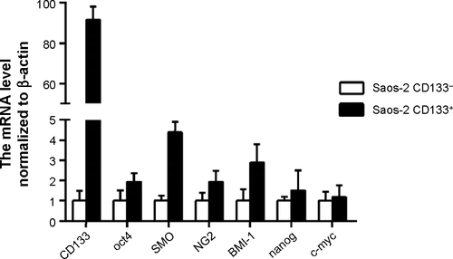
Figure S2 In vitro cellular uptake of nanoparticles evaluated by confocal studies in Saos-2 CD133− cells.
Abbreviations: Ap-C6-NP, coumarin 6-loaded PLGA nanoparticles conjugated with CD133 aptamers; C6-NP, coumarin 6-loaded nanoparticles; DAPI, 4′,6-diamidino-2-phenylindole dihydrochloride; PLGA, poly(lactic-co-glycolic acid).
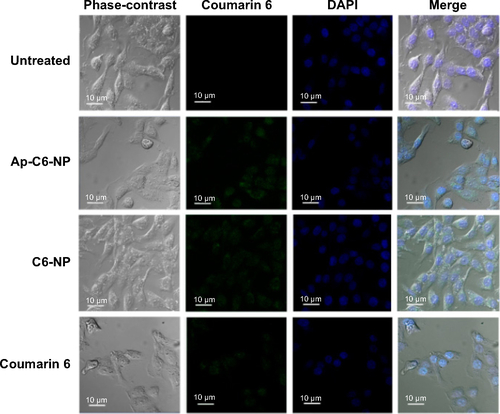
Figure S3 The cell proliferation assay of nanoparticles at 72 hours.
Notes: Briefly, CD133+ and CD133− osteosarcoma cells were seeded in 96-well plates with a density of 5×10³ cells per well overnight; The cells were incubated with a series of concentrations of the nanoparticles or free salinomycin; After 72 hours, the cytotoxicity was evaluated by the CCK-8 method; (A) Saos-2 CD133+; (B) Saos-2 CD133−; (C) U-2 OS CD133+; (D) U-2 OS CD133−; (E) MG-63 CD133+; (F) MG-63 CD133−; Data are expressed as mean ± SD (n=3).
Abbreviations: Ap-NP, PLGA nanoparticles conjugated with CD133 aptamers; Ap-SAL-NP, salinomycin-loaded PLGA nanoparticles conjugated with CD133 aptamers; CCK-8, Cell Counting Kit-8; PLGA, poly(lactic-co-glycolic acid); SAL, salinomycin; SAL-NP, salinomycin-loaded PLGA nanoparticles; SD, standard deviation.
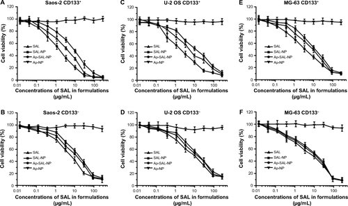
Figure S4 The weight change of the mice during the treatment.
Notes: The body weight of the mice was monitored once every 5 days; Data are expressed as mean ± SD (n=8).
Abbreviations: Ap-NP, PLGA nanoparticles conjugated with CD133 aptamers; Ap-SAL-NP, salinomycin-loaded PLGA nanoparticles conjugated with CD133 aptamers; PLGA, poly(lactic-co-glycolic acid); SAL, salinomycin; SAL-NP, salinomycin-loaded PLGA nanoparticles; SD, standard deviation.
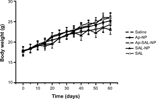
Table S1 The sequence of the primers used in the RT-PCR
Table S2 The in vivo tumorigenic potential of CD133+ and CD133− Saos-2 cellsTable Footnotea
Acknowledgments
This work was supported by the Young Project of the Shanghai Health and Family Planning Commission (2014-676).
Disclosure
The authors report no conflicts of interest in this work.