

Abstract
Lycopene (LP), an important functional compound in tomatoes, and gold nanoparticles (AN), have received considerable attention as potential candidates for cancer therapy. However, the extreme instability and poor bioavailability of LP limits its in vivo application. This study intends to develop a nanoemulsion system incorporating both LP and AN, and to study the possible synergistic effects on the inhibition of the HT-29 colon cancer cell line. LP–nanogold nanoemulsion containing Tween 80 as an emulsifier was prepared, followed by characterization using transmission electron microscopy (TEM), dynamic light scattering (DLS) analysis, ultraviolet spectroscopy, and zeta potential analysis. The particle size as determined by TEM and DLS was 21.3±3.7 nm and 25.0±4.2 nm for nanoemulsion and 4.7±1.1 nm and 3.3±0.6 nm for AN, while the zeta potential of nanoemulsion and AN was −32.2±1.8 mV and −48.5±2.7 mV, respectively. Compared with the control treatment, both the combo (AN 10 ppm plus LP 12 μM) and nanoemulsion (AN 0.16 ppm plus LP 0.4 μM) treatments resulted in a five- and 15-fold rise in early apoptotic cells of HT-29, respectively. Also, the nanoemulsion significantly reduced the expressions of procaspases 8, 3, and 9, as well as PARP-1 and Bcl-2, while Bax expression was enhanced. A fivefold decline in the migration capability of HT-29 cells was observed for this nanoemulsion when compared to control, with the invasion-associated markers being significantly reversed through the upregulation of the epithelial marker E-cadherin and downregulation of Akt, nuclear factor kappa B, pro-matrix metalloproteinase (MMP)-2, and active MMP-9 expressions. The TEM images revealed that numerous nanoemulsion-filled vacuoles invaded cytosol and converged into the mitochondria, resulting in an abnormally elongated morphology with reduced cristae and matrix contents, demonstrating a possible passive targeting effect. The nanoemulsion containing vacuoles were engulfed and internalized by the nuclear membrane envelop for subsequent invasion into the nucleoli. Taken together, LP–nanogold nanoemulsion could provide synergistic effects at AN and LP doses 250 and 120 times lower than that in the combo treatment, respectively, demonstrating the potential of nanoemulsion developed in this study for a possible application in colon cancer therapy.
Supplementary materials
Figure S1 Representative microphotographs of MRC-5 cells exposed to AN (10 ppm), LP (12 μM), AN+LP (10 ppm, 12 μM), nanoemulsion (AN 0.16 ppm + LP 0.4 μM), and blank E (1.4 μL).
Notes: Magnification: 200×; scale bar: 50 μm.
Abbreviations: AN, gold nanoparticles; LP, lycopene; E, emulsion.
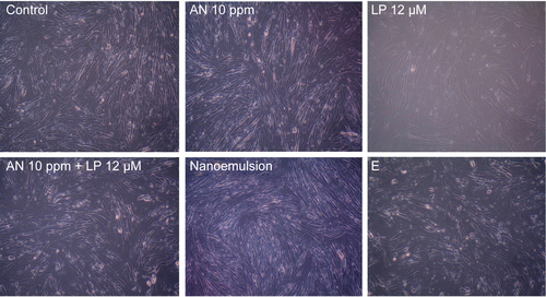
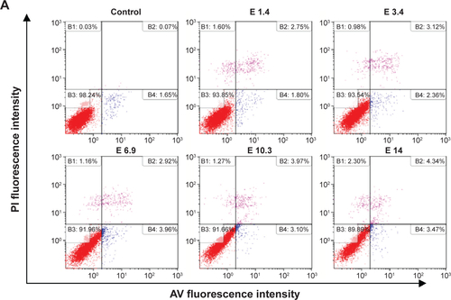
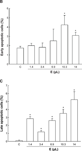
Figure S2 Effects of the blank emulsion particles on the apoptotic death of HT-29 cells.
Notes: Cells were treated with various doses of emulsions (1.4 μL/mL, 3.4 μL/mL, 6.9 μL/mL, 10.3 μL/mL, and 14 μL/mL medium) without AN and LP for 24 hours. A dose of 1.4 μL of emulsion was the same volume as the nanoemulsion containing AN at 0.16 ppm and LP at 0.4 μM. (A) Representative quarterly plots of apoptotic and necrotic cell death and (B) quantitative analysis of early apoptotic (AV-positive/PI-negative) and (C) late apoptotic cells (AV-positive/PI-positive). Apoptotic death was measured by AV and PI staining following the flow cytometric analysis. Values are presented as the mean ± SD (3 -9 independent experiments). *P<0.05, the blank nanoemulsion group compared to the control group.
Abbreviations: PI, propidium iodide; E, emulsion; C, control; AN, gold nanoparticles; LP, lycopene; AV, Annexin V; SD, standard deviation.
Acknowledgments
The authors wish to thank Mr Yen-Sheng Wu from Tzong Jao Hang’s Electron Microscope Laboratory, School of Medicine, Fu Jen Catholic University, Taipei, Taiwan for technical assistance in recording the transmission electron microscopic images.
Disclosure
The authors report no conflicts of interest in this work.