

Abstract
Fully dispersible, cationic ultrasmall (7 nm diameter) superparamagnetic iron oxide nanoparticles, exhibiting high relaxivity (178 mM−1s−1 in 0.47 T) and no acute or subchronic toxicity in Wistar rats, were studied and their suitability as contrast agents for magnetic resonance imaging and material for development of new diagnostic and treatment tools demonstrated. After intravenous injection (10 mg/kg body weight), they circulated throughout the vascular system causing no microhemorrhage or thrombus, neither inflammatory processes at the mesentery vascular bed and hepatic sinusoids (leukocyte rolling, adhesion, or migration as evaluated by intravital microscopy), but having been spontaneously concentrated in the liver, spleen, and kidneys, they caused strong negative contrast. The nanoparticles are cleared from kidneys and bladder in few days, whereas the complete elimination from liver and spleen occurred only after 4 weeks. Ex vivo studies demonstrated that cationic ultrasmall superparamagnetic iron oxide nanoparticles caused no effects on hepatic and renal enzymes dosage as well as on leukocyte count. In addition, they were readily concentrated in rat thigh by a magnet showing its potential as magnetically targeted carriers of therapeutic and diagnostic agents. Summarizing, cationic ultrasmall superparamagnetic iron oxide nanoparticles are nontoxic and efficient magnetic resonance imaging contrast agents useful as platform for the development of new materials for application in theranostics.
Supplementary materials
Comparison of rats treated with 10 mg/kg of cat-USPIOs in the presence and absence of a magnet in the left thigh
The suitability of ethylamine-functionalized cationic ultrasmall superparamagnetic iron oxide nanoparticles (cat-USPIOs) as magnetic resonance imaging (MRI) contrast agent after intravenous (IV) administration of 50 mg/kg body weight of them dispersed in 1 mL of acetate buffer (10 mM pH 4.5) containing 50 mM of NaCl was described in the manuscript. The same procedures used in those experiments were adopted here, but administering a lower dose of 10 mg/kg body weight in control (without a magnet) and experimental (with a magnet in the left thigh) groups, in order to compare the efficiency as MRI contrast and the clearance time. The MRI images were collected from about 2 hours up to 28 days after IV administration.
The contrast at liver and kidneys of control group of Wistar rats started to become brighter 14 days and 1 day () after administration, respectively, whereas the same started to happen with the experimental group after 7 days and 2 hours (). This result was confirmed by following the density of white pixels in MRI, where the experimental group exhibited higher density than the control group. This result was expected since in the controls, the total amount of cat-USPIOs was just distributed to liver, spleen, and kidneys, whereas a significant amount was retained in the thigh by a magnet in the experimental group. Considering the fact that this magnetically concentrated nanoparticles are cleared more slowly than those accumulated in the internal organs, and that all experimental conditions are identical except for the magnet, it is possible to conclude that the clearance time from the internal organs is dose-dependent. In other words, the clearance rate is dependent on the dose of cat-USPIOs.
Intravital microscopy of rat mesentery: leukocyte–endothelium interactions
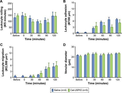
Male Wistar rats (about 200 g) were treated with saline (0.9% NaCl) and 10 mg/kg cat-USPIOs. After subcutaneous anesthesia and subsequent insertion of a cannula into the left femoral vein, a single 1 mL dose of each treatment was administered IV at an infusion rate of 500 µL/min.
Medial laparotomy was carried out and the mesentery exposed for in situ microscopic examination according to Zweifach.Citation1 The protocols described by Macedo et alCitation2 and de Lima et alCitation3 were employed for animal surgery and the maintenance of mesentery microcirculation. Data were collected before, just after, and every 30 minutes (until up to 2 hours) after injection. Three different vessels were evaluated and averaged for each animal to avoid sampling associated variability.
Injection of cat-USPIOs caused no change in the number of rolled, adhered, and migrated leukocytes (), as compared to the saline control, within 2 hours of observation. No significant difference was observed in the venular diameter (µm) measured under the same conditions and time ().
Figure S1 MRI monitoring the chest of a male Wistar rat just before (control), and 2 hours, 1, 7, 14, and 28 days after IV administration of 10 mg/kg of cat-USPIOs.
Notes: Images from (A) the control group (without a magnet) and (B) the experimental group (with a magnet in the left thigh). A yellow rectangle indicates the region where the liver and the spleen are localized while yellow circles indicate the kidneys.
Abbreviations: MRI, magnetic resonance imaging; IV, intravenous; cat-USPIOs, cationic ultrasmall superparamagnetic iron oxide nanoparticles.
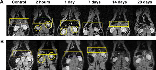
Figure S2 Effect of cat-USPIOs on leukocyte–endothelium interactions evaluated in male Wistar rat mesentery microcirculation by intravital microscopy.
Notes: Number of (A) rolled, (B) adhered, and (C) migrated leukocytes along the mesentery vessel walls before and 30–120 minutes after treatment. (D) Venular diameter (µm) measured under the same conditions and time. Values express the average data obtained from 4 to 7 animals in each group. No significant difference was observed.
Abbreviation: cat-USPIOs, cationic ultrasmall superparamagnetic iron oxide nanoparticles.
References
- DaldrupHELinkTMBlasiusSMonitoring radiation-induced changes in bone marrow histopathology with ultra-small superpara-magnetic iron oxide (USPIO)-enhanced MRIJ Magn Reson Imaging19999564365210331759
- ZweifachBWMicroscopic observations of circulation in rat mesoappendix and dog omentum; use in study of vasotropic substancesMethods Med Res1948113113918888762
- MacedoSMLourencoELBorelliPEffect of in vivo phenol or hydroquinone exposure on events related to neutrophil delivery during an inflammatory responseToxicology20062202–312613516427181
- de LimaCBTamuraEKMontero-MelendezTActions of translocator protein ligands on neutrophil adhesion and motility induced by G-protein coupled receptor signalingBiochem Biophys Res Commun2012417291892322209795
Acknowledgments
The authors would like to thank Prof Dr Edivaldo Sabadini and Dr Delmarcio Gomes da Silva for the measurement of cat-USPIOs relaxivity, as well as the financial support granted by Fundação de Amparo a Pesquisa do Estado de São Paulo (FAPESP 2009/08584-6), Conselho Nacional de Desenvolvimento Científico e Technológico (CNPq), and Petrobras. MKU and ALBS are doctoral fellows (projects 2010/50072-0 and 2011/09677-8, respectively), and SFR is a postdoctoral fellow (2011/02438-8) of FAPESP.
Disclosure
The authors report no conflicts of interest. The authors alone are responsible for the content and writing of this paper. The authors have no relevant affiliations or financial involvement with any organization or entity with a financial interest in or financial conflict with the subject matter or materials discussed in the manuscript. This includes employment, consultancies, honoraria, stock ownership or options, expert testimony, grants or patents received or pending, or royalties. No writing assistance was utilized in the production of this manuscript.