

Abstract
Periodontal regeneration is an important part of regenerative medicine, with great clinical significance; however, the effects of nanotopography on the functions of periodontal ligament (PDL) stem cells (PDLSCs) and on PDLSC sheet based periodontal regeneration have never been explored. Titania nanotubes (NTs) layered on titanium (Ti) provide a good platform to study this. In the current study, the influence of NTs of different tube size on the functions of PDLSCs was observed. Afterward, an ectopic implantation model using a Ti/cell sheets/hydroxyapatite (HA) complex was applied to study the effect of the NTs on cell sheet based periodontal regeneration. The NTs were able to enhance the initial PDLSC adhesion and spread, as well as collagen secretion. With the Ti/cell sheets/HA complex model, it was demonstrated that the PDLSC sheets were capable of regenerating the PDL tissue, when combined with bone marrow mesenchymal stem cell (BMSC) sheets and HA, without the need for extra soluble chemical cues. Simultaneously, the NTs improved the periodontal regeneration result of the ectopically implanted Ti/cell sheets/HA complex, giving rise to functionally aligned collagen fiber bundles. Specifically, much denser collagen fibers, with abundant blood vessels as well as cementum-like tissue on the Ti surface, which well-resembled the structure of natural PDL, were observed in the NT5 and NT10 sample groups. Our study provides the first evidence that the nanotopographical cues obviously influence the functions of PDLSCs and improve the PDLSC sheet based periodontal regeneration size dependently, which provides new insight to the periodontal regeneration. The Ti/cell sheets/HA complex may constitute a good model to predict the effect of biomaterials on periodontal regeneration.
Supplementary materials
Figure S1 Isolation and characterization of human PDLSCs.
Notes: (A) Single PDLSC formed cell colonies after 14 days of culture. (B) Calcified nodules stained with Alizarin Red S after 4 weeks of osteogenic induction. (C) Oil Red O–positive lipid droplets formed after 3 weeks of adipogenic induction. (D) Flow cytometry analysis on the surface markers of PDLSCs.
Abbreviation: PDLSC, periodontal ligament stem cell.
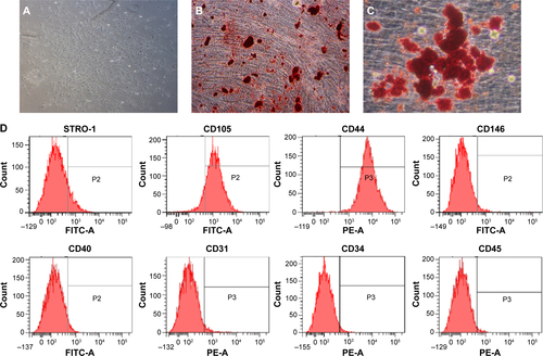
Figure S2 Isolation and characterization of human BMSCs.
Notes: (A) Single BMSC formed cell colonies after 14 days of culture. (B) Calcified nodules stained with Alizarin Red S after 4 weeks of osteogenic induction. (C) Oil Red O–positive lipid droplets formed after 3 weeks of adipogenic induction; (D) Flow cytometry analysis on the surface markers of BMSCs.
Abbreviation: BMSC, bone marrow mesenchymal stem cell.
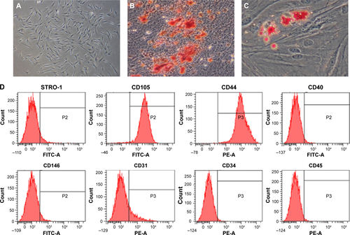
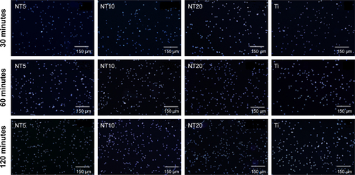
Figure S3 The initial PDLSC adhesion on the Ti samples displayed by DAPI-staining followed by observation under the fluorescence microscopy, after 30, 60, and 120 minutes of incubation.
Note: Scale bars are 150 μm.
Abbreviations: DAPI, 4′,6′-diamidino-2-phenylindole; PDLSC, periodontal ligament stem cell; Ti, titanium.
Acknowledgments
This work was supported by grants from the National Natural Science Foundation of People’s Republic of China (grant numbers 31030033, 31401255, 81470710, and 31200716), the National Major Scientific Research Program of People’s Republic of China (grant number 2010CB944800), the Foundation for the Author of National Excellent Doctoral Dissertation of People’s Republic of China (grant number 201483), the National High Technology Research and Development Program of People’s Republic of China (grant number SS2015AA020921), and the Self-Selected Topic Project of State Key Laboratory of Military Stomatology (grant number 2014ZB04). LZ Zhao also appreciates the grant from the School of Stomatology, The Fourth Military Medical University.
Disclosure
The authors report no conflicts of interest in this work.