

Abstract
For many years, novel strategies for cancer detection and treatment using nanoparticles (NPs) have been developed. Esophageal adenocarcinoma is the sixth leading cause of cancer-related deaths in Western countries, and despite recent advances in early detection and treatment, its prognosis is still very poor. This study investigated the use of fluorescent organic NPs as potential diagnostic tool in an experimental in vivo model of Barrett’s esophageal adenocarcinoma. NPs were made of modified polysaccharides loaded with [4-(dicyanomethylene)-2-methyl-6-(4-dimethylaminostyryl)-4H-pyran] (DCM), a well-known fluorescent dye. The NP periphery might or might not be decorated with ASYNYDA peptide that has an affinity for esophageal cancer cells. Non-operated and operated rats in which gastroesophageal reflux was surgically induced received both types of NPs (NP-DCM and NP-DCM-ASYNYDA) by intravenous route. Localization of mucosal NPs was assessed in vivo by confocal laser endomicroscopy, a technique which enables a “real time” and in situ visualization of the tissue at a cellular level. After injection of NP-DCM and NP-DCM-ASYNYDA, fluorescence was observed in rats affected by esophageal cancer, whereas no signal was observed in control non-operated rats, or in rats with simple esophagitis or Barrett’s esophagus mucosa. Fluorescence was observable in vivo 30 minutes after the administration of NPs. Interestingly, NP-DCM-ASYNYDA induced strong fluorescence intensity 24 hours after administration. These observations suggested that NPs could reach the tumor cells, likely by enhanced permeability and retention effect, and the peptide ASYNYDA gave them high specificity for esophageal cancer cells. Thus, the combination of NP platform and confocal laser endomicroscopy could play an important role for highlighting esophageal cancer conditions. This result supports the potential of this strategy as a targeted carrier for photoactive and bioactive molecules in esophageal cancer diagnosis and treatment.
Supplementary materials
Figure S1 Fluorescence excitation (blue line) and emission (red line) spectra of NP-DCM nanoparticles in saline buffer, and solid-state emission spectrum of the dry powder (orange line).
Notes: For emission spectra, λex =488 nm. For excitation spectra, λem =620 nm.
Abbreviations: DCM, 4-(dicyanomethylene)-2-methyl-6-(4-dimethylaminostyryl)-4H-pyran; NP, nanoparticle.
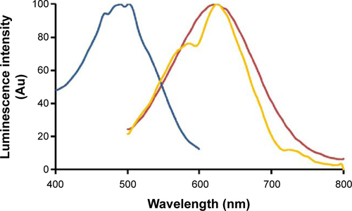
Figure S2 NP-DCM-ASYNYDA binds to human esophageal carcinoma cells.
Notes: (A) NP-DCM fluorescent signal was observed in OE19 cells, but it was qualitatively less robust compared with the fluorescent signal given by NP-DCM-ASYNYDA. (B) A very weak fluorescent signal was observed in BAR-T cells with both NPs formulations. (C) NP-DCM-ASYNYDA did not show fluorescent signal in Caco-2 cells.
Abbreviations: DCM, 4-(dicyanomethylene)-2-methyl-6-(4-dimethylaminostyryl)-4H-pyran; NP, nanoparticle.

Video S1 CLE upon intravenous administration of NP-DCM acquired after 24 hours.
Abbreviations: CLE, confocal laser endomicroscopy; DCM, 4-(dicyanomethylene)-2-methyl-6-(4-dimethylaminostyryl)-4H-pyran; NP, nanoparticles.
Video S2 CLE upon intravenous administration of NP-DCM-ASYNYDA acquired after 24 hours.
Abbreviations: CLE, confocal laser endomicroscopy; DCM, 4-(dicyanomethylene)-2-methyl-6-(4-dimethylaminostyryl)-4H-pyran; NP, nanoparticles.
Acknowledgments
The research was funded by EuroNanoMed (JTC-2011, FONDIAG project), AIRC (Italian Association for Cancer Research, project no 10761), and Ricerca Corrente IOV (2014). We are grateful to L Vidotto for technical contribution to histology. The funding agency had no role in the design and conduct of the study.
Disclosure
The authors report no conflicts of interest in this work.