

Abstract
Zinc oxide nanoparticles (ZnO NPs) have been widely used in consumer products, therapeutic agents, and drug delivery systems. However, the fate and behavior of ZnO NPs in living organisms are not well described. The purpose of this study was to develop a physiologically based pharmacokinetic model to describe the dynamic interactions of 65ZnO NPs in mice. We estimated key physicochemical parameters of partition coefficients and excretion or elimination rates, based on our previously published data quantifying the biodistributions of 10 nm and 71 nm 65ZnO NPs and zinc nitrate (65Zn(NO3)2) in various mice tissues. The time-dependent partition coefficients and excretion or elimination rates were used to construct our physiologically based pharmacokinetic model. In general, tissue partition coefficients of 65ZnO NPs were greater than those of 65Zn(NO3)2, particularly the lung partition coefficient of 10 nm 65ZnO NPs. Sensitivity analysis revealed that 71 nm 65ZnO NPs and 65Zn(NO3)2 were sensitive to excretion and elimination rates in the liver and gastrointestinal tract. Although the partition coefficient of the brain was relative low, it increased time-dependently for 65ZnO NPs and 65Zn(NO3)2. The simulation of 65Zn(NO3)2 was well fitted with the experimental data. However, replacing partition coefficients of 65ZnO NPs with those of 65Zn(NO3)2 after day 7 greatly improved the fitness of simulation, suggesting that ZnO NPs might decompose to zinc ion after day 7. In this study, we successfully established a potentially predictive dynamic model for slowly decomposed NPs. More caution is suggested for exposure to 65ZnO NPs <10 nm because those small 65ZnO NPs tend to accumulate in the body for a relatively longer time than 71 nm 65ZnO NPs and 65Zn(NO3)2 do.
Supplementary materials
Figure S1 Raman spectra of ZnO NPs.
Notes: Raman spectra of (A) 10 nm and (B) 71 nm ZnO NPs.
Abbreviation: NP, nanoparticle.
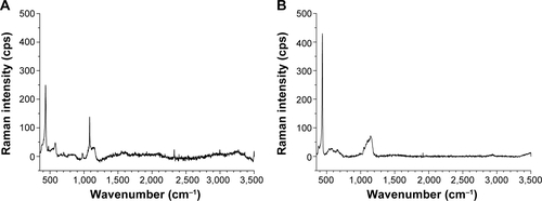
Figure S2 XRD patterns of ZnO NPs.
Notes: XRD patterns of (A) 10 nm and (B) 71 nm ZnO NPs.
Abbreviations: XRD, X-ray diffraction; NP, nanoparticle.
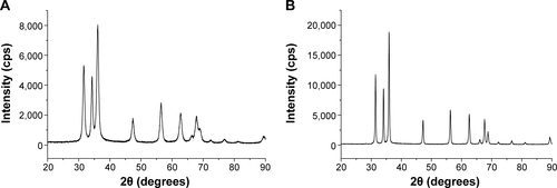
Table S1 Partition coefficients of 10 nm 65ZnO NPs in each organ
Table S2 Partition coefficients of 71 nm 65ZnO NPs in each organ
Table S3 Partition coefficients of 65Zn(NO3)2 in each organ
Table S4 Estimated excretion and elimination rates (h−1) used in PBPK model for 65ZnO NPs and 65Zn(NO3)2 in mice
Acknowledgments
This study was supported by a grant from the Ministry of Science and Technology of Taiwan (MOST 102-2932-I-400 -001-MY3). This work is a subproject of the collaborative project of the Seventh Framework Programme (FP7), funded by the European Union under project number 310715 (MOD-ENP-TOX). The authors are grateful for the technical consultation provided by Miss Jui-Ping Li and Nai-Chun Huang of the Institute of Biomedical Engineering and Nanomedicine (BN-104-PP-27) and physicochemical characterization support provided by the Institute of Bio medical Engineering and Nanomedicine Service, NHRI, Zhunan, Taiwan.
Author contributions
All authors contributed toward data analysis, drafting and critically revising the paper, and agree to be accountable for all aspects of the work.
Disclosure
The authors report no conflicts of interest in this work.