

Abstract
Nanoparticles have been widely used for nonviral gene delivery. Recently, cationic hybrid nanoparticles consisting of two different materials were suggested as a promising delivery vehicle. In this study, nanospheres with a poly(d,l-lactic-co-glycolic acid) (PLGA) core and cationic lipid shell were prepared, and the effect of cationic lipid concentrations on the properties of lipid polymer hybrid nanocarriers investigated. Lipid–polymer hybrid nanospheres (LPHNSs) were fabricated by the emulsion-solvent evaporation method using different concentrations of cationic lipids and characterized for size, surface charge, stability, plasmid DNA-binding capacity, cytotoxicity, and transfection efficiency. All LPHNSs had narrow size distribution with positive surface charges (ζ-potential 52–60 mV), and showed excellent plasmid DNA-binding capacity. In vitro cytotoxicity measurements with HEK293T, HeLa, HaCaT, and HepG2 cells also showed that LPHNSs exhibited less cytotoxicity than conventional transfection agents, such as Lipofectamine and polyethyleneimine–PLGA. As cationic lipid concentrations increased, the particle size of LPHNSs decreased while their ζ-potential increased. In addition, the in vitro transfection efficiency of LPHNSs increased as lipid concentration increased.
Supplementary materials
Figure S1 Representative dynamic light-scattering (DLS) graphs.
Notes: Influence of cationic lipid concentration on LPHNS size and surface changes. The concentration-dependent size reduction and surface-charge changes are shown in the representative DLS images.
Abbreviations: LPHNS, lipid–polymer hybrid nanosphere; PLGA, poly(d,l-lactic-co-glycolic acid); DOTAP, 1,2-di-(9Z-octadecenoyl)-3-trimethylammonium-propane (chloride salt).
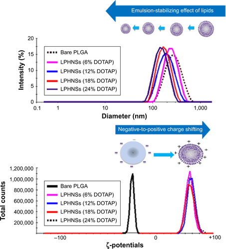
Figure S2 Microscopic analysis. EFTEM analysis of LPHNSs and LPHNS–pDNA complex. Scale bar represents 0.5 µm.
Abbreviations: EFTEM, energy-filtered transmission electron microscopy; LPHNSs, lipid–polymer hybrid nanospheres; pDNA, plasmid DNA; DOTAP, 1,2-di-(9Z-octadecenoyl)-3-trimethylammonium-propane (chloride salt); w/w, weight/weight.
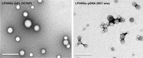
Figure S3 Effect of NS:pDNA complex concentration on cell viability.
Notes: Effect of LPHNS:pDNA complex concentrations (15, 30, 60, and 90:1 w/w) on HEK293 cell viability compared with PEI–PLGA:pDNA complex concentration (90:1 w/w). Error bars represent standard error of mean; n=3.
Abbreviations: LPHNS, lipid–polymer hybrid nanosphere; pDNA, plasmid DNA; DOTAP, 1,2-di-(9Z-octadecenoyl)-3-trimethylammonium-propane (chloride salt); PEI, polyethyleneimine; PLGA, poly(d,l-lactic-co-glycolic acid); w/w, weight/weight; NS, nanosphere.

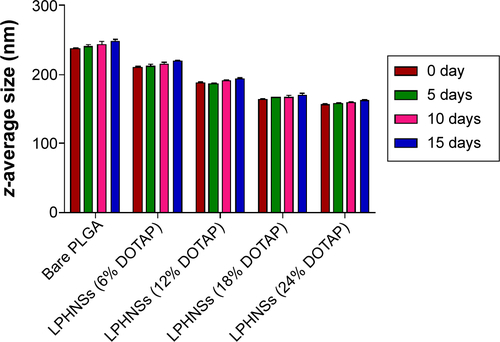
Figure S4 Short-term stability studies of LPHNSs by dynamic light scattering (DLS).
Notes: The particle sizes of the LPHNSs were used to determine the stability of LPHNSs by DLS (Malvern Nano ZS), and measurements were taken at 5-day intervals.
Abbreviations: LPHNSs, lipid–polymer hybrid nanospheres; PLGA, poly(d,l-lactic-co-glycolic acid); DOTAP, 1,2-di-(9Z-octadecenoyl)-3-trimethylammonium-propane (chloride salt).
Acknowledgments
This research was supported by the National Research Foundation of Korea (NRF), funded by the Ministry of Science, ICT and Future Planning (NRF-2013R1A2A1A09013980). This research was also supported by the Korea Health Technology R&D Project through the Korea Health Industry Development Institute (KHIDI), funded by the Ministry of Health and Welfare, Republic of Korea (HI14C3484).
Disclosure
The authors report no conflicts of interest in this work.