
Abstract
Background
Heart function tests performed with myocardial stress, or “cardiac stress tests”, may be beneficial for detection of cardiovascular disease. Women who have been diagnosed with breast cancer are more likely to develop cardiovascular diseases than the general population, in part due to the direct toxic effects of cancer treatment on the cardiovascular system. The aim of this review was to determine the utility of cardiac stress tests for the detection of cardiovascular disease after cardiotoxic breast cancer treatment.
Design
Systematic review.
Methods
Medline and Embase were searched for studies utilizing heart function tests in breast cancer survivors. Studies utilizing a cardiac stress test and a heart function test performed at rest were included to determine whether stress provided added benefit to identifying cardiac abnormalities that were undetected at rest within each study.
Results
Fourteen studies were identified. Overall, there was a benefit to utilizing stress tests over tests at rest in identifying evidence of cardiovascular disease in five studies, a possible benefit in five studies, and no benefit in four studies. The most common type of stress test was myocardial perfusion imaging, where reversible perfusion defects were detected under stress in individuals who had no defects at rest, in five of seven studies of long-term follow-up. Two studies demonstrated the benefit of stress echocardiography over resting echocardiography for detecting left ventricular dysfunction in anthracycline-treated breast cancer survivors. There was no benefit of stress cardiac magnetic resonance imaging in one study. Two studies showed a potential benefit of stress electrocardiography, whereas three others did not.
Conclusion
The use of cardiac stress with myocardial perfusion imaging and echocardiography may provide added benefit to tests performed at rest for detection of cardiovascular disease in breast cancer survivors, and merits further research.
Introduction
Adjuvant treatment for breast cancer, particularly some chemotherapy regimens and left-sided radiotherapy, are well-recognized to cause direct adverse effects on the cardiovascular system.Citation1 Thanks to progressively effective antineoplastic therapies, women diagnosed with breast cancer are increasingly living longer, which increases the age-related risk of cardiovascular disease, independent of breast cancer treatment.Citation2 An important determinant of the increased risk of cardiovascular disease with age in women is the occurrence of menopause;Citation3 chemotherapy for breast cancer will induce menopause in 21 to 100% of women, depending on age.Citation4 In light of this combination of risk factors, it is not surprising then that women who have been treated for breast cancer are more likely to die of cardiovascular disease than women who have not had breast cancer.Citation5,Citation6
The cardiovascular toxicity of breast cancer treatment can occur as an early effect, within the first year of treatment, or as a late effect, occurring years to decades after treatment.Citation7,Citation8 In both instances, individuals may often be asymptomatic before progression to overt disease.Citation9 Therefore, there is great interest in sensitive heart function tests for detection of occult cardiovascular disease, both during treatment and in long-term follow-up in this population. The utility of resting echocardiography, resting electrocardiography (ECG), resting nuclear imaging techniques, and serum cardiac biomarkers for early detection of cardiovascular disease in cancer survivors has been reviewed extensively.Citation10–Citation14 Although these diagnostic techniques do appear to have some sensitivity for cancer treatment-induced cardiac abnormalities, cardiac stress tests could be an effective technique for uncovering occult cardiovascular disease related to breast cancer treatment, including coronary artery disease and left ventricular dysfunction.
The human body is a complex, adaptive system with incredible capacity for compensation. Compensatory patterns may maintain homeostasis at rest; however, the introduction of stress challenges the stability of the system, and occult dysfunction may be unmasked. For example, the myocardium may be adequately perfused at rest, despite sub-total occlusion of a major epicardial coronary artery.Citation15 However, with the heightened metabolic demands of an increased physical workload, and corresponding myocardial blood flow requirements, there is insufficient coronary reserve, resulting in diminished myocardial perfusion and ischemia.Citation15 Similarly, stress may also uncover systolic dysfunction that may not be apparent at rest. Cardiac stress testing or, more generally, heart function tests performed under myocardial stress, may reveal occult coronary artery disease or left ventricular dysfunction that is not apparent with heart function tests performed at rest, due to compensatory mechanisms. In addition to exercise, a number of pharmacological agents or, less commonly, a cold pressor test may be used to impose myocardial stress.
The purpose of this systematic review was to determine the utility of myocardial stress in combination with heart function tests, or “cardiac stress tests”, in detection of evidence of cardiovascular disease among breast cancer survivors. To our knowledge, this is the first review to compare cardiac stress tests against tests performed at rest, for the identification of cardiovascular disease among a population treated for cancer.
Methods
Search strategy
The search strategy was formulated in consultation with a research librarian. Medline (1946 to present) and Embase (1947 to present) were searched up to 21 November 2013. The Ovid interface was used to search the Medline and Embase databases. The detailed search for Medline is provided in . The general search strategy involved (breast cancer) AND (keywords/MeSH [medical subject heading] terms related to cardiac imaging/heart function tests) AND (keywords/MeSH terms related to cardiotoxicity OR cardiotoxic cancer treatments OR related cardiac issues). The search strategy included search terms for all types of cardiac imaging techniques and heart function tests, with or without stress. The search was limited to human studies and English-language papers published in peer-reviewed journals. Experimental, observational, prospective, and retrospective study designs were included; reviews, editorials, letters to the editor, and comments were excluded. Other inclusion criteria involved: study participants diagnosed with any stage of breast cancer; any age range; at least ten study participants; and count data of heart function test results, or statistical analysis of results between a group who received cardiotoxic treatment and a group who did not.
Table 1 Ovid Medline search strategy
No studies involving breast cancer survivors were identified that compared cardiac stress tests to the gold standard for diagnosis of cardiotoxicity, endomyocardial biopsy, likely because this technique is invasive and rarely performed. Therefore, in lieu of a reference standard, an alternative technique for assessing the utility of cardiac stress tests in breast cancer survivors was required. An inclusion criterion was added that studies must include a cardiac stress test and at least one heart function test performed at rest. The outcome of the cardiac stress test was compared to that of a test at rest to determine whether the use of stress identified evidence of occult cardiovascular disease that was not detected at rest.
The reference lists of all studies meeting inclusion criteria and related studies were hand searched for additional relevant publications. Studies with relevant titles but no abstract were included in full text review. Studies with abstracts that reported the use of two or more heart function tests, without enough information to determine whether they were used under resting conditions, were included in full text review. Upon full text review, the methods section of these articles was searched to determine the use of stress with at least one of the tests, in addition to at least one test performed at rest, for comparison. If the methods section did not describe the use of stress, it was assumed that all tests were performed at rest. Authors were not contacted to acquire additional information or data.
Data collection
The following data were extracted from each study: sample size; breast cancer treatments received by study participants; dosage and timing of delivery of treatment; timing of heart function tests with respect to treatment receipt; type of each test used with stress; type of stress used; type of each test used at rest; parameters of each test measured; and results for each parameter.
Determination of utility of cardiac stress tests in detection of cardiovascular disease
The findings of the cardiac stress tests were compared to the tests at rest to determine the utility of the stress test within each individual study. Studies were labeled as “no benefit”, “possible benefit”, or “benefit”, based on whether the cardiac stress tests identified evidence of cardiovascular disease in the study participants that was not identified by the heart function tests performed at rest in that study. Where applicable, the reported thresholds for determining cardiovascular dysfunction (eg, LVEF [left ventricular ejection fraction] lower limits of normal) from each individual paper were used. In other cases, the identification of abnormalities was taken as evidence of occult cardiovascular disease (eg, identification of perfusion defects with myocardial perfusion imaging). The study was labeled “no benefit” if: 1) the cardiac stress test did not identify additional study participants with evidence of cardiovascular disease who were not identified with tests at rest, or 2) the cardiac stress tests and the tests at rest were either both statistically different or both not different between treated and non-treated groups. The study was labeled “benefit” if: 1) the cardiac stress test identified study participants with evidence of cardiovascular disease who were not identified by the techniques at rest, or 2) there was a statistically significant difference in cardiac function for the cardiac stress tests, but not for the tests performed at rest. The label “possible benefit” was assigned to studies where the cardiac stress test identified evidence of cardiovascular disease, but it could not be determined from the reported data whether the cases identified with tests performed at rest occurred in the same study participants as the cases identified with stress tests.
Results
The process for selection of papers for this review is outlined in . Fourteen studiesCitation16–Citation29 met the inclusion criteria for the review and are summarized in .
Figure 1 Preferred Reporting Items for Systematic Reviews and Meta-Analyses (PRISMA) flow diagram of study selection.
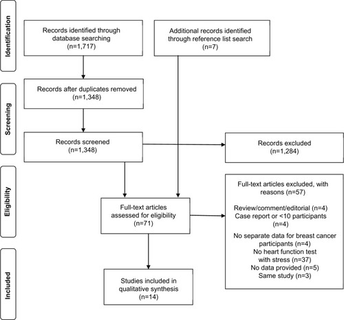
Table 2 Summary of articles using cardiac stress testing in breast cancer survivors
Utility of cardiac stress tests
Overall, five studiesCitation16,Citation22,Citation23,Citation25,Citation26 showed a benefit, fiveCitation18,Citation19,Citation24,Citation27,Citation28 showed possible benefit, and fourCitation17,Citation20,Citation21,Citation29 showed no benefit to utilizing cardiac stress tests in identifying evidence of cardiovascular disease in breast cancer survivors.
Cardiac stress test types
Heart function tests that were performed with stress in the selected studies were scintigraphy (n=5),Citation17,Citation19,Citation20,Citation28,Citation29 single photon emission computed tomography (SPECT) (n=4),Citation18,Citation22,Citation24,Citation25 echocardiography (n=2),Citation16,Citation23 cardiac magnetic resonance imaging (cMRI) (n=1),Citation21 nuclear ventriculography (n=1),Citation26 and ECG (n=5).Citation17,Citation19,Citation20,Citation22,Citation27 The utility of stress among each technique is shown in .
Figure 2 Distribution of utility (ie, “benefit”, “possible benefit”, or “no benefit”) of stress.
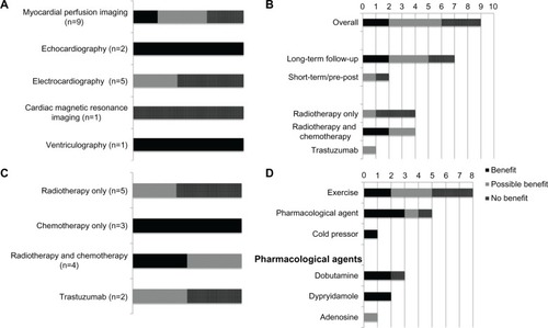
Stress myocardial perfusion imaging techniques (ie, scintigraphy or SPECT) were the most commonly used type of cardiac stress test, used in nine studies. The timing and treatment type distributions (ie, chemotherapy, radiotherapy, or trastuzumab) of the effects are shown in . The most common use of myocardial perfusion imaging was for long-term follow-up to radiotherapy, in seven studies, showing a benefitCitation22,Citation25 or possible benefitCitation18,Citation19,Citation24 in five of these. Stress echocardiography was used in two studies that both demonstrated a benefit in detecting anthracycline-induced left ventricular dysfunction.Citation16,Citation23 The timing of imaging in one study was before, during, and after (ie, before each cycle, then 2, 4, 7, and 12–18 months after) chemotherapy,Citation16 while, in the other study, imaging was a one-time assessment in long-term follow-up to chemotherapy treatment (mean follow-up of 34 months).Citation23 Stress ECG was utilized in five studies: one demonstrated a benefit,Citation22 although the benefit in this study was attributed to stress SPECT, not ECG; two demonstrated possible benefit,Citation19,Citation27 and two no benefit.Citation17,Citation20 Stress ventriculography was used in one study and demonstrated a benefit, compared to testing performed at rest;Citation26 while stress cMRI was also used in one study and failed to show a benefit, relative to resting imaging.Citation21
Breast cancer treatments
The adjuvant breast cancer treatments included in the studies were chemotherapy, radiotherapy, and a targeted, humanized, monoclonal antibody (trastuzumab). The overall utility of cardiac stress tests among the different treatment types is displayed in . Cardiac stress tests appear to be most beneficial for detecting cardiovascular disease following chemotherapy, or chemotherapy and radiotherapy combined treatment.
Method of inducing myocardial stress
Exercise was used to induce stress in eight studies,Citation17–Citation20,Citation23,Citation25,Citation27,Citation29 pharmacological agents were used in five studiesCitation16,Citation21,Citation22,Citation24,Citation25 and the cold pressor test in one study.Citation26 The utility of the use of exercise, pharmacological agents, or the cold pressor test to induce myocardial stress within the studies is displayed in . Exercise and pharmacological agents demonstrated a benefit or possible benefit in 63% and 80% of the studies they were used in to induce stress, respectively. The one study to use the cold-pressor test to induce stress demonstrated a benefit. No adverse reactions to stress were reported.
Discussion
This review demonstrated that cardiac stress testing may be beneficial in detecting evidence of cardiovascular disease in breast cancer survivors that was missed by heart function tests performed at rest. The primary findings of this review are that, in general, cardiac stress tests appear to be most beneficial in chemotherapy-treated populations (with or without radiotherapy), while, specifically, stress myocardial perfusion imaging appears to be beneficial for long-term follow-up to radiotherapy, and stress echocardiography appears to be beneficial in detecting left ventricular dysfunction following anthracycline treatment.
With an approximate 5-year survival rate of 80% or greater for all stages of breast cancer combined in North America and Europe,Citation30–Citation32 cancer is beginning to be considered a manageable disease.Citation33 Short and long-term cardiac effects of cancer treatment are significant determinants of all-cause mortality rates for cancer survivors.Citation9 As such, identification of the optimal screening strategies for cardiovascular effects of cancer therapies is a high priority.Citation9
Heart function tests, in combination with exercise or pharmacological stress, are commonly used for diagnostic and prognostic cardiac care,Citation34,Citation35 and have been used to a limited extent for long-term follow-up in childhood cancer survivors treated with chemotherapy.Citation36–Citation43 In this review, two studiesCitation16,Citation23 demonstrated the benefit of parameters of global systolic function measured via stress echocardiography in identifying left ventricular dysfunction in breast cancer survivors who have received a cardiotoxic systemic treatment. Khouri et alCitation23 demonstrated that the change in global longitudinal strain from rest to peak exercise was significantly different between anthracycline-treated breast cancer survivors and healthy controls. In addition to long-term cardiac effects, anthracyclines are also associated with acute or early cardiac effects, defined as occurring between treatment initiation and 1 year later.Citation8 Early identification of left ventricular dysfunction is critical, as it allows for earlier intervention in the form of dose reduction or initiation of therapeutic measures. Civelli et alCitation16 monitored patients for early cardiotoxicity during ongoing anthracycline chemotherapy treatment, and demonstrated the ability of dobutamine echocardiography to identify subclinical left ventricular dysfunction with a reduction in left ventricular contractile reserve (LVCR) (stress–rest LVEF) before the third chemotherapy cycle. This study also demonstrated the predictive value of LVCR for traditional diagnostic parameters of cardiotoxicity (resting LVEF decrease to <50% and ≥10% drop from baseline) at 12–18 months post-chemotherapy.
Radiation to the breast can cause ultrastructural damage to myocardial capillaries and valves, atherosclerosis in coronary arteries, valvular abnormalities, and myocardial fibrosis leading to diastolic and systolic dysfunction, pericardial inflammation, or effusion.Citation44–Citation47 There is a 1.27 excess rate ratio of heart disease in women treated with radiation for breast cancer relative to those who were not.Citation48 Mortality from coronary artery disease and risk of cardiovascular disease has decreased for breast cancer survivors diagnosed after 1980,Citation6,Citation49 although more follow-up data is need to determine long-term risk associated with more modern treatment techniques. The cardiac injury associated with radiotherapy for breast cancer is typically a “late effect”, occurring years to decades after treatment.Citation7 Long-term cardiac follow-up is suggested for breast cancer survivors who receive radiotherapy.Citation50 In this review, myocardial perfusion imaging was used in several studies of long-term follow-up to radiotherapy. To perform this type of imaging, a radioactive tracer substance is injected, and its accumulation in the myocardium, as detected by a gamma camera, is considered proportional to the myocardial blood flow.Citation15 A perfusion defect can be identified on the image produced as an area of decreased perfusion or injured heart tissue resulting from a diseased coronary artery. A perfusion defect that is detected at rest and does not change during stress imaging is considered fixed or irreversible, and suggests non-viable myocardium; while a perfusion defect that is absent at rest but detected during stress is considered reversible, and indicates ischemic myocardium.Citation51 Five studiesCitation18,Citation19,Citation22,Citation24,Citation28 in this review identified reversible defects with stress tests that would not have been discovered with tests performed at rest alone in breast cancer survivors treated with left-sided radiotherapy and other systemic treatments. This suggests that stress myocardial perfusion imaging could be an effective method of long-term follow-up for detection of coronary artery disease in radiation-treated breast cancer survivors. Gyenes et alCitation20 reported several fixed perfusion defects, but no reversible defects at 1 year post-radiotherapy. An incidence of fixed perfusion defects of 60% has been reported 6 months following radiotherapy for left-sided breast cancer.Citation52 It may be that the utility of stress myocardial perfusion imaging for screening purposes is limited to long-term follow-up only.
It is unclear from this review whether stress ECG is beneficial for identifying cardiovascular disease in breast cancer survivors. Two studies demonstrated possible benefit. In one of these,Citation27 ECG was used under stress and at rest, and stress did appear to better detect specific abnormalities, namely ectopic beats and, not surprisingly, minor ST depressions that developed 6 months after radiotherapy to the sternum. However, numerous arrthymias and more T-wave items were detected only at rest and there was unknown overlap between abnormalities detected under each condition. A similar case of unknown overlap between abnormalities detected with stress ECG and resting echocardiography occurred in the other study of long-term follow-up.Citation19 The lack of ability to detect occult cardiovascular disease may be attributed to the temporally late occurrence of electrocardiographic changes in the cascade of occurrences in the development of ischemia.Citation53
Baseline and periodic monitoring of cardiac function is recommended in individuals treated with trastuzumab.Citation50 Haykowsky et alCitation21 used cMRI, at rest and with dobutamine stress, at baseline and 4 months into trastuzumab treatment. This study did not demonstrate the utility of cardiac stress, as the cMRI findings were the same at rest and with stress. However, left ventricular contractile reserve was not reported, which is a more sensitive measure of early left ventricular dysfunction, as evidenced by Civelli et al.Citation16 Kirthi et alCitation28 used scintigraphy at rest and with stress, in addition to resting cMRI, following trastuzumab treatment. One additional case of cardiovascular dysfunction was identified with stress scintigraphy, compared against scintigraphy at rest, demonstrating a benefit for this technique alone; however, cMRI at rest also identified four cases of cardiovascular dysfunction.Citation28
Myocardial stress can be induced by exercise, a number of pharmacological agents, or a cold pressor test. Pharmacological-induced stress minimizes factors such as movement artifact that may degrade cardiac imaging quality and decrease accuracy, but incurs the additional cost of the pharmacological agent.Citation53 The cold pressor test would likely carry similar advantages, but does not incur additional costs. However, the clinical significance of a cold pressor-induced change in cardiovascular function is unclear. Dynamic exercise tends to be the preferred method to induce myocardial stress, because it is less expensive, perceived to be safer, and the equipment (ie, cycle ergometer or treadmill) is widely available in clinical settings.Citation54 Exercise stress also provides additional ancillary prognostic, diagnostic, and therapeutic information, such as peak oxygen consumption, heart rate, and blood pressure response, and variables required for exercise prescription. However, the inability to reach maximal exercise capacity will limit the sensitivity of stress tests, in which case pharmacological stress is a suitable alternative.Citation54
There is additional cost involved in cardiac stress tests with either exercise or pharmacological stress. For example, stress echocardiography is approximately double the cost of resting echocardiography,Citation55 but is reported to potentially enhance overall cost-effectiveness for diagnosis and management of suspected cardiovascular disease.Citation56 Stress myocardial perfusion imaging is often performed in addition to imaging at rest; however, the use of stress myocardial perfusion imaging alone would decrease the cost, radiation exposure, and time of testing, and is not associated with differences in prognosis or mortality outcomes.Citation57
In ten of the 14 studies in this review, evidence of cardiovascular disease was missed by heart function tests performed at rest, but detectable with cardiac stress tests. This information is relevant to those involved in the treatment and care of breast cancer survivors, including family physicians, medical and radiation oncologists, and cardiologists. Lack of change in the most common oncological clinical parameter of cardiac function, resting LVEF, is often equated to a lack of cardiotoxicity, despite convincing evidence in the literature that substantial myocyte damage can occur before a change in LVEF results.Citation58 However, when exercise stress is used to evaluate change in LVEF, sensitivity for detection of anthracycline-induced left ventricular dysfunction in cancer survivors is dramatically increased.Citation59 Similarly, in this review, the sensitivity of LVEF, perfusion defects, and global longitudinal strain is increased by cardiac stress tests.Citation16,Citation18,Citation19,Citation22–Citation26 This preliminary evidence that cardiac stress tests may be more sensitive for detection of evidence of cardiovascular disease and may also be used to inform the type of heart function tests utilized to address cardiovascular safety in future clinical trials of antineoplastic therapies.
The parameters of cardiac stress tests that were shown to be beneficial in this review were perfusion defects, measured during stress myocardial perfusion imaging, as well as global longitudinal strain reserve and LVCR measured during stress echocardiography. These parameters are associated with negative clinical outcomes. Perfusion defects are reported to be a significant predictor of cardiac events and cardiac death in cancer survivors, and those with reversible perfusion defects (ie, those detected only with stress tests) have a lower probability of 3-year cardiac event-free survival than those with fixed perfusion defects.Citation60 Further investigation into the utility of stress myocardial perfusion imaging as a cardiovascular surveillance tool for radiation-treated breast cancer survivors is warranted, given the demonstrated benefit of stress tests in identifying prognostically valuable reversible perfusion defects.
The parameters assessed by stress echocardiography that were beneficial in identifying cardiovascular disease in breast cancer survivors in this review were peak LVEF,Citation16 LVCR,Citation16 and global longitudinal strain.Citation23 Strain imaging is an advanced echocardiographic technique that is new to evaluation with stress.Citation61 Strain or strain rate has prognostic and diagnostic value in cardiac disease and surgery,Citation61 but its value in the evaluation of anthracycline-induced left ventricular dysfunction is unknown. Additionally, the absence of LVCR (ie, no change or a decrease in LVEF when stress is applied) is associated with decreased myocardial blood flow and oxygen consumption, at rest and under stress,Citation62 a maladaptive response predictive of heart failure patient survival.Citation63 Although there has been limited research on the use of stress echocardiography for detection of chemotherapy-associated left ventricular dysfunction in breast cancer survivors, the two initial studiesCitation16,Citation23 highlight the potential for this technique to be used both for prognosis and follow-up in this population. Future studies are required, investigating the use of sensitive stress echocardiographic parameters, including LVCR, strain, and diastolic function for identification of left ventricular dysfunction associated with anthracyclines and other chemotherapeutic agents.
This review is subject to some limitations. Firstly, no studies involving breast cancer survivors were identified that compared a cardiac stress test to the gold standard for diagnosis of cardiotoxicity, endomyocardial biopsy, which required that a nontraditional method of synthesizing results be developed. Additionally, there was considerable variability in the format of data reported within each study, which made data synthesis difficult. The small number of available studies that fit the inclusion criteria, as well as the small sample sizes (8 to 38) within these studies limit the interpretation and generalization of the results. Lastly, the findings of this review only provide information as to whether or not cardiac stress tests are beneficial, but do allow for specific recommendations on the optimal timing of this technique with specific breast cancer treatments. However, despite the shortcomings of this review, it demonstrates that there appears to be some benefit to the use of cardiac stress testing of breast cancer survivors to uncover occult cardiovascular disease, and that this technique merits further investigation.
In summary, this is the first review to evaluate the utility of cardiac stress tests in breast cancer survivors. Although few studies of breast cancer survivors have employed cardiac stress tests to monitor cardiac function, and no studies have compared stress tests to the reference standard, the available evidence suggests that myocardial stress may be beneficial in identifying evidence of cardiovascular disease in breast cancer survivors. In particular, stress myocardial perfusion imaging appears to be beneficial in long-term follow-up to left-sided radiotherapy for breast cancer, and stress echocardiography may be beneficial in early and late follow-up to anthracycline treatment. Further research is needed to determine the optimal heart function tests and parameters to be measured during a stress test, the optimal timing of stress tests with different treatments, and the cost-to-benefit ratio of performing a stress test over a heart function test at rest in this population.
Disclosure
The authors declare that there is no conflict of interest.
References
- BirdBRSwainSMCardiac toxicity in breast cancer survivors: Review of potential cardiac problemsClin Cancer Res2008141142418172247
- EwerMSGlückSA woman’s heart: The impact of adjuvant endocrine therapy on cardiovascular healthCancer200911591813182619235248
- World Heart Federation Cardiovascular disease risk factorsGenevaWorld Heart Federation2013
- MintonSEMunsterPNChemotherapy-induced amenorrhea and fertility in women undergoing adjuvant treatment for breast cancerCancer Control20029646647212514564
- RiihimakiMThomsenHBrandtASundquistJHemminkiKDeath causes in breast cancer patientsAnn Oncol201223360461021586686
- HooningMJBotmaAAlemanBMLong-term risk of cardiovascular disease in 10-year survivors of breast cancerJ Natl Cancer Inst200799536537517341728
- MarksLBYuXProsnitzRGThe incidence and functional consequences of RT-associated cardiac perfusion defectsInt J Radiat Oncol Biol Phys200563121422316111592
- JonesRLSwantonCEwerMSAnthracycline cardiotoxicityExpert Opin Drug Saf20065679180917044806
- CarverJRShapiroCLNgAAmerican Society of Clinical Oncology clinical evidence review on the ongoing care of adult cancer survivors: cardiac and pulmonary late effectsJ Clin Oncol200725253991400817577017
- DolciADominiciRCardinaleDSandriMTPanteghiniMBiochemical markers for prediction of chemotherapy-induced cardiotoxicity: systematic review of the literature and recommendations for useAm J Clin Pathol2008130568869518854260
- Fallah-RadNWalkerJRWassefAThe utility of cardiac biomarkers, tissue velocity and strain imaging, and cardiac magnetic resonance imaging in predicting early left ventricular dysfunction in patients with human epidermal growth factor receptor II-positive breast cancer treated with adjuvant trastuzumab therapyJ Am Coll Cardiol201157222263227021616287
- GanzWISridharKSGanzSSReview of tests for monitoring doxorubicin-induced cardiomyopathyOncology19965364614708960141
- GoethalsIDe WinterODe BondtPDe SutterJDierckxRVan De WieleCThe clinical value of nuclear medicine in the assessment of irradiation-induced and anthracycline-associated cardiac damageAnn Oncol20021391331133912196357
- PlanaJCChemotherapy and the heartRev Esp Cardiol (Engl Ed)2011645409415
- ManoucheriMKarunaratneHBThe role of imaging techniques in stress testingPrim Care19942135355559132757
- CivelliMCardinaleDMartinoniAEarly reduction in left ventricular contractile reserve detected by dobutamine stress echo predicts high-dose chemotherapy-induced cardiac toxicityInt J Cardiol2006111112012616242796
- CowenDGonzague-CasbiancaLBrenot-RossiIThallium-201 perfusion scintigraphy in the evaluation of late myocardial damage in left-side breast cancer treated with adjuvant radiotherapyInt J Radiat Oncol Biol Phys19984148098159652842
- GalluciGCoccaroMStortoGThe clinical impact of a cardiologic follow-up in breast cancer aurvivors: an observational studyInt J Immunopathol Pharmacol20102341221122721244771
- GyenesGFornanderTCarlensPRutqvistLEMorbidity of ischemic heart disease in early breast cancer 15–20 years after adjuvant radiotherapyInt J Radiat Oncol Biol Phys1994285123512418175411
- GyenesGFornanderTCarlensPDetection of radiation-induced myocardial damage by technetium-99m sestamibi scintigraphyEur J Nucl Med19972432862929143466
- HaykowskyMJMackeyJRThompsonRBJonesLWPatersonDIAdjuvant trastuzumab induces ventricular remodeling despite aerobic exercise trainingClin Cancer Res200915154963496719622583
- HøjrisISandNPAndersenJMyocardial perfusion imaging in breast cancer patients treated with or without post-mastectomy radiotherapyRadiother Oncol200055216317210799728
- KhouriMGHornsbyWEVelazquezEJJonesLWDouglasPSExercise stress testing with strain echocardiography is superior to resting echocardiography in identifying doxorubicin-induced preclinical LV dysfunction in breast cancer patientsCirculation2011124A16399
- SeddonBCookAGothardLDetection of defects in myocardial perfusion imaging in patients with early breast cancer treated with radiotherapyRadiother Oncol2002641536312208576
- SiokaCExarchopoulosTTasiouIMyocardial perfusion imaging with 99mTc-tetrofosmin SPECT in breast cancer patients that received postoperative radiotherapy: a case-control studyRadiat Oncol2011615122067743
- CassidyJMerrickMVSmythJFLeonardRCCardiotoxicity of mitozantrone assessed by stress and resting nuclear ventriculographyEur J Cancer Clin Oncol19882459359383169098
- LindahlJStrenderLELarssonLEUnsgaardAElectrocardiographic changes after radiation therapy for carcinoma of the breast. Incidence and functional significanceActa Radiol Oncol19832264334406328873
- KirthiVSchultzCDaviesSWCardiac imaging of trastuzumab-induced cardiomyopathyEur J Heart Fail Suppl201110Suppl 1S18
- GustavssonABendahlPOCwikielMEskilssonJThapperKLPahlmONo serious late cardiac effects after adjuvant radiotherapy following mastectomy in premenopausal women with early breast cancerInt J Radiat Oncol Biol Phys199943474575410098429
- SantMAllemaniCCapocacciaRStage at diagnosis is a key explanation of differences in breast cancer survival across EuropeInt J Cancer2003106341642212845683
- Canadian Cancer Society’s Steering Committee on Cancer StatisticsCanadian Cancer Statistics 2012TorontoCanadian Cancer Society2012
- American Cancer SocietyCancer Facts and Figures 20132013 edAtlantaAmerican Cancer Society2013
- YehETTongATLenihanDJCardiovascular complications of cancer therapy: diagnosis, pathogenesis, and managementCirculation2004109253122313115226229
- CorreaCRDasIJLittHIAssociation between tangential beam treatment parameters and cardiac abnormalities after definitive radiation treatment for left-sided breast cancerInt J Radiat Oncol Biol Phys200872250851618339489
- KohliPGulatiMExercise stress testing in women: going back to the basicsCirculation2010122242570258021156655
- De WolfDSuysBMaurusRDobutamine stress echocardiography in the evaluation of late anthracycline cardiotoxicity in childhood cancer survivorsPediatr Res19963935045128929873
- ElblLHrstkovaHChaloupkaVNovotnyJMichalekJThe evaluation of left ventricular function in childhood cancer survivors by pharmacological stress echocardiographyNeoplasma200350319119712937852
- Guimaraes-FilhoFVTanDMBragaJCRodriguesAWaibPHMatsubaraBBVentricular systolic reserve in asymptomatic children previously treated with low doses of anthracyclines: a longitudinal, prospective exercise echocardiography studyPediatr Blood Cancer201159354855222970439
- JarfeltMKujacicVHolmgrenDBjarnasonRLanneringBExercise echocardiography reveals subclinical cardiac dysfunction in young adult survivors of childhood acute lymphoblastic leukemiaPediatr Blood Cancer200749683584017610264
- KlewerSEGoldbergSJDonnersteinRLBergRAHutterJJJrDobutamine stress echocardiography: a sensitive indicator of diminished myocardial function in asymptomatic doxorubicin-treated long-term survivors of childhood cancerJ Am Coll Cardiol19921923944011732369
- SieswerdaEKremerLCVidmarSExercise echocardiography in asymptomatic survivors of childhood cancer treated with anthracyclines: a prospective follow-up studyPediatr Blood Cancer201054457958420014238
- LanzariniLBossiGLaudisaMLKlersyCAricòMLack of clinically significant cardiac dysfunction during intermediate dobutamine doses in long-term childhood cancer survivors exposed to anthracyclinesAm Heart J2000140231532310925349
- YildirimASedef TunaogluFPinarliFGEarly diagnosis of anthracycline toxicity in asymptomatic long-term survivors: dobutamine stress echocardiography and tissue Doppler velocities in normal and abnormal myocardial wall motionEur J Echocardiogr2010111081482220562426
- BeckmanJAThakoreAKalinowskiBHHarrisJRCreagerMARadiation therapy impairs endothelium-dependent vasodilation in humansJ Am Coll Cardiol200137376176511693749
- CornBWTrockBJGoodmanRLIrradiation-related ischemic heart diseaseJ Clin Oncol1990847417502179483
- GayaAMAshfordRFCardiac complications of radiation therapyClin Oncol (R Coll Radiol)200517315315915900998
- VirmaniRFarbACarterAJJonesRMPathology of radiation-induced coronary artery disease in human and pigCardiovasc Radiat Med1999119810111272363
- ClarkeMCollinsRDarbySEffects of radiotherapy and of differences in the extent of surgery for early breast cancer on local recurrence and 15-year survival: an overview of the randomised trialsLancet200536695032087210616360786
- GiordanoSHKuoYFFreemanJLBuchholzTAHortobagyiGNGoodwinJSRisk of cardiac death after adjuvant radiotherapy for breast cancerJ Natl Cancer Inst200597641942415770005
- BovelliDPlataniotisGRoilaFESMO Guidelines Working GroupCardiotoxicity of chemotherapeutic agents and radiotherapy-related heart disease: ESMO Clinical Practice GuidelinesAnn Oncol201021Suppl 5v277v28220555097
- BaileyIKGriffithLSRouleauJStraussWPittBThallium-201 myocardial perfusion imaging at rest and during exercise. Comparative sensitivity to electrocardiography in coronary artery diseaseCirculation19775517987830222
- HardenberghPHMunleyMTBentelGCCardiac perfusion changes in patients treated for breast cancer with radiation therapy and doxorubicin: preliminary resultsInt J Radiat Oncol Biol Phys20014941023102811240243
- SicariRNihoyannopoulosPEvangelistaAStress Echocardiography Expert Consensus Statement – Executive Summary: European Association of Echocardiography (EAE) (a registered branch of the ESC)Eur Heart J200930327828919001473
- CerqueiraMDPharmacologic stress versus maximal-exercise stress for perfusion imaging: which, when, and why?J Nucl Cardiol199636 Pt 2S10S148989681
- PicanoEEconomic and biological costs of cardiac imagingCardiovasc Ultrasound200531315916702
- MarwickTHShawLCaseCVaseyCThomasJDClinical and economic impact of exercise electrocardiography and exercise echocardiography in clinical practiceEur Heart J200324121153116312804930
- ChangSMNabiFXuJRazaUMahmarianJJNormal stress-only versus standard stress/rest myocardial perfusion imagingJ Am Coll Cardiol201055322123019913381
- EwerMSLenihanDJLeft ventricular ejection fraction and cardiotoxicity: is our ear really to the ground?J Clin Oncol20082681201120318227525
- McKillopJHBristowMRGorisMLBillinghamMEBockemuehlKSensitivity and specificity of radionuclide ejection fractions in doxorubicin cardiotoxicityAm Heart J19831065 Pt 1104810566637763
- ChandraSLenihanDJWeiWYusufSWTongATMyocardial perfusion imaging and cardiovascular outcomes in a cancer populationTex Heart Inst J200936320521319568389
- ArgyleRARaySGStress and strain: double trouble or useful tool?Eur J Echocardiogr200910671672219525297
- LeeHHDávila-RománVGLudbrookPADependency of contractile reserve on myocardial blood flow: implications for the assessment of myocardial viability with dobutamine stress echocardiographyCirculation1997969288428919386153
- RamahiTDobutamine-induced augmentation of left ventricular ejection fraction predicts survival of heart failure patients with severe non-ischaemic cardiomyopathyEur Heart J2001221084985611350094