
Abstract
A 3-year-old boy was referred with suspected leukocoria in the right eye, detected in all smartphone photographs taken by his parents. His medical and family history was unremarkable. The visual acuity was 20/20 in both eyes. Eye examination revealed full motility and normal pupils. The ocular fundi and ultrasonography appeared normal. The child was looking to the left side in his photographs, away from the camera, and illuminating the nasal retina. In this circumstance, the optic nerve head acts as a diffuse reflector, reflecting the light out of the eye through the pupil. In the case of normal clinical findings in a child presenting leukocoria in smartphone photographs (photoleukocoria), the ophthalmologist should suspect the possibility of the described phenomenon avoiding other studies.
Introduction
Leukocoria is the medical term for the white eye reflex common in several childhood eye diseases affecting any of the structures that form the visual axis, including the cornea, the lens, the vitreous, and the retina.Citation1 The majority of these conditions are significant for visual impairment and other conditions, as retinoblastoma can be life-threatening.Citation2–Citation5 Leukocoria, which requires urgent evaluation by an ophthalmologist, represents a great challenge in ophthalmology.Citation4,Citation6 Leukocoria can be detectable through simple flash photographyCitation7,Citation8 and the popularization of smartphone cameras enables parents to look for early diagnosis, seeing a white eye instead of the typical red eye in photographs, a phenomenon known as photoleukocoria.Citation9 Any process that prevents the flash light of a camera from reaching the retina will produce photoleukosis.Citation4 We report a case of a 3-year-old boy who presented with leukocoria in the right eye, noted in all his smartphone photographs, but with normal ocular examination.
Clinical case
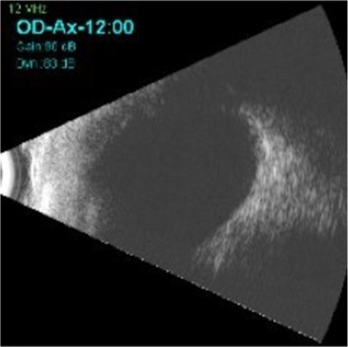
A 3-year-old male infant with his alarmed parents were referred to our emergency department for a 1-month history of leukocoria in the right eye, which was detected in all smartphone photographs taken at different times (). Birth history: 36-week gestation and normal vaginal delivery. His mother denied smoking, drug use, and alcohol intake during pregnancy. His parents had no specific concerns on history. On examination, his visual acuity was 20/20 in both eyes without glasses. No significant finding was observed on anterior segment examination. Ocular alignment and motility were normal. No nystagmus was observed. He had no afferent pupillary defect. Dilated fundus examination appeared normal with a clear vitreous and no masses in the retina. The macula showed a normal reflex, and the disc margins were distinct (). B-scan ultrasonography on the patient did not show any underlying retinal pathology (). The child was scheduled for a 6-month review and, at the follow-up visit, his vision was 20/20 in both eyes with clear ocular media and normal fundi.
Figure 1 Four different smartphone photographs. In all pictures the child is not fixing directly on the camera, which resulted in a white pupillary reflex. Leukocoria is due to reflection off the optic nerve head.
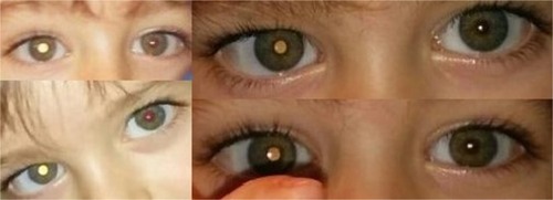
Figure 2 Color fundus photograph composition of this patient’s right eye. Optic nerve, vessels, and macula are normal. Notice the retinal nerve fiber layer, especially detectable around the fovea and vasculature, a normal finding in children.
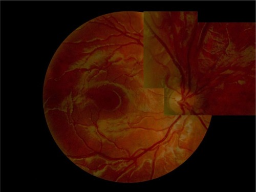
Figure 3 B-scan ultrasonography confirming normal posterior pole of the right eye.
Discussion
Leukocoria, from the greek “leucos” (white) and “korê” (pupil), means “white pupil” and describes the clinical finding of a white pupillary reflex.Citation3,Citation4,Citation10,Citation11 Several eye conditions can cause white pupil as a first sign, such as congenital cataract, retinoblastoma, retinopathy of prematurity, nematode endophthalmitis, persistent hyperplastic primary vitreous, Coats’ disease, ocular toxocariasis, retinal hamartomas, severe refractive error, organized vitreous hemorrhage, ocular albinism, myelinated fibers, and the presenting sign of abusive head trauma.Citation2–Citation4,Citation11 A white pupil is often noticed by parents or relatives on flash photography, this is the first reason for consultation, as in our patient, where a consistent leukocoria appeared in the right eye, similar to a cat’s eye reflection. The presence of an apparent leukocoria in a normal eye in a photograph taken from a temporal side has been reported previously.Citation12,Citation13 This finding can occur repeatedly in the same patient, but always in one eye only. In our patient, the apparent leukocoria was always evident in the right eye. This phenomenon has been reported when photographs are taken with an amateur camera with flash coaxially to the camera lens in eyes with undilated pupils and when the child’s eye is turned toward the nose.Citation12,Citation13 The retina, at the point of the optic disc, contains no rod or cone cells, so it cannot process light. When a camera flash hits the optic disc directly, light is reflected back, causing the pupil to appear white, even though the eye is healthy. This reflection most often occurs when the child’s eye is turned about 15° toward the nose and the flash light hits the optic disc directly.Citation12,Citation13 In our child, each picture (shown in ) was looking in the opposite direction: right leukocoria was evident in left gaze. Each photograph was an outdoor photo taken with a smartphone camera at different times and at different distances of the camera and was fixating off axis. Marshall and GoleCitation12 reported an observational case series of three children who presented with unilateral leukocoria seen in flash photographs and normal ocular exploration. The photographs were all found to be approximately 15° off axis, with the leukocoria seen in the eyes where the flash illuminated the nasal retina. This is known as photoleukocoria and it is normal. Batra et alCitation13 described seven children referred due to unilateral leukocoria detected in amateur photographs. Each child had a normal ocular examination. In keeping with the findings reported in our patient, leukocoria was unilateral and in the eye in which the nasal retina was illuminated. They consider that leukocoria in normal eyes detected by flash photography only occurs in off-axis fixation.Citation13 Different camera angles reflect light differently and can sometimes show a false leukocoria, especially in children looking directly at the camera. For reasons unknown, photos taken with smartphones can also create sporadic false positives in the same eye. As smartphones frequently capture false leukocoria, they are not the best tool for red reflex screening photos. However, false leukocoria can only be confirmed by an ophthalmologist through a red reflex eye exam. All children with newly discovered leukocoria should be referred to an ophthalmologist to exclude life- or sight-threatening diseases.
Acknowledgments
The authors confirm that written informed consent has been provided by the patient’s parents to have the case details and any accompanying images published.
Disclosure
The authors report no conflicts of interest in this work.
References
- DamascoVCDireDJA child with leukocoriaPediatr Emerg Care201127121170117422158277
- TuliSYGiordanoBPKellyMFillippsDTuliSSNewborn with an absent red reflexJ Pediatr Health Care2013271515522197583
- HaiderSQureshiWAliALeukocoria in childrenJ Pediatr Ophthalmol Strabismus200845317918018524199
- McLaughlinCLevinAVThe red reflexPediatr Emerg Care200622213714016481935
- PatelNSalchowDJMaterinMDifferentials and approach to leukocoriaConn Med201377313314023589950
- AbramsonDHFrankCMSusmanMWhalenMPDunkelIJBoydNWIIIPresenting signs of retinoblastomaJ Pediatr19981323 Pt 15055089544909
- MorganKSKennemerJCOff-axis photorefractive eye screening in childrenJ Cataract Refract Surg1997234234289159688
- TongPY1BassinREEnke-MiyazakiEScreening for amblyopia in preverbal children with photoscreening photographs: sensitivity and specificity of the MTI photoscreenerOphthalmology200010791623162910964818
- RecchiaFMRetinoblastomaHoACBrownGCArch McNamaraJRecchiaFMRegilloCDVanderJFRetina Color Atlas & Synopsis of Clinical OphthalmologyNew York, NYMcGraw-Hill2003180182
- SimonJWKawPCommonly missed diagnoses in the childhood eye examinationAm Fam Physician200164462362811529261
- PatelNSalchowDJMaterinMDifferentials and approach to leukocoriaConn Med201377313314023589950
- MarshallJGoleGAUnilateral leukocoria in off axis flash photographs of normal eyesAm J Ophthalmol2003135570971112719083
- BatraRRoweFRowlandsANoonanCUnilateral leucocoria in clinically normal eyesBr J Ophthalmol200993455655719321484