
Abstract
Giant ureteral calculi are defined as stones greater than 5 cm in length or circumference. These giant calculi can cause blockage of the ureter, dilation of the kidney and also decreased kidney function if not treated in time. The patient in this report presented with complaints of bilateral episodic pain of the bilateral lumbar region. Kidney, ureter and bladder (KUB) X-ray test showed a large bilateral ureteral stone about 14 cm in length and 106 g weight in the left ureter and 3 cm longitudinal diameter in the right ureter and also a staghorn stone in the left upper collecting system. Thereafter, the ureteric calculi were managed successfully using the combination of open and endoscopic techniques.
Introduction
Ureteral stones are a common complaint in primary care centers. Renal colic and hematuria are the classic symptoms, while abdominal or flank pain, urinary urgency, nausea, frequency and difficulty of urinating, testicular or penile pain are atypical symptoms of the ureteral stones.Citation1 Eighty percent of the stones are composed of calcium oxalate or calcium phosphate. Others may be Struvite (magnesium ammonium phosphate), uric acid, and cysteine stones.Citation2
The likelihood of spontaneous passage of the ureteral stones is associated with both location (proximal, mid, and distal ureter) and size of the stone.Citation3 Ureteric calculi are usually small, but can grow larger in size gradually. Two factors exist that increase the stone diameters: first, stone impaction and aggregation of crystals, and the second cause is distal ureteral obstruction.Citation4 If stones grow to sufficient size of 3 mm or more, they can cause blockage of the ureter which is the main cause of clinical symptoms.Citation5 Ureter blockage can also lead to dilation of the kidney and decreased kidney function.Citation6 The term giant ureteric calculus has been used for ureteric stones, which exceed 5 cm in length, circumference or weighing more than 50 g.Citation7
Based on the prior studies, the spontaneous passage rate as a function of stone size was 87% for stones 1 mm in diameter; 76% for stones 2–4 mm; 60% for stones 5–7 mm; 48% for stones 7–9 mm; and 25% for stones larger than 9 mm.Citation8,Citation9 In addition, spontaneous passage rate was more likely to be for distal and ureterovesical junction stones than stones in the proximal ureter or mid-ureteral stones.Citation9
In this study, we report a case of large bilateral ureteral stones with 14 cm length and 106 g weight in the left side and 3 cm longitudinal diameter in the right side with a complete staghorn stone in the left upper collecting system.
Case report
A 32-year-old male patient presented on March 14, 2015 with complaints of bilateral episodic pain of the bilateral lumbar region for 6 months. The history of his illness indicated that the patient used opium to reduce his pain. No significant medical or family history was mentioned and the laboratory findings were as follows: Hemoglobin: 14.6 g/dL, Blood urea nitrogen: 18 mg/dL, Creatinine: 1.3 mg/dL, Na: 142 m mol/L, K: 3.9 mmol/L, serum calcium: 9 mg/dL, and serum phosphorus: 3.5 mg/dL. Results of his physical examination on admission were unremarkable.
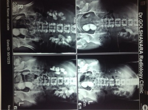
According to the clinical symptoms, the kidney, ureter and bladder (KUB) X-ray test was ordered. Findings illustrated a huge left distal ureteral calculus with 14 cm length and 106 g weight, a large right ureteral calculus with 3 cm longitudinal diameter and also an opaque staghorn stone in the left kidney (). The total weight of these stones was more than 300 g. Although excretion was reduced slightly in the right kidney, his overall renal function was normal and acceptable excretion was observed on the left side.
Figure 1 Kidney, ureter and bladder X-ray showing bilateral ureteric calculi; a huge distal ureteral calculus and an opaque staghorn stone in the left side and also a large right ureteral calculus.
We employed the combination of open and endoscopic technique for treatment of this special case of ureteral stone. The patient underwent right side transurethral lithotripsy (TUL) and left open ureterolithotomy through the Gibson incision to remove the giant uretric calculus. Longitudinal incision was performed on the ureteral wall to remove the stone from the left ureter and the stone was extracted with difficulty due to its angle and impaction to the ureteral wall. Thereafter, the distal part of the left ureter was resected due to distal stenosis and then uretroscopy was performed through the resected ureter with advancement to the proximal part of the ureter; as we reached to the renal pelvis stone, it was fragmented. In fact, the retrograde intrarenal surgery (RIRS) was performed for three main goals: reducing the bulk of the stone, applying ureteral stents to ensure the patency of the ureter and facilitating percutaneous nephrolithotomy (PCNL) for the next surgery.
Two weeks after the first surgery, in the second session, PCNL was conducted to remove the remaining kidney stones and then the patient was transferred to the ward for subsequent observation and management. The patient was discharged a few days later.
Discussion
To the best of our knowledge, only 13 case reports of ureteral calculi measuring 12 cm or more have been previously published. Therefore, giant ureteric stones are rare. Another rarity of its incidence was due to the normal renal function of the patient, whereas in most cases, the kidney above the stone is functionless.Citation7,Citation10
Although the longest stone was reported by Taylor in 1934 which was 21.5 cm in length, the stone reported in the present article was one of the largest ureteral calculi so far reported in the world ().Citation11 Before the present document, Mayer indicated the largest ureteral stone (11 × 5.5 × 5 cm), which weighed 286 g.Citation7,Citation12 Another study also reported a giant stone with 13 cm of length and 90 g of weight.Citation13
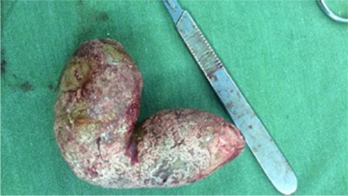
Figure 2 Extracted giant ureteral stones measuring 14 cm in length and 106 g in weight in the left ureter.
Most of these giant stones were distal ureteral calculi. This may be due to their role in distal ureteral obstruction, which is a factor that increases the diameter of the stone.Citation14 The majority of small stones with relatively mild hydronephrosis (HDN) can be managed with acetaminophen.Citation15 However, according to the size and location of the stones, about 20% of the stones require surgical operation.Citation16 There are various techniques in the management of ureteral stones, including extracorporeal shock wave lithotripsy (ESWL), open surgery, medical expulsive therapy (MET), ureteroscopy (URS), laparoscopy (LAP), and PCNL.Citation17 Drainage with an internal ureteral stent is also common due to its positive effects on morbidity.Citation18 In this regard, we introduced the combination of open and endoscopic techniques for treatment of the patient. Nowadays, ESWL and PCNL are the two most commonly performed treatments since they are minimally invasive surgical methods that significantly decrease the morbidity of stone removal.Citation19 In these techniques, success is determined by fragmentation rates and the size of the remaining stone fragments.Citation20
In the diagnosis of ureteral stones, the clinical diagnosis should be supported by an appropriate imaging procedure. Computed tomography (CT) is now the first line imaging technique to confirm the diagnosis of a urinary stone. In addition, the KUB study is an X-ray procedure that assesses the organs of the urinary system. It can also identify kidney stones and certain types of gallstones.Citation21
Ethics statement
We declare that our study was approved by the ethics committee of Hormozgan University of Medical Sciences, Hormozgan, Iran. Additionally, written informed consent was provided by the patients to have the case details and any accompanying images published, the safety of our procedure, and its probable complications.
Author contributions
All authors contributed to data analysis, drafting or revising the article, gave final approval of the version to be published, and agree to be accountable for all aspects of the work.
Acknowledgments
The authors would like to thank Shiraz University of Medical Sciences, Shiraz, Iran and also Center for Development of Clinical Research of Nemazee Hospital and Dr Nasrin Shokrpour for editorial assistance.
Disclosure
The authors report no conflicts of interest in this work.
References
- MatlagaBRJansenJPMeckleyLMByrneTWLingemanJETreatment of ureteral and renal stones: a systematic review and meta-analysis of randomized, controlled trialsJ Urol2012188113013722591962
- HammadyAGamalWMZakiMHusseinMAbuzeidAEvaluation of ureteral stent placement after retroperitoneal laparoscopic uretero-lithotomy for upper ureteral stone: randomized controlled studyJ Endourol201125582583021457084
- Perez CastroEOstherPJSJingaVCROES Ureteroscopy Global Study GroupDifferences in ureteroscopic stone treatment and outcomes for distal, mid-, proximal, or multiple ureteral locations: the clinical research office of the Endourological Society Ureteroscopy Global StudyEur Urol201466110210924507782
- LidénMAnderssonTBroxvallMThunbergPGeijerHUrinary stone size estimation: a new segmentation algorithm-based CT methodEur Radiol201222473173722160167
- WangY-HZhuY-SYangJ-QApplication of ultrasonography in the diagnostic of ureteris obstruction of transplanted kidneysActa Academiae Medicinae CPAF200812016
- SwensonDWDargeKZinielSIChowJSCharacterizing upper urinary tract dilation on ultrasound: a survey of North American pediatric radiologists’ practicesPediatr Radiol201545568669425421301
- RathodRBansalPGuttaSA giant ureteric calculusIndian J Urol201329326324082453
- OsorioLLimaEAutorinoRMarceloFEmergency management of ureteral stones: recent advancesIndian J Urol200824446119468497
- CollDMVaranelliMJSmithRCRelationship of spontaneous passage of ureteral calculi to stone size and location as revealed by unenhanced helical CTAJR Am J Roentgenol2002178110110311756098
- BanerjiJSDevasiaAGiant ureteric calculusANZ J Surg2011819653654
- TaylorWMLarge ureteral calculi. Report of a caseThe Journal of Urology193432193102
- MayersMMA Giant Ureteral Calculus (Weight: 286 Grams)The Journal of Urology19404414753
- SabnisRBDesaiRMBradooAMPunekarSVBapatSDGiant ureteral stoneJ Urol199214838618621512840
- BeliaevAMBeavenTKoyaMPSpontaneous passage of a very large ureteral stoneANZ J Surg201686121063106425113144
- FalahatkarSKhosropanahIAllahkhahAJafariAOpen surgery, laparoscopic surgery, or transureteral lithotripsy—which method? Comparison of ureteral stone management outcomesJ Endourol2011251313420977370
- SeminsMJShoreADMakaryMAThe association of increasing body mass index and kidney stone diseaseJ Urol2010183257157520018330
- CuiYCaoWShenHComparison of ESWL and ureteroscopic holmium laser lithotripsy in management of ureteral stonesPLoS One201492e8763424498344
- BerentACWeisseCWToddKBagleyDHTechnical and clinical outcomes of ureteral stenting in cats with benign ureteral obstruction: 69 cases (2006–2010)J Am Vet Med Assoc2014244555957624548231
- OberlinDTFlumASBachrachLMatulewiczRSFlurySCContemporary surgical trends in the management of upper tract calculiJ Urol2015193388088425219700
- TuruncTKuzgunbayBGulUFactors affecting the success of ureteroscopy in management of ureteral stone diseases in childrenJ Endourol20102481273127720420550
- BerkovitzNSimanovskyNKatzRSalamaSHillerNCoronal reconstruction of unenhanced abdominal CT for correct ureteral stone size classificationEur Radiol20102051047105119890646