
Abstract
Here we reported a rare case of the implantation of a dexamethasone intravitreal implant (DEX) in which decreased retinal vessel density (VD) was found by optical coherence tomography angiography (OCTA). A 74-year-old male with diabetes mellitus presented with bilateral macular edema. The best-corrected visual acuity (BCVA) was 0.6 in the right eye. Diabetic macular edema (DME) was diagnosed. A DEX for the right eye was planned, and the preoperative evaluation showed a superficial VD of 48.74 percent, a deep VD of 53.12 percent, and a foveal avascular zone (FAZ) 0.165 mm2 in size by OCTA. The BCVA in the right eye recovered to 0.8, and a notably lower superficial VD of 45.97 percent and a deep VD of 45.40 percent were observed with an enlarged FAZ of 0.294 mm2 one month postoperatively. Moreover, BCVA in the right eye was maintained at 0.8, while further reductions in both superficial (40.07 percent) and deep (40.91 percent) VD were noted with a FAZ measured at 0.305 mm2 two months postoperatively. In conclusion, retinal superficial and deep VD decreased, while the FAZ increased, after the implantation of the DEX in a patient with DME.
Introduction
Diabetic macular edema (DME) will lead to the progressive reduction in central vision and poor quality of life.Citation1 The dys-regulation of vessel growth and inflammatory cytokines are the major pathophysiological mechanisms of DME and lead to increased central macular thickness (CMT).Citation2 Concerning the retinal image, both fluorescein angiography and optical coherence tomography angiography (OCTA) revealed the loss of a regular pattern in the retinal capillary plexus.Citation3,Citation4 The conventional treatments for DME, including intravitreal corticosteroid and intravitreal anti-angiogenesis agent injections, have different outcomes.Citation1
Dexamethasone intravitreal implant (DEX, brand name: Ozurdex) is a biodegradable steroid delivery system that has been widely applied to patients with DME with favorable morphological and functional outcomes.Citation5 In clinical practice, the DEX was commonly implanted and with a pro re nata treatment approximately five months after the first implantation, and the long-term efficiency and safety was guaranteed in repeated DEX implantation.Citation6 In a previous study, DEX reduces the CMT more effectively than anti- angiogenesis agents with significant improvement in retinal vascular caliber.Citation7 Moreover, the DEX can be used to treat macular edema that shows a poor response to anti- angiogenesis agents with satisfactory results.Citation8
Although the main theory of the use of DEX for treating DME is its anti-inflammatory effect,Citation2 vascular endothelial growth factor is also suppressed by corticosteroids.Citation2,Citation5 Moreover, the different response of each retinal vascular layer to anti- angiogenesis therapy indicates that similar vascular alterations may also occur in patients receiving DEX implantation.Citation9 Nevertheless, only one study demonstrated a change in the OCTA vasculature in patients who received DEX implantation with DME with insignificant alterations.Citation10 In this study, we aimed to report a case of DME in which decreased retinal vessel density (VD) was found by OCTA after DEX implantation.
Case report
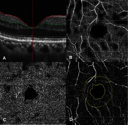
A 74-year-old Taiwanese male with diabetes mellitus presented with progressive blurred vision of both eyes. On examination, the best-corrected visual acuity (BCVA) was 0.6 in the right eye and 0.7 in the left eye with an intraocular pressure of 11 mmHg in both eyes. Bilateral macular edema was observed using an indirect fundoscopy. Color fundus photography and fluorescein angiography revealed an edematous macula and vessel leakage; thus, DME was diagnosed (). For the DME in the right eye, DEX injection was planned for the right eye, and the preoperative evaluation showed a CMT of 385 μM, and a superficial VD of 48.74 percent, a deep VD of 53.12 percent and a foveal avascular zone (FAZ) of 0.165 mm2 (). The surgery was performed smoothly. One month after surgery, BCVA in the right eye recovered to 0.8, and the CMT decreased to 292 μM. Moreover, a considerably lower superficial VD of 45.97 percent and a deep VD of 45.40 percent were observed with an enlarged FAZ of 0.294 mm2 (). In the latest follow-up visit two months postoperatively, BCVA in the right eye was maintained at 0.8 with a CMT of 281 μM. VD further declined in both superficial (40.07 percent) and deep (40.91 percent) areas, while the FAZ measured 0.305 mm2, which was larger than previously observed (). In addition, the VD of the choriocapillaris in the right eye decreased from 2.103 to 1.853 mm2.
Figure 1 Photograph of the fundus in the case with diabetic macular edema. (A) Color fundus photography. (B) Fluorescein angiography.
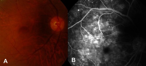
Figure 2 Macular conditions before dexamethasone implantation via optical coherence tomography angiography device. (A) Foveal thickness. (B) Superficial vessel density. (C) Deep vessel density. (D) Foveal avascular zone.
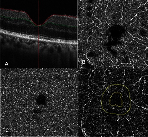
Figure 3 Macular conditions one month after dexamethasone implantation assessed an optical coherence tomography angiography device. (A) Foveal thickness. (B) Superficial vessel density. (C) Deep vessel density. (D) Foveal avascular zone.
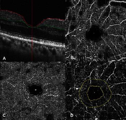
Figure 4 Macular conditions two months after dexamethasone implantation assessed using an optical coherence tomography angiography device. (A) Foveal thickness. (B) Superficial vessel density. (C) Deep vessel density. (D) Foveal avascular zone.
Discussion
In our patient, we used the same OCTA device (Angiovue, Optovue Inc., Bayview, CA, USA) in all OCTA examination, and the vessel layers was segmented manually by one ophthalmologist (HC Chen) then the VD and FAZ were calculated by the software in the same OCTA device. The results showed a decrement of VD and increment of the FAZ area after the DEX implantation. OCTA has been used to evaluate the retinal vessel for years, and its accuracy is similar to that of fluorescein angiography.Citation3,Citation11 Moreover, cystoid macular edema and DME were examined by OCTA in previous studies with a significant alteration of macular VD.Citation12,Citation13 The distribution of VD in the superficial and deep retinal layers was observed in a patient with DME who received intravitreal anti-angiogenesis agent injection via OCTA.Citation14 As a result, it is reasonable to use OCTA to evaluate the changes in the macular vasculature in layers after receiving DEX intravitreal therapy in patients diagnosed with DME. Still, the segmentation artifacts would become more prominent in patients with macular edema and results in poorer interpretability of OCTA image,Citation15 which can occur in the current study. Further effort to decrease such artifact in OCTA device is necessary.
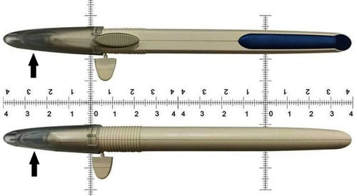
DEX is a long-term steroid-release system that has a pen-like appearance that consists of the delivery device and dexamethasone medication (). Upon injection, the dexamethasone-contain polymer device (arrow, ) would be delivered into the vitreous cavity and release the dexamethasone continuously. The previous experimental study revealed that the tumor-induced angiogenesis diminished after dexamethasone application,Citation16 implying the possibility of a steroid-induced anti-vascular growth effect in the eye. Only one study evaluated the change in the vasculature in patients with DME after DEX implantation without significant changes among the superficial VD, deep VD and choriocapillaris capillary density, and the difference was within two percent.Citation10 In addition, another study also revealed non-significant alteration of vascular distribution in patients with retinal venous occlusions after the implantation of DEX.Citation17 However, a decrease in the superficial and deep VDs of approximately 20 percent after DEX implantation was observed in the current study, while the enlargement of the FAZ found in the current study was not reported in previous studies. The prominent difference in vessel alteration and the FAZ indicated that the retinal vasculature may change in some patients with DME after DEX implantation. Although diabetic retinopathy would uneventfully enlarge the FAZ, which progressed with the disease severity,Citation18 no severe complications, such as vitreous hemorrhage or tractional retinal detachment, were found during the two-month period. In addition, the blood sugar level was 212 mg/dL and 171 mg/dL before and two months after the DEX implantation, and the glycosylated hemoglobin was 7.3 percent and 7.0 percent before and two months after the DEX implantation, implied that the progression of diabetes mellitus was not likely. Moreover, the enlarged FAZ area in diabetic retinopathy and retinal venous occlusion are associated with worse visual outcome according to previous research,Citation19 while the recovered visual acuity is discordant to the enlarged FAZ area in our patient, which further demonstrated that the enlarged FAZ would probably result from the anti-angiogenesis effect of DEX, rather than the influence of diabetic retinopathy. As a consequence, we speculated that the anti-angiogenesis effect of DEX, which proven in experimental study,Citation16 retarded the angiogenesis process and lead to the decrement of retinal vasculature in our patient which presented as decreased VD and enlarged FAZ compared to the minimal change in the fellow eye. In addition, the effect of anti-angiogenesis persisted for months which may due to the long-term effect of DEX. Conversely, the mean CMT and the mean initial superficial VD in the study by Toto et al were grossly different from those of the patient in the current study,Citation10 while the initial visual acuity was worse in the study by Mastropasqua et al compared with our patient.Citation17 The above differences indicate that the initial severity of microvasculopathy and the visual performance in retinal vascular diseases might influence the anti-angiogenesis effect of DEX.
Figure 5 The appearance of dexamethasone intravitreal implant. Arrow: the dexamethasone-contain polymer device in the plastic cap.
The effectiveness of DEX in patients with DME has been illustrated in previous studies.Citation5,Citation7,Citation8 For those patients with recalcitrant DME unresponsive to anti-angiogenesis agent therapy, DEX still may slow the DME with morphological improvement.Citation8 Although our patient was a new case and had not previously received anti-angiogenesis agent therapy, the edematous status progressed rapidly in three months thus DEX is theoretically more suitable than anti-angiogenesis agent therapy to retard the macular edema in our patient. In addition, the patient had previously received phacoemulsification; therefore, the steroid-induced cataract formation which can be induced by dexamethasone in the DEX was beyond our consideration. Also, the preoperative IOP was in the low-to-normal range so the chance to develop steroid-induced ocular hypertension after DEX implantation was unlikely in our patient. Since no contraindication was found and the edematous status was progressed, we selected DEX as the first-line treatment rather than anti-angiogenesis agent therapy. The decrease in vasculature with the improvement of both BCVA and CMT were observed for at least two months which might result from the anti-inflammatory effect of DEX in our opinion, and our finding may demonstrate the possibility that DEX can be the first treatment for DME in certain situations.
Conclusion
In conclusion, retinal VD decreased after the implantation of DEX in our patient with DME. Further large-scale studies are warranted to investigate the correlation between a decrease in VD and the recovery of visual parameters.
Ethics approval and informed consent
The study adhered to the declaration of Helsinki and was approved by the Institutional Review Board at the Show Chwan Memorial Hospital. In addition, written informed consent was obtained from the patient for publication of this case report including any accompanying images.
Abbreviations
DEX, dexamethasone intravitreal implant; DME, diabetic macular edema; OCTA, optical coherence tomography angiography; BCVA, best-corrected visual acuity; CMT, central macular thickness; VD, vessel density.
Author contributions
All authors contributed to data analysis, drafting and revising the article, gave final approval of the version to be published, and agree to be accountable for all aspects of the work.
Data availability
Since the data included in the current report may contain some privacy information, the data will only be provided upon the request of editorial board.
Disclosure
The authors report no conflicts of interest in this work.
References
- Relhan N, Flynn HW Jr. The early treatment diabetic retinopathy study historical review and relevance to today’s management of diabetic macular edema. Curr Opin Ophthalmol. 2017;28(3):205–212. doi:10.1097/ICU.000000000000036228151747
- Whitcup SM, Cidlowski JA, Csaky KG, Ambati J. Pharmacology of corticosteroids for diabetic macular edema. Invest Ophthalmol Vis Sci. 2018;59(1):1–12. doi:10.1167/iovs.17-2225929297055
- Peres MB, Kato RT, Kniggendorf VF, et al. Comparison of optical coherence tomography angiography and fluorescein angiography for the identification of retinal vascular changes in eyes with diabetic macular edema. Ophthalmic Surg Lasers Imaging Retina. 2016;47(11):1013–1019. doi:10.3928/23258160-20161031-0527842196
- Mane V, Dupas B, Gaudric A, et al. Correlation between cystoid spaces in chronic diabetic macular edema and capillary nonperfusion detected by optical coherence tomography angiography. Retina. 2016;36:S102–S110. doi:10.1097/IAE.000000000000128928005668
- Mastropasqua R, Toto L, Borrelli E, et al. Morphology and function over a one-year follow up period after intravitreal dexamethasone implant (ozurdex) in patients with diabetic macular edema. PLoS One. 2015;10(12):e0145663. doi:10.1371/journal.pone.014566326720268
- Bucolo C, Gozzo L, Longo L, Mansueto S, Vitale DC, Drago F. Long-term efficacy and safety profile of multiple injections of intravitreal dexamethasone implant to manage diabetic macular edema: a systematic review of real-world studies. J Pharmacol Sci. 2018;138(4):219–232. doi:10.1016/j.jphs.2018.11.00130503676
- Wickremasinghe SS, Fraser-Bell S, Alessandrello E, Mehta H, Gillies MC, Lim LL. Retinal vascular calibre changes after intravitreal bevacizumab or dexamethasone implant treatment for diabetic macular oedema. Br J Ophthalmol. 2017;101(10):1329–1333. doi:10.1136/bjophthalmol-2016-30988228228411
- Iacono P, Parodi MB, Scaramuzzi M, Bandello F. Morphological and functional changes in recalcitrant diabetic macular oedema after intravitreal dexamethasone implant. Br J Ophthalmol. 2017;101(6):791–795. doi:10.1136/bjophthalmol-2016-30872627625164
- Gill A, Cole ED, Novais EA, et al. Visualization of changes in the foveal avascular zone in both observed and treated diabetic macular edema using optical coherence tomography angiography. Int J Retina Vitreous. 2017;3:19. doi:10.1186/s40942-017-0074-y28642823
- Toto L, D’Aloisio R, Di Nicola M, et al. Qualitative and quantitative assessment of vascular changes in diabetic macular edema after dexamethasone implant using optical coherence tomography angiography. Int J Mol Sci. 2017;18(6):1181. doi:10.3390/ijms18061181
- Vangipuram G, Rezaei KA. Optical coherence tomography angiography as an imaging modality for evaluation of diabetic macular edema. J Ophthalmic Vis Res. 2017;12(4):359–360. doi:10.4103/jovr.jovr_175_1729090042
- Lupidi M, Coscas F, Cagini C, Coscas G. Optical coherence tomography angiography in macular edema. Dev Ophthalmol. 2017;58:63–73. doi:10.1159/00045526928351045
- Chetrit M, Bonnin S, Mane V, et al. Acute pseudophakic cystoid macular edema imaged by optical coherence tomography angiography. Retina. 2017;38(10):2073–2080. doi:10.1097/IAE.0000000000001829
- Ghasemi Falavarjani K, Iafe NA, Hubschman JP, Tsui I, Sadda SR, Sarraf D. Optical coherence tomography angiography analysis of the foveal avascular zone and macular vessel density after anti-VEGF therapy in eyes with diabetic macular edema and retinal vein occlusion. Invest Ophthalmol Vis Sci. 2017;58(1):30–34. doi:10.1167/iovs.16-2057928114569
- Enders C, Lang GE, Dreyhaupt J, et al. Quantity and quality of image artifacts in optical coherence tomography angiography. PLoS One. 2019;14(1):e0210505. doi:10.1371/journal.pone.021050530682050
- Fan Z, Sehm T, Rauh M, et al. Dexamethasone alleviates tumor-associated brain damage and angiogenesis. PLoS One. 2014;9(4):e93264. doi:10.1371/journal.pone.009326424714627
- Mastropasqua R, Toto L, Di Antonio L, et al. Optical coherence tomography angiography microvascular findings in macular edema due to central and branch retinal vein occlusions. Sci Rep. 2017;7:40763. doi:10.1038/srep4076328098203
- Khadamy J, Abri Aghdam K, Falavarjani KG. An update on optical coherence tomography angiography in diabetic retinopathy. J Ophthalmic Vis Res. 2018;13(4):487–497. doi:10.4103/jovr.jovr_57_1830479720
- Balaratnasingam C, Inoue M, Ahn S, et al. Visual acuity is correlated with the area of the foveal avascular zone in diabetic retinopathy and retinal vein occlusion. Ophthalmology. 2016;123(11):2352–2367. doi:10.1016/j.ophtha.2016.07.00827523615