
Abstract
Introduction
The objective of this report is to describe the first patient presenting clinical features of trisomy 13 in association with a sacrococcygeal teratoma.
Case presentation
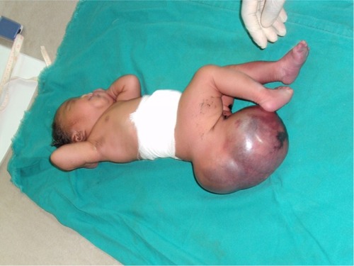
We present the case of a Congolese female infant born with bilateral cleft lip and palate, hypotelorism, microcephaly, and capillary hemangioma on her face. She presented with a large sacrococcygeal mass (15.0 cm ×12.0 cm ×5.0 cm) with a cystic consistency and a positive transillumination.
Conclusion
This observation suggests that overexpression of certain genes on chromosome 13 may lead to tumor formation from remnant cells of Hensen’s node.
Introduction
Sacrococcygeal teratoma is a rare tumor with a birth prevalence of 1 in 21,700 births.Citation1 In a relatively large proportion (20%), this tumor can have a malignant development, most commonly a papillary adenocarcinoma.Citation2 Previously, teratome of the liver, umbilical cord, and oral region have been described in three patients with trisomy 13 syndrome (Patau syndrome), but thus far, no sacrococcygeal teratoma has been observed in this syndrome. Trisomy 13 has an incidence of 1 in 10,000–20,000 with variable expression and is characterized by the clinical triad of cleft lip and palate, microphthalmia/anophthalmia, and postaxial polydactyly.Citation3 However, other anomalies are frequently associated.Citation4,Citation5 The objective of this report is to describe a Congolese newborn presenting with a sacrococcygeal teratoma and the clinical features of trisomy 13.
Case presentation
A female patient was born in Fungurume (mining area in southern Katanga Province, Democratic Republic of Congo) at 40 weeks of gestation via lower segment cesarean section delivery with a birth weight of 4,580 g (+2.48 standard deviation [SD], World Health Organization [WHO] growth charts 2006Citation6) and a length of 51 cm (0.9 SD, WHO growth charts 2006Citation6). Her mother was 40 years old and her father 51 years old, both were healthy and unrelated. The mother did not use medications during pregnancy. No prenatal ultrasound scan was performed. She was referred on day 2. On clinical examination, we noted a bilateral cleft lip and palate, hypotelorism, microcephaly (28 cm, −5.06 SD, WHO growth chartsCitation6), and capillary hemangioma on her face. No scalp defect has been observed. The infant had low-set ears and a short neck (). No organized ocular tissues could be seen or palpated. The infant had five normal fingers in both hands and feet. She had a large sacrococcygeal mass (15.0 cm ×12.0 cm ×5.0 cm) with a cystic consistency and a positive transillumination (). However, abdominal and pelvic urtrasonography could not be performed, and thus, the extent of the sacrococcygeal teratoma could not be graded according to the American Academy of Pediatrics classification. The genitalia or rectum was not involved. The anal sphincter and limb tone were normal. Since genetic diagnosis was not available in this part of the world when the infant was born, we were not able to assess genetic confirmation of the suspected diagnosis of trisomy 13. The child was hypertonic, had experienced feeding difficulties, and died on day 8 from severe sepsis and acute anemia.
Figure 1 Craniofacial abnormalities observed in our patient.
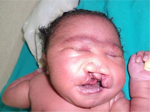
Figure 2 Large sacrococcygeal teratoma.
Discussion
Dysmorphology is a major challenge in low-income countries, where imaging and genetic technologies are not available. Since comparative genomic hybridization array and karyotyping are not available in this part of the world, the diagnosis of trisomy 13 in this patient relied on clinical grounds. Two signs of the characteristic clinical triad were present in our patient (cleft lip and palate and microphthalmia/anophthalmia), but no postaxial polydactyly has been observed.Citation3 shows clinical features in trisomy 13 with molecular confirmation versus our patient.Citation7–Citation11 As we can see in as well as in Caba et al’s paper, full classical triad of trisomy 13 is not constant, which is an indirect evidence supporting the argument that our patient likely had trisomy 13.Citation3
Table 1 Clinical features in trisomy 13 with molecular confirmation versus our patient
Whereas in most industrialized countries, trisomy 13 is diagnosed prenatally,Citation12 ~24% of pregnant women have no access to prenatal care services, and the vast majority of them currently do not have access to prenatal ultrasound follow-up in the Democratic Republic of Congo.Citation11
The identification of cell-free DNA in maternal blood and the recent development of polymerase chain reaction-based noninvasive DNA hold enormous promise for diagnosis of fetal trisomies 21, 18, and 13 in general pregnant populations. However, the lack of teaching, and thus interest and knowledge, in human genetics might be barriers to this testing in Democratic Republic of Congo.
Upon reviewing the literature, the relationship of aneuploidy and chromosomal instability to neoplasia has been discussed over the past decades. Three patients with trisomy 13 disorders have been reported with teratoma. In Dische and Gardner’s paper, a fetus with trisomy 13 syndrome and teratomas of liver and neck has been described.Citation13 Yapar et al have presented a case of epignathus (oral cavity teratoma) detected prenatally in a fetus with trisomy 13 syndrome.Citation14 More recently, an unusual solid and cystic mass was diagnosed at 17 weeks fetus with trisomy 13. Fetopathological examination revealed an umbilical cord teratoma.Citation15 These three cases suggest the association between trisomy 13 and teratoma. However, the mechanism of origin of these tumors is probably not the same. Extragonadal teratomas are thought to arise from germ cells that migrate aberrantly during embryogenesis, whereas sacrococcygeal teratoma is believed to arise from totipotent cells derived from the remnant of Hensen’s node located in the coccyx region, the caudal eminence.Citation16
As we can see in , sacrococcygeal teratoma has already been described in some chromosomal disorders syndrome such as partial trisomy 1q, partial trisomy 3q, and mosaic r (21),Citation13–Citation15,Citation17–Citation24 and genomic gain in 1q and 12p has been observed in sacrococcygeal teratoma. Furthermore, it has been shown that some genes in individuals with trisomy 21 may inhibit the generation and growth of tumors originating from the remnant of Hensen’s node.Citation16
Table 2 Overview of reported teratoma in patients with chromosomal disorder syndromes
Trisomies as a gain-of-function mechanism by selection of a gain-of-function mutation or by gene dosage have been also implicated in hematologic malignancies as well as solid tumors through duplication and amplification of mutated alleles (RUNX1 or MET oncogene).Citation25
It is not excluded that this represents a chance of association of two pathogenetically unrelated conditions, and the lack of genetic testing is a weakness of this report. Alternatively, given the previous reports of sacral appendageCitation26 and terato-mas with other localizations () in other patient with trisomy 13, it is not excluded that overexpression of certain genes on chromosome 13 may lead to tumor formation from remnant cells of Hensen’s node.
Consent
Written informed consent was obtained from the patient’s legal guardians for publication of this case report and any accompanying images. The Hôpital Général De Référence de Kamalondo Ethics Committee advised ethical approval was not required.
Author contributions
All authors were responsible for the study conception, data retrieval, and drafting the manuscript. All authors read and approved the final manuscript.
Acknowledgments
The authors are grateful to the parents of the reported infant for their kind collaboration.
Disclosure
The authors report no conflicts of interest in this work.
References
- GucciardoLUyttebroekADe WeverIPrenatal assessment and management of sacrococcygeal teratomaPrenat Diagn20113167868821656530
- ListerJSacro-coccygeal teratomaArch Dis Child195530666914350823
- CabaLRusuCButnariuLPhenotypic variability in Patau syndromeRev Med Chir Soc Med Nat Iasi201311732132724340511
- DuarteACMenezesAIDevensESRothJMGarciasGLMartino-RothMGPatau syndrome with a long survival. A case reportGenet Mol Res2004328829215266400
- CareyJCPerspectives on the care and management of infants with trisomy 18 and trisomy 13: striving for balanceCurr Opin Pediatr20122467267823044555
- WHO Multicentre Growth Reference Study GroupWHO Child Growth Standards: Length/height-for-age, Weight-for- age, Weight-for-length, Weight-for-height and Body mass index-for-age: Methods and DevelopmentGenevaWorld Health Organization2006 Available from: http://www.who.int/childgrowth/standards/technical_report/en/Accessed September 10, 2015
- PatauKSmithDWThermanEInhornSWagnerHMultiple congenital anomaly caused by an extra autosomeLancet196027579079314430807
- LinHYLinSPChenYJClinical characteristics and survival of trisomy 18 in a medical center in Taipei, 1988–2004Am J Med Genet2006140A94595116528742
- PetryPPolliJBMattosVFClinical features and prognosis of a sample of patients with trisomy 13 (Patau syndrome) from BrazilAm J Med Genet2013161A1278128323613355
- QuelinCSpaggiariEKhung-SavatovskySDupontCPasquierLLoeuilletLInversion duplication deletions involving the long arm of chromosome 13: phenotypic description of additional three fetuses and genotype-phenotype correlationAm J Med Genet2014164A2504250924975584
- Mbuyi-MusanzayiSLumakaAZYogoleloHBPreaxial poly-dactyly of the foot: variable expression of trisomy 13 in a case from Central AfricaCase Rep Genet2014201436503125254124
- KroesIJanssensSDefoortPUltasound features in trisomy 13 (Patau syndrome) and trisomy 18 (Edwards syndrome) in a consecutive series of 47 casesFacts Views Vis Obgyn2014624524925593701
- DischeMRGardnerHAMixed teratoid tumors of the liver and neck in trisomy 13Am J Clin Pathol197869631637665584
- YaparEGEkiciEGökmenOSonographic diagnosis of epignathus (oral teratoma), prosencephaly, meromelia and oligohydramnios in a fetus with trisomy 13Clin Dysmorphol199542662717551166
- HargitaiBCsabaiLBánZRare case of exomphalos complicated with umbilical cord teratoma in a fetus with trisomy 13Fetal Diagn Ther20052052853316260890
- KobayashiTSakemiYYamashitaHIncreased incidence of ret-roperitoneal teratomas and decreased incidence of sacrococcygeal teratomas in infants with Down syndromePediatr Blood Cancer20146136336523904199
- WaxJRBennPSteinfeldJDIngardiaCJPrenatally diagnosed sac-rococcygeal teratoma. A Unique Expression of Trisomy 1qCancer Genet Cytogenet2000117848610700874
- DundarMUzakAErdoganMPartial trisomy 3q in a child with sacrococcygeal teratoma and Cornelia de Lange syndrome phenotypeGenet Couns20112219920521848013
- ChenCPChangSDLeeYXRing chromosome 21 presenting with sacrococcygeal teratoma: prenatal diagnosis, molecular cytogenetic characterization and literature reviewGene201352211111623545316
- López GinésCGilRPellinAMartorellMVilarFLlombart-BoschATrisomy 12 and translocation (7;9) in an ovarian immature teratomaInt J Gynecol Pathol198982772852767876
- SchwartzSRaffelLJSunCCWatersEAn unusual mosaic karyo-type detected through prenatal diagnosis with duplication of 1q and 19p and associated teratoma developmentTeratology1992463994041384156
- MertensFKullendorffCMHjorthLAlumetsJMandahlNTrisomy 3 as the sole karyotypic change in a pediatric immature teratomaCancer Genet Cytogenet199810283859530347
- Le CaignecCWinerNBocenoMPrenatal diagnosis of sacrococcygeal teratoma with constitutional partial monosomy 7q/trisomy 2pPrenat Diagn20032398198414663834
- BatukanCOzgunMTBasbugMCaglayanODundarMMuratNSacrococcygeal teratoma in a fetus with prenatally diagnosed partial trisomy 10q (10q24. 3→qter) and partial monosomy 17p (p13. 3→ pter)Prenat Diagn20072736536817295347
- DickerFHaferlachCKernWHaferlachTSchnittgerSTrisomy 13 is strongly associated with AML1/RUNX1 mutations and increased FLT3 expression in acute myeloid leukemiaBlood200711041308131617485549
- PachajoaHEscobarLEMMosaic trisomy 13 and a sacral appendageBMJ Case Rep20132013bcr2012008150
- ScheresJMJCDe PaterJMStoutenbeekPWijmengaCRosenbergCPearsonPLIsochromosome 1q as the sole chromosomal abnormality in two fetal teratomas: possible trisomic or tetrasomic zygote rescue in fetal teratoma with an additional isochromosome 1qCancer genetics and cytogenetics1999115111010565292