
Abstract
Hepatocellular carcinoma (HCC) is one of the most common primary hepatic malignancies and one of the fastest-growing causes of cancer-related mortality in the United States. The molecular basis of HCC carcinogenesis has not been clearly identified. Among the molecular signaling pathways implicated in the pathogenesis of HCC, the Wnt/β-catenin signaling pathway is one of the most frequently activated. A great effort is under way to clearly understand the role of this pathway in the pathogenesis of HCC and its role in the transition from chronic liver diseases, including viral hepatitis, to hepatocellular adenomas (HCAs) and HCCs and its targetability in novel therapies. In this article, we review the role of the β-catenin pathway in hepatocarcinogenesis and progression from chronic inflammation to HCC, the novel potential treatments targeting the pathway and its prognostic role in HCC patients, as well as the imaging features of HCC and their association with aberrant activation of the pathway.
Introduction
Hepatocellular carcinoma (HCC) is the most common primary hepatic malignancy, constituting around 85–90% of primary liver cancers. The incidence of HCC is on the rise, and HCC is now the fastest-growing cause of cancer-related mortality in the United States.Citation1–Citation3 HCC is also the third leading cause of cancer-related mortality in the world,Citation4,Citation5 causing approximately 750,000 deaths worldwide in 2012.Citation3 It is the sixth most common malignancy.Citation4,Citation6 The number of cases reported annually reaches more than half a million worldwide.Citation7,Citation8
HCC frequently develops in the setting of underlying chronic liver disease. Viral hepatitis (infection with either hepatitis C virus [HCV] or hepatitis B virus [HBV]) is thought to be the most common etiology of HCC.Citation9 However, other factors may contribute to the development of HCC. The interaction of aflatoxin B1 (a contaminant found in food) with HBV infection is believed to increase the prevalence of HCC.Citation10 Cirrhosis due to non-alcoholic steatohepatitis is also associated with the development of HCC.Citation7 In addition to underlying liver disease, lifestyle factors such as tobacco use, alcohol use, and obesity may also contribute to the development of HCC.Citation6,Citation7,Citation11
It is known that genetic mutations and abnormal activation of signal transduction pathways are involved in the development of HCC.Citation1,Citation6,Citation7,Citation11–Citation13 However, it has been difficult to elucidate the specific molecular pathophysiology that leads to HCC tumor development.Citation3,Citation12 Unfortunately, this lack of understanding has limited the development of targeted molecular therapies to treat HCC,Citation3 and the prognosis of HCC has remained poor.Citation12,Citation14,Citation15 Signaling pathways involved in cell proliferation, apoptosis, metabolism, splicing, and the cell cycle are believed to play a vital role in the formation of HCC. The signaling pathways known to be activated in HCC tissue include the Wnt/β-catenin pathway (up to 50% of HCC), the phosphatidylinositol-3-kinase and protein kinase B (PI3K/Akt) pathway (40–60% of HCC), the Myc pathway (30–60%), the Hedgehog pathway (50–60%), and the MET pathway (30–40%).Citation2,Citation7,Citation12,Citation15–Citation17 The Wnt/β-catenin pathway regulates multiple cellular processes that are involved in the initiation, growth, survival, migration, differentiation, and apoptosis of HCC.Citation2,Citation6,Citation8,Citation13,Citation18 The Wnt/β-catenin pathway has shown significant promise as a potential target for novel molecular therapies. Also, β-catenin mutation has been shown to affect the prognosis of HCC, which warrants more understanding of the role of this pathway in developing HCC.
Materials and methods
In this article, we review the role of the β-catenin pathway in hepatocarcinogenesis and progression from chronic liver disease to HCC, the novel potential treatments targeting the pathway and its prognostic role in HCC patients, as well as the imaging features of HCC on magnetic resonance imaging (MRI) and computed tomography (CT) associated with aberrant activation of the pathway.
CTNNB1 and β-catenin
Previous attempts to understand the pathogenesis of HCC have identified the β-catenin gene (CTNNB1) as one of the primary oncogenes involved in HCC development.Citation1,Citation6,Citation10 CTNNB1 encodes β-catenin, a protein that serves multiple important cellular functions. β-catenin is found in the adherens junctions and plays a key role in intercellular adhesion and communication.Citation1,Citation9,Citation13,Citation19–Citation23 In particular, deletions or missense mutations in exon 3 of this gene are thought to be the most common genetic abnormality leading to the development of HCC.Citation1,Citation12,Citation16,Citation18,Citation23–Citation26 Previous studies have shown a correlation between CTNNB1 missense mutations and increased nuclear and cytoplasmic expression of β-catenin, suggesting that these mutations cause aberrant activation of the Wnt pathway and play a role in the development of HCC.Citation1,Citation7,Citation18,Citation22,Citation26 shows a summary of the clinical studies on β-catenin and HCC with their clinical significance.
Table 1 Summary of different studies concerning β-catenin in HCC and their clinical significance
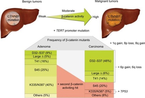
HCC can develop in healthy livers without the presence of fibrosis; in fact, most HCC tumors that contain β-catenin mutation develop from normal liver tissue. Some HCC tumors develop from the malignant transformation of hepatocellular adenomas (HCAs) (). These HCC tumors typically have mutations in the β-catenin gene. Notably, pre-malignant liver lesions, such as cirrhotic nodules, have never been found to have mutations in the β-catenin gene.Citation2,Citation3,Citation27
Figure 1 Carcinogenesis in HCC with β-catenin mutation with different β-catenin mutants identified, leading to different levels of β-catenin activation depending on the state of tumor progression. Reproduced with permission from Rebouissou S, Franconi A, Calderaro J, et al. Genotype-phenotype correlation of CTNNB1 mutations reveals different β-catenin activity associated with liver tumor progression. Hepatology. 2016;64(6):2047–2061. Copyright John Wiley and Sons.Citation40
Immunohistochemical staining reveals that β-catenin is localized primarily in the hepatocyte membrane in normal liver tissue, as shown by Li et al using clear linear brown staining.Citation1 However, in cirrhosis and HCC, β-catenin shows significantly decreased expression in the membrane and increased expression in the cytoplasm and nucleus. This abnormal β-catenin protein expression was significantly greater in HCC tissue (72.94% [62/85]) compared with para-carcinoma tissues and hepatic cirrhosis tissues (22.35% [19/85] and 26.67% [4/15], respectively; P < 0.001).Citation1,Citation7,Citation21
CTNBB1 and Axin1 mutation
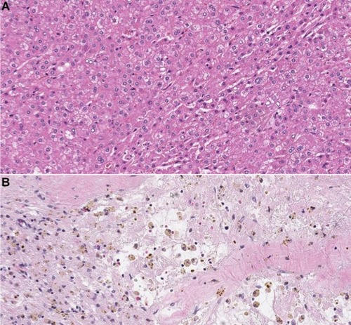
CTNNB1 mutation leading to activation of the Wnt pathway and the resulting accumulation of β-catenin have been associated with well-to-moderately differentiated tumors with small size and good overall prognosis. demonstrates the immunohistochemical staining of β-catenin protein in moderately differentiated HCC with β-catenin mutation. However, the nuclear expression of β-catenin due to its mutation in HCC has been associated with an increased incidence of microvascular and macrovascular invasion (78% in HCC with β-catenin mutation vs. 38% in HCC without β-catenin mutation), doubled tumor size (compared with tumors without β-catenin mutation), and multiplicity of nodules.Citation28
Figure 2 Hematoxylin-eosin staining showing moderately-differentiated HCC (A). At immunohistochemical staining (B), the tumor shows expression of β-catenin protein in the nucleus and/or cytoplasm. Magnification ×200.
There is strong evidence that in human HCC, gain-of-function β-catenin mutations are greatly different from inactivating mutations of Axin1. Axin mutations (and, rarely, β-catenin mutations) are found mainly in chromosomally unstable HCCs that are associated with HBV infection, while β-catenin mutations are found mainly in non-HBV, well-differentiated, chromosomally stable HCCs.Citation8,Citation29
WNT signaling
WNT signaling in normal tissue and HCC development
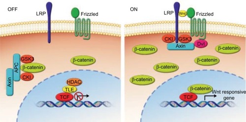
The WNT signaling pathway serves many vital functions in embryonic patterning, cell proliferation, differentiation, and angiogenesis. It is heavily involved in the development of embryonic tissues before birth and is also believed to play a role in the maintenance of stem cells in adult tissues.Citation1,Citation12,Citation24 The Wnt pathway is categorized into a canonical pathway (β-catenin dependent) and a non-canonical pathway (β-catenin independent).Citation9,Citation12,Citation14 Both categories of Wnt signaling begin when extracellular Wnt ligands bind to receptors on the cell membrane. In the canonical pathway, binding of Wnt ligands to the transmembrane receptor’s Wnt/FZD-related protein and to the low-density lipoprotein receptor results in the dissolution of the proteasome complex (APC protein, axin, and glycogen synthase kinase 3 [GSK-3]), and this complex is usually responsible for the destruction of β-catenin. Destruction of this complex results in the accumulation of β-catenin in the cytoplasm and its subsequent relocation into the nucleus, where β-catenin forms a binding complex with the transcription factor, i.e., lymphoid enhancer factor/T cell factor (LEF/TCF) proteins. This complex then regulates the transcription of target genes that are involved in cell proliferation, migration, cell cycle regulation, and metastasis ().Citation1,Citation3,Citation7,Citation9,Citation14,Citation15,Citation19–Citation21,Citation23,Citation24,Citation27,Citation30,Citation31
Figure 3 Overview of Wnt/β-catenin signaling.
Abbreviations: CKI, cyclin-dependent kinase inhibitor; GSK-3, glycogen synthase kinase 3; TCF/TLE, T cell-factor proteins/transducin like enhancer of split; HDAC, histone deacetylases; LRP, low-density-lipoprotein receptor-related protein; APC, adenomatous polyposis coli.
Wnt/β-catenin signaling plays an important role in hepatocyte development and function.Citation1,Citation6,Citation32 In normal hepatocytes, levels of β-catenin are low owing to the presence of the abovementioned β-catenin destruction complex (APC, axin, and GSK-3). However, there are multiple ways in which the Wnt/β-catenin pathway can become aberrantly activated and cause the development and progression of HCC. Mutations that involve the β-catenin gene and the AXIN1/2 gene result in sustained aberrant activation of the Wnt/β-catenin pathway and thus dysregulate multiple cellular functions, including proliferation, apoptosis, and cell motility.Citation1,Citation3,Citation7,Citation19,Citation31–Citation33 Mutations in CTNNB1 can result in the production of β-catenin proteins that are resistant to degradation by the proteasome.Citation3,Citation7,Citation14,Citation20,Citation31 In addition, loss-of-function mutations in APC and axin, which are members of the destruction complex, lead to the accumulation of β-catenin and allow for oncogenic transcription.Citation9,Citation14 β-catenin mutation is reportedly present in approximately 20–40% of all cases of HCC.Citation3,Citation9,Citation27 AXIN1 mutation is seen in 3–16% of all HCC cases, and AXIN2 mutation is seen in 3% of all HCC cases.Citation3,Citation19,Citation33,Citation34 However, it is important to note that up to 40–60% of HCC tumors do not have mutations in CTNNB1, AXIN1, or AXIN2.Citation3
Mutations that affect CTNNB1 and correlate with the amount of nuclear β-catenin are considered functionally important with regard to the development of HCC.Citation3,Citation35 However, a previous study using glutamine synthase as an immunohistochemical marker of β-catenin activity suggested that mutations in Axin1 may also exert their oncogenic effects through non-Wnt pathway mechanisms.Citation29,Citation35,Citation36 In addition, other studies have demonstrated that factors affecting E-cadherin production can also lead to the accumulation of β-catenin in the cytosol and subsequent translocation of β-catenin to the nucleus. Some pathogens are able to stimulate β-catenin signaling by disrupting intercellular junctions and thus mobilizing β-catenin from complexes with cadherins.Citation1,Citation9,Citation13,Citation19–Citation21,Citation23,Citation37
Hypoxia and activation of Wnt/β-catenin signaling
In addition to genetic mutations in Wnt signaling components, aberrant Wnt/β-catenin signaling in HCC tissue may be stimulated by hypoxia. Experiments have demonstrated crosstalk between Wnt/β-catenin and hypoxia signaling pathways.Citation38 B-cell lymphoma 9 (BCL9) is an important co-activator of β-catenin–mediated transcription. Hypoxia-inducible factors, including HIF-1α, regulate the expression of BCL9 and thus can increase transcriptional activity of β-catenin. This increased transcriptional activity of β-catenin can occur in the absence of other mutations affecting Wnt components. Evidence suggests that hypoxia-inducible factors play a role in HCC development, likely through crosstalk between hypoxia signaling pathways and the Wnt/β-catenin signaling pathway. Increased expression of BCL9 in HCC tumors has been shown to be a poor prognostic indicator.Citation14
Wnt/β-catenin pathway in the progression from chronic HCV/HBV to HCC
HCV is one of the most common causes of HCC worldwide. The mechanism by which HCV affects the development of HCC is not well understood, but it is believed that proteins that make up the HCV virus, including the HCV core protein and nonstructural protein 5A, independently stimulate the Wnt/β-catenin signaling pathway.Citation9,Citation37 It has also been shown that HCV-related HCC tumors have a significantly increased frequency of CTNNB1 mutations compared with HBV-related and non-viral HCC tumors. Notably, HCV is an RNA virus that exerts its effects on cells without going through a DNA intermediate phase. Thus, HCV’s effect on the hepatocyte genome is likely through an indirect mechanism.Citation9,Citation12 As chronic hepatitis infection progresses to cirrhosis, genetic mutations accumulate gradually.
In patients with HCC associated with HBV infection, aberrant WNT/β-catenin signaling appears to be more frequently stimulated by Axin1-inactivating mutations rather than by CTNNB1 mutations.Citation29,Citation30,Citation34 However, CTNNB1 mutations are also present in some HBV-associated HCC. Single-nucleotide polymorphisms in AXIN1 and CTNNB1 in HBV-associated HCC were correlated with overall patient survival.Citation30
Prognostic role of b-catenin mutation
β-catenin is involved in the development and progression of HCC, but its role in prognostication is less clear.Citation6 There have been conflicting reports as to whether CTNNB1 mutations typically occur late or early in the development of HCC.Citation3,Citation23,Citation24 Mutation in the β-catenin gene has been associated with less aggressive tumors, which are less likely to invade the vasculature and which contain more highly differentiated cells. β-catenin mutation is also more frequently seen in earlier stages of HCC (I and II) than in more advanced stages (III and IV).Citation6,Citation18,Citation20,Citation39 Patients with HCC tumors containing only β-catenin mutation also have lower serum α-fetoprotein levels. Higher α-fetoprotein levels are seen in tumors that contain mutations in other genes, such as p53 and cytokeratin 19 (CK19), and are associated with decreased patient survival.Citation20,Citation25,Citation27 Also, patients over the age of 60 years are more likely to have CTNNB1 mutation than younger patients are.Citation18–Citation20 According to one study, HCC tumors that contain mutations in CTNNB1 are associated with older age, male sex, low serum α-fetoprotein levels, well-differentiated cells, large tumors, high alcohol intake, microscopic vascular invasion, tumor capsule invasion, and intra-tumor cholestasis.Citation40 Some of the aforementioned disease characteristics are associated with good prognosis, like old age, low serum α-fetoprotein levels and well-differentiated cells, while others are associated with poorer prognosis, like microscopic vascular invasion and tumor capsule invasion.
β-catenin mutation is associated with increased nuclear and cytoplasmic β-catenin expression,Citation2,Citation3,Citation19–Citation21,Citation31 and while nuclear β-catenin expression is associated with a relatively good prognosis, cytoplasmic and membranous β-catenin expression is associated with a poorer prognosis.Citation19,Citation20,Citation31 Of note, it appears that prognosis is affected by whether the β-catenin that accumulates in the nucleus is mutated. Tumors with an accumulation of mutated β-catenin in the nucleus are associated with a better 5-year survival than tumors with increased levels of wild-type β-catenin in the nucleus.Citation19,Citation20 However, in contrast, a study by Inagawa et al showed that nuclear β-catenin expression in poorly differentiated HCCs is associated with increased tumor progression, recurrence, and poor prognosis.Citation37
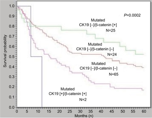
Previous studies have shown that 2.8–23.8% of patients with early-stage HCC have mutations in exon 3 of CTNNB1, which encodes the portion of the β-catenin protein that is involved in phosphorylation and degradation by the proteasome. In early HCC, these exon 3 mutations are associated with better prognosis in patients undergoing curative surgical resection of HCC tumors.Citation10,Citation19 According to one study showing the relation between β-catenin mutation and cytokeratin 19 (CK19) expression and their influence on HCC recurrence and prognosis, the frequency of vascular invasion and early tumor recurrence in HCCs with CK19 expression but not β-catenin mutation was higher than in HCCs without either β-catenin mutation or CK19 expression. HCCs with β-catenin mutation but not CK19 expression had the lowest frequencies of vascular invasion and early tumor recurrence.Citation39 Additionally, the overall 5-year survival of HCCs with β-catenin mutation alone was significantly better than the HCCs without β-catenin mutation with CK19 expression ().Citation39
Figure 4 HCCs with β-catenin mutation but not CK19 expression had the best 5-year survival rate, while HCCs with CK19 expression but not β-catenin mutation had the worst 5-year survival rate (P = 0.0002).
Abbreviations: HCC, hepatocellular carcinoma; CK19, cytokeratin 19.
In one recent study, it was observed that the mutation rate of CTNNB1 in patients with advanced HCC was not higher than that in early-stage HCC tumors, suggesting that CTNNB1 mutation did not significantly affect prognosis.Citation18
Wnt/b-catenin in predicting response to transarterial chemoembolization
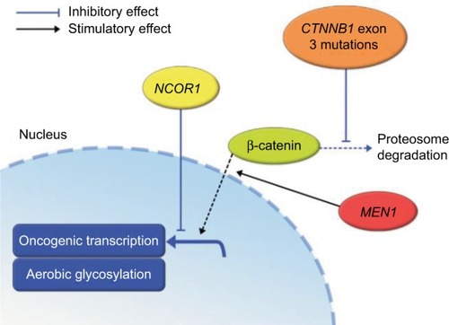
An important goal of ongoing research is to predict the response of HCC tumors to embolization therapies. It has been demonstrated that there is no statistically significant difference in the overall survival of HCC patients treated with embolization and chemoembolization therapies, suggesting that the efficacy of embolization therapy is primarily mediated by hypoxia. However, hypoxia results in a wide variety of signaling interactions that ultimately may lead to cell death or even promote cell survival. HCC tumors with mutations in CTNNB1 and MEN1 (a promoter of Wnt signaling) have been shown to have better response to embolization therapy than those with wild-type CTNNB1 and MEN1. Conversely, tumors with mutations in NCOR1 (a negative regulator of β-catenin transcriptional target genes) do not respond as well to embolization therapy ().Citation14,Citation41
Figure 5 Relationship between β-catenin, MEN1, and NCOR1 mutations and the Wnt signaling pathway summarized.Citation41
Imaging features of HCC associated with aberrant activation of Wnt/b-catenin
As discussed previously, the presence of β-catenin mutation in HCC tumors may provide important information regarding prognosis and treatment response. As a result, there have been attempts to determine whether non-invasive imaging methods can identify tumors that have β-catenin mutation and those that do not. Contrast-enhanced CT is one of the most common imaging modalities used for the detection and monitoring of HCC in patients. Overall, the sensitivity of CT for HCC has been reported to be approximately 68%, but triphasic multidetector CT may have a sensitivity of up to 100% for lesions greater than 2 cm and 96% for lesions between 1 and 2 cm in size.Citation2 A study by Kitao et al demonstrated that the attenuation and enhancement during the arterial phase of dynamic CT images could not differentiate between tumors with β-catenin mutation and those without.Citation27 The quantitative CT imaging features of HCC with β-catenin mutation were investigated at our institution to determine whether they could provide quantitative biomarkers to differentiate HCC with proven β-catenin mutation from those without mutation; this preliminary study demonstrated the feasibility of quantitative imaging feature extraction from contrast-enhanced CT imaging to differentiate HCC with proven β-catenin gene mutation and HCC without such mutation.Citation42 However, future work is needed to verify those findings.
MRI is also an extremely common imaging modality used for the evaluation of HCC. MRI sensitivities for detecting HCC have been reported to be around 81%.Citation2 On T1- and T2-weighted images, there appears to be no significant difference between HCC with and without β-catenin mutation. However, on diffusion-weighted imaging, HCC tumors with β-catenin mutation were shown to have lower contrast-to-noise ratios and higher apparent diffusion coefficients.Citation27 A higher apparent diffusion coefficient on diffusion-weighted imaging correlates with more tumors that are well differentiated.
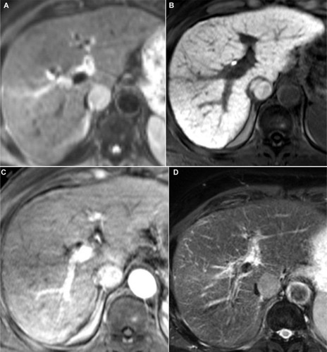
HCC tumors with β-catenin mutation were also shown to have higher contrast-to-noise ratios and higher enhancement ratios during the hepatobiliary phase (HBP) of gadoxetic acid-enhanced MRI ().Citation27,Citation43
Figure 6 Axial images in a 72-year-old man with HCC.
Abbreviation: HCC, hepatocellular carcinoma.
Gadoxetic acid (Gd-EOB-DTPA) is a contrast agent that is specific to hepatocytes and is useful for the identification of focal hepatic lesions. It can also be used for HBP imaging. Most HCC tumors appear hypointense compared with surrounding liver tissue on HBP images. This hypointensity is believed to be primarily due to a decrease in the expression of the organic anion transporting polypeptide 1B3 (OATP1B3), which is a transporter that takes up Gd-EOB-DTPA into hepatocytes. Expression of this transporter is usually decreased during HCC carcinogenesis. However, up to 10–20% of HCC tumors are either hyperintense or isointense on HBP images. HCC tumors that are hypointense on HBP () have lower expression of OATP1B3, while HCC tumors that are hyperintense on HBP () have high expression of OATP1B3. Interestingly, increased expression of OATP1B3 is strongly associated with increased Wnt/β-catenin signaling ().Citation43,Citation44 The presence of tumor enhancement on HBP images can accurately predict Wnt/β-catenin–activated HCC with a sensitivity of 78.9% and specificity of 81.7%.Citation27,Citation44 Furthermore, HCC tumors with increased expression of OATP1B3, in addition to hyperintensity on HBP imaging and low serum α-fetoprotein levels, have been associated with a better prognosis.Citation27,Citation43,Citation45
Figure 7 Axial image in HCV-related hepatitis showing HCC without β-catenin mutation.
Abbreviations: HCV, hepatitis C virus; HCC, hepatocellular carcinoma.
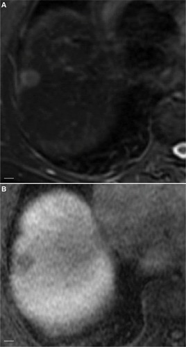
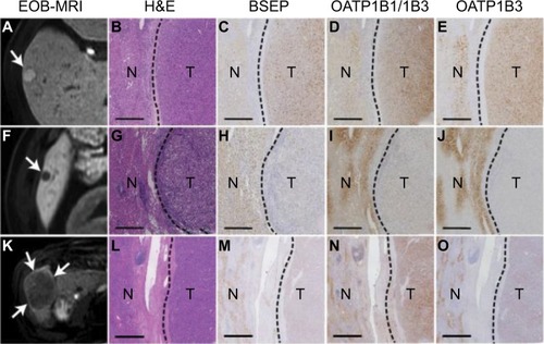
Figure 8 Relationship between expression of different genes and the tumor enhancement patterns in hepatobiliary phase images of EOB-MRI.Citation44
Abbreviation: EOB-MRI, gadoxetic acid-enhanced magnetic resonance imaging; H&E, hematoxylin and eosin staining; N, non-tumoural liver; T, tumor; HCC, hepatocellular carcinoma.
Therapies targeting the Wnt/β-catenin pathway and their anti-tumor activity
There is ongoing research focused on developing specific therapies that target molecular mechanisms involved in the pathogenesis and progression of HCC. Currently, sorafenib (a VEGF-targeted therapy) is the standard therapy that provides a small prolongation of overall survival for patients with advanced HCC. Other agents (regorafenib and nivolumab) were recently approved by the US Food and Drug Administration (USFDA) for the treatment of advanced HCC in patients who have been previously treated with sorafenib.Citation46,Citation47 However, there is evidence that sorafenib and other VEGF- targeted therapies may actually increase tumor cell invasion and metastasis.Citation12
Aberrant Wnt/β-catenin signaling has been shown to be common in HCC tumors and to have significant clinical impact on tumor behavior, prognosis, and response to treatment. As a result, Wnt/β-catenin may be a promising target for future HCC therapies.Citation48
Combretastatin A-1 phosphate (CA1P) is a microtubule polymerization inhibitor that is currently being studied as a promising treatment for HCC. CA1P has been shown to have significant antitumor activity in HCC tumors both in vitro and in vivo. CA1P is believed to exert its antitumor effects through inactivation of AKT, which in turn activates GSK-3B and inhibits the Wnt/β-catenin pathway.Citation48
FH535 is another small-molecule targeted therapy that is currently being studied in the treatment of HCC and hepatoblastoma. FH535 inhibits both peroxisome proliferator-activated receptor and β-catenin/LEF/TCF. This agent inhibits the proliferation of both HCC and hepatoblastoma cell lines. When combined with sorafenib, FH535 causes synergistic inhibition of HCC and hepatoblastoma cells. However, additional studies are still needed to determine the efficacy and side effects of FH535 in HCC patients.Citation12,Citation15
Currently, there are two clinical phase I/II trials studying the use of agents (such as PRI-724 and OMP-18R5) that specifically target the β-catenin signaling pathway to treat solid tumors and myeloid malignancies.Citation12
Risk stratification of hepatic adenomas expressing beta catenin biomarkers
HCAs transforming into HCCs is a rare occurrence with a reported frequency of 4.2%.Citation49 However, β-catenin mutation and the consequent Wnt signaling pathway activation play a big role in the proliferation of hepatic cells and the progression to malignancy.Citation49 Zucman-Rossi et al analyzed the genotype–phenotype correlation of hepatic adenomas and their relationship with HCC and found out that among the β-catenin-activated adenomas (10–15% of all adenomas), around 46% progressed to HCC which was a rare occurrence in HCAs with other types of mutations.Citation50 Glutamine synthase expression was associated with β-catenin activating mutations.Citation40,Citation50 Rebouissou et al found that in benign tumors, there are three levels of β-catenin activation depending on specific mutations: (1) weak activity by S45, K335, and N387 mutations; (2) moderate activity by T41 mutations; and (3) high activity through exon 3 deletions. High activity mutations were associated with malignancy and strong GS expression. Weak mutations were more frequent in HCA except for S45 mutation which was identified in 20% of mutated HCAs and HCCs.Citation40 Mutations in CTNNB1 exon 7 or 8 were associated with mild activations of Wnt/β-catenin signaling and without increased risk of malignant transformation unlike CTNNB1 exon 3 mutations.Citation51 HCAs with high/moderate or S45 mutations should be followed up for potential HCC transformation and may necessitate more aggressive treatment options.Citation40
Conclusion
Mutations of the CTNNB1 gene are thought to be the most common genetic abnormality leading to the development of HCC. Multiple studies suggest that the presence of β-catenin mutation in HCC tumors is associated with a more favorable prognosis and provides useful information about treatment response and tumor behavior. However, the accumulation of β-catenin in the nucleus and cytoplasm may also be associated with increased vascular invasion, cell proliferation, and more poorly differentiated tumors. It has also been shown that HCV-related HCC tumors have a significantly increased frequency of CTNNB1 mutations compared with HBV-related and non-viral HCC tumors. While most HCC tumors appear hypointense compared with surrounding liver tissue on HBP images, the presence of tumor enhancement on HBP images can accurately predict Wnt/β-catenin-activated HCC.
More research is needed to determine the effect of β-catenin mutation on HCC tumor pathogenesis, prognosis, and behavior. Nowadays, small molecular therapy targeting the Wnt/β-catenin pathway is being investigated for treatment of HCC. Improving our understanding of the clinical and pathologic significance of the molecular mechanisms that lead to Wnt/β-catenin signaling activation in HCC tumors will guide the development of new targeted therapies for HCC.
Although HCA progression into HCC is a rare event, the β-catenin-activated subtype of HCAs was the most frequent to turn malignant, warranting more aggressive follow-up and treatment options such as complete resection.
Acknowledgments
The University of Texas is supported in part by the National Institutes of Health through Cancer Center Support Grant P30CA016672. The authors would also like to thank the department of scientific publication at The University of Texas, MD Anderson Cancer Center for their contribution.
Disclosure
The authors report no conflicts of interest in this work.
References
- LiPCaoYLiYZhouLLiuXGengMExpression of Wnt-5a and B-catenin in primary hepatocellular carcinomaInt J Clin Exp Pathol2014763190319525031739
- ShanbhogueAKPrasadSRTakahashiNVikramRSahaniDVRecent advances in cytogenetics and molecular biology of adult hepatocel-lular tumors: implications for imaging and managementRadiology2011258367369321339346
- WaisbergJSabaGTWnt−/−β-catenin pathway signaling in human hepatocellular carcinomaWorld J Hepatol20157262631263526609340
- DahmaniRJustP-APerretCThe Wnt/β-catenin pathway as a therapeutic target in human hepatocellular carcinomaClin Res Hepatol Gastroenterol2011351170971321778132
- OishiNWangXWNovel therapeutic strategies for targeting liver cancer stem cellsInt J Biol Sci20117551753521552419
- WangZShengYGaoXβ-catenin mutation is correlated with a favorable prognosis in patients with hepatocellular carcinomaMol Clin Oncol20153493694026171210
- LeeJMYangJNewellPβ-Catenin signaling in hepatocellular cancer: implications in inflammation, fibrosis, and proliferationCancer Lett20143431909724071572
- LiuLJXieSXChenYTXueJLZhangCJZhuFAberrant regulation of WNT signaling in hepatocellular carcinomaWorld J Gastroenterol201622337486749927672271
- WangWPanQFuhlerGMSmitsRPeppelenboschMPAction and function of Wnt/β-catenin signaling in the progression from chronic hepatitis C to hepatocellular carcinomaJ Gastroenterol201752441943128035485
- de La CosteARomagnoloBBilluartPSomatic mutations of the beta-catenin gene are frequent in mouse and human hepatocellular carcinomasProc Natl Acad Sci U S A19989515884788519671767
- FriemelJRechsteinerMFrickLIntratumor heterogeneity in hepatocellular carcinomaClin Cancer Res20152181951196125248380
- VilchezVTurciosLMartiFGedalyRTargeting Wnt/B-catenin pathway in hepatocellular carcinoma treatmentWorld J Gastroenterol201622282383226811628
- WangHZhangJFengWPIN1 gene overexpression and ?-catenin gene mutation/expression in hepatocellular carcinoma and their significanceJ Huazhong Univ Sci Technol20072715457
- XuWZhouWChengMHypoxia activates Wnt/β-catenin signaling by regulating the expression of BCL9 in human hepatocellular carcinomaSci Rep201774044628074862
- GedalyRGaluppoRDailyMFTargeting the Wnt/β-catenin signaling pathway in liver cancer stem cells and hepatocellular carcinoma cell lines with FH535PLoS One201496e9927224940873
- KondoYKanaiYSakamotoMβ-catenin accumulation and mutation of exon 3 of the β-catenin gene in hepatocellular carcinomaJpn J Cancer Res199990121301130910665646
- PolakisPWnt signaling and cancerGenes Dev200014151837185110921899
- LuLCShaoYYLeeYHβ-Catenin (CTNNB1) mutations are not associated with prognosis in advanced hepatocellular carcinomaOncology201487315916625012536
- HsuHCJengYMMaoTLChuJSLaiPLPengSYBeta-catenin mutations are associated with a subset of low-stage hepatocellular carcinoma negative for hepatitis B virus and with favorable prognosisAm J Pathol2000157376377010980116
- MaoTLChuJSJengYMLaiPLHsuHCExpression of mutant nuclear beta-catenin correlates with non-invasive hepatocellular carcinoma, absence of portal vein spread, and good prognosisJ Pathol200119319510111169521
- NhieuJTRenardCAWeiYCherquiDZafraniESBuendiaMANuclear accumulation of mutated β-catenin in hepatocellular carcinoma is associated with increased cell proliferationAm J Pathol1999155370371010487827
- PolakisPThe oncogenic activation of beta cateninCurr Opin Genet Dev199991152110072352
- SuzukiTYanoHNakashimaYNakashimaOKojiroMBeta-catenin expression in hepatocellular carcinoma: a possible participation of beta-catenin in the dedifferentiation processJ Gastroenterol Hepatol2002179994100012167121
- MacDonaldBTTamaiKHeXWnt/β-catenin signaling: components, mechanisms, and diseasesDev Cell200917192619619488
- PengSYChenWJLaiPLJengYMSheuJCHsuHCHigh α-fetoprotein level correlates with high stage, early recurrence and poor prognosis of hepatocellular carcinoma: significance of hepatitis virus infection, age, p53 and β-catenin mutationsInt J Cancer20041121445015305374
- TerrisBPineauPBregeaudLClose correlation between β-catenin gene alterations and nuclear accumulation of the protein in human hepatocellular carcinomasOncogene199918476583658810597262
- KitaoAMatsuiOYonedaNHepatocellular carcinoma with β-catenin mutation: imaging and pathologic characteristicsRadiology2015275370871725668519
- TienLTItoMNakaoMExpression of beta-catenin in hepatocellular carcinomaWorld J Gastroenterol200511162398240115832407
- Zucman-RossiJBenhamoucheSGodardCDifferential effects of inactivated Axin1 and activated β-catenin mutations in human hepatocellular carcinomasOncogene200726577478016964294
- KimSSChoHJLeeH-YGenetic polymorphisms in the Wnt/B-catenin pathway genes as predictors of tumor development and survival in patients with hepatitis B virus-associated hepatocellular carcinomaClin Biochem20164910–1179280126968103
- WongCMFanSTNgIOLβ-catenin mutation and overexpression in hepatocellular carcinoma: clinicopathologic and prognostic significanceCancer200192113614511443619
- WangWXuLLiuPBlocking Wnt secretion reduces growth of hepatocellular carcinoma cell lines mostly independent of β-catenin signalingNeoplasia2016181271172327851986
- HuangHFujiiHSankilaABeta-catenin mutations are frequent in human hepatocellular carcinomas associated with hepatitis C virus infectionAm J Pathol199915561795180110595907
- LevreroMZucman-RossiJMechanisms of HBV-induced hepatocellular carcinomaJ Hepatol2016641 SupplS84S10127084040
- Zucman-RossiJVillanuevaANaultJCLlovetJMGenetic landscape and biomarkers of hepatocellular carcinomaGastroenterology201514951226123926099527
- AustinatMDunschRWittekindCTannapfelAGebhardtRGaunitzFCorrelation between β-catenin mutations and expression of Wnt-signaling target genes in hepatocellular carcinomaMol Cancer2008712118282277
- InagawaSItabashiMAdachiSExpression and prognostic roles of beta-catenin in hepatocellular carcinoma: correlation with tumor progression and postoperative survivalClin Cancer Res20028245045611839663
- BogaertsEHeindryckxFVandewynckelYPVan GrunsvenLAVan VlierbergheHThe roles of transforming growth factor-β, Wnt, Notch and hypoxia on liver progenitor cells in primary liver tumours (review)Int J Oncol20144441015102224504124
- YuanRHJengYMHuRHRole of p53 and β-catenin mutations in conjunction with CK19 expression on early tumor recurrence and prognosis of hepatocellular carcinomaJ Gastrointest Surg201115232132921061181
- RebouissouSFranconiACalderaroJGenotype-phenotype correlation of CTNNB1 mutations reveals different β-catenin activity associated with liver tumor progressionHepatology20166462047206127177928
- ZivEYarmohammadiHBoasFEGene signature associated with upregulation of the Wnt/beta-catenin signaling pathway predicts tumor response to transarterial embolizationJ Vasc Interv Radiol2017283349.e1355.e128126478
- KhalafAMFuentesDTAhmedKQuantitative CT imaging features for hepatocellular carcinoma (HCC) with b-catenin (CTNNB1) gene mutationJ Clin Oncol2017354 Suppl25325328056207
- JooILeeJMRecent advances in the imaging diagnosis of hepatocellular carcinoma: value of gadoxetic acid-enhanced MRILiver Cancer201551678726989660
- UenoAMasugiYYamazakiKOATP1B3 expression is strongly associated with Wnt/B-catenin signalling and represents the transporter of gadoxetic acid in hepatocellular carcinomaJ Hepatol20146151080108724946283
- VilgrainVVan BeersBEPastorCMInsights into the diagnosis of hepatocellular carcinomas with hepatobiliary MRIJ Hepatol201664370871626632635
- El-KhoueiryABSangroBYauTNivolumab in patients with advanced hepatocellular carcinoma (CheckMate 040): an open-label, non-comparative, phase 1/2 dose escalation and expansion trialLancet2017389100882492250228434648
- KimKJhaRPrinsPARegorafenib in advanced hepatocellular carcinoma (HCC): considerations for treatmentCancer Chemother Pharmacol201780594595428932966
- MaoJWangDWangZCombretastatin A-1 phosphate, a microtubule inhibitor, acts on both hepatocellular carcinoma cells and tumor-associated macrophages by inhibiting the Wnt/β-catenin pathwayCancer Lett2016380113414327349166
- StootJHMBCoelenRJSDe JongMCDejongCHCMalignant transformation of hepatocellular adenomas into hepatocellular carcinomas: a systematic review including more than 1600 adenoma casesHPB (Oxford)201012850952220887318
- Zucman-RossiJJeannotEVan NhieuJTGenotype-phenotype correlation in hepatocellular adenoma: new classification and relationship with HCCHepatology200643351552416496320
- NaultJCCouchyGBalabaudCMolecular classification of hepatocellular adenoma associates with risk factors, bleeding, and malignant transformationGastroenterology2017152488089427939373