Abstract
Purpose
The purpose of this work was to determine the pro-and anti-inflammatory properties of the single-cell organism Euglena gracilis (EG) and various fractions of its whole biomass.
Methods
Heterotrophically grown EG was tested, along with its aqueous fraction (E-AQ), the intact linear β-glucan paramylon granules (PAR), and alkaline-solubilized paramylon. Peripheral blood mononuclear cell cultures were treated with the test products and analyzed for a variety of cellular responses. Immune cell activation was evaluated by flow cytometry detection of CD69 levels on CD3−CD56+ NK cells, CD3+CD56+ NKT cells, and monocytes, and cytokines were analyzed from the cell culture supernatants. Antioxidant capacity was measured by Folin–Ciocalteu assay and cellular antioxidant protection and MTT assays.
Results
EG and E-AQ were the most effective in driving immune cell responses as measured by CD69 upregulation on NK and NKT cells and proinflammatory (tumor necrosis factor, IL-6, IL-1β) cytokine production. None of the test products effectively stimulated monocyte. EG and PAR inhibited reactive oxygen species under conditions of oxidative stress. E-AQ contained antioxidants capable of providing cellular antioxidant protection from oxidative damage and protection of mitochondrial function under inflammatory conditions.
Conclusion
The effects of EG on immune function are only partially attributable to the content of the β-glucan, paramylon. The regulation of additional cellular responses, such a reactive oxygen species production and resistance to oxidative stress, is likely mediated by currently unknown molecules found in the EG cell.
Acknowledgments
The authors would like to thank Kelli Herrlinger, Laura Wonderling, Sally Moore, and Jon Rubach of Kemin Industries (Des Moines, IA, USA) for their careful review of this manuscript, Katie-Jo Galayda of Kemin Industries (Des Moines, IA, USA) for her molecular weight analysis of β-glucan, and Tracey Stewart of Iowa State University (Ames, IA, USA) biotechnology service facility for her assistance with the scanning electron microscope.
Disclosure
This study was sponsored by Kemin Industries, Des Moines, IA, USA. RL serves as a consultant for Kemin Industries. FCP, RT, and GH are employed by Kemin Industries. GSJ holds a patent for the CAP-e assay (patent number 8465988) and is employed by NIS Labs, Klamath Falls, OR, USA. The other authors report no other conflicts of interest in this work.
Supplementary material
Figure S1 Physical characteristics of E. gracilis and paramylon.
Notes: (A) Chemical structure of 1,3-β-glucan. (B) Light microscopy image of live E. gracilis filled with paramylon granules. (C) Dried E. gracilis material for EG and E-AQ fractions. (D) Purified paramylon powder for PAR and PAR-S fractions. (E) Scanning electron microscopy images of paramylon material. 40× magnification.
Abbreviations: E-AQ, aqueous fraction of EG; EG, Euglena gracilis whole algae; PAR, granular paramylon; PAR-s, alkaline-solubilized paramylon.
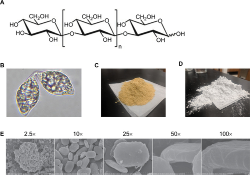
Figure S2 CD69 expression on human PBMCs stimulated with pathogen-associated molecular patterns is modulated by EG, but not E-AQ, Par, or PAR-S.
Notes: Human PBMCS from three healthy donors were stimulated in vitro for 24 hours with LPS (10 ng/ml) (A–C) or poly I:C (2.5 μg/ml) (D–F), plus each of the four test products (EG, E-AQ, PAR, and PAR-S), stained and analyzed by flow cytometry for expression of CD69, which is represented as a fold increase in MFI over the LPS control (=1) (A) monocytes, (B) NK cells, and (C) NKT cells were stimulated with LPS. (D) Monocytes, (E) NK cells, and (F) NKT cells were stimulated with poly I:C. All samples were analyzed in triplicate. Symbols represent mean ± SD.
Abbreviations: E-AQ, aqueous fraction of EG; EG, Euglena gracilis whole algae; LPS, lipopolysaccharide; MFI, mean fluorescence intensity; PAR, granular paramylon; PAR-S, alkaline-solubilized paramylon; PBMCs, peripheral blood mononuclear cells; poly I:C, polyinosinic–polycytidylic acid.
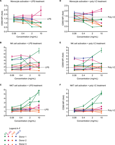
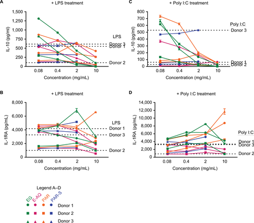
Figure S3 anti-inflammatory cytokine production by human PBMCs stimulated with pathogen-associated molecular patterns may be regulated by EG and PAR.
Notes: Human PBMCs from three healthy donors were stimulated in vitro for 24 hours with LPS (10 ng/ml) (A, B) or poly I:C (2.5 μg/ml) (C, D), plus each of the four test products (EG, E-AQ, PAR, and PAR-S). Cell culture supernatants were analyzed by luminex multiplex for (A, C) IL-10 and (B, D) IL-1RA. All samples were analyzed in triplicate. Symbols represent mean ± SD.
Abbreviations: E-AQ, aqueous fraction of EG; EG, Euglena gracilis whole algae; IL-IRA, IL-1 receptor antagonist; LPS, lipopolysaccharide; PAR, granular paramylon; PAR-S, alkaline-solubilized paramylon; PBMCs, peripheral blood mononuclear cells; poly I:C, polyinosinic–polycytidylic acid.