Abstract
Background
Parkinson’s disease (PD) patients frequently present gastrointestinal (GI) dysfunction that, in many cases, predates the onset of motor symptoms. In PD, the presynaptic protein alpha-synuclein (α-syn) undergoes pathological changes, including phosphorylation and aggregation leading to the formation of Lewy bodies, which can be found in neurons of the enteric nervous system (ENS). Inflammation has been proposed as a possible trigger of α-syn pathology. Interestingly, patients with inflammatory bowel disease and irritable bowel syndrome, conditions associated with GI inflammation, are at higher risk of developing PD. Captive common marmosets (Callithrix jacchus) develop colitis, providing a natural platform to assess the relationship between α-syn pathology and GI inflammation.
Materials and Methods
Sections of proximal colon from marmosets with colitis (n=5; 5.3±2.3 years old; 4 male) and normal controls (n=5; 4.1±1.6 years old; 1 male) were immunostained against protein gene product 9.5 (PGP9.5), human leukocyte antigen DR (HLA-DR), cluster of differentiation 3 (CD3), cluster of differentiation 20 (CD20), glial fibrillary acidic protein (GFAP), 8-hydroxy-2’-deoxyguanosine (8-OHdG), α-syn, and serine 129 phosphorylated α-syn (p-α-syn). Immunoreactivity of each staining in the myenteric plexus was quantified using NIH ImageJ software.
Results
Marmosets with colitis had significantly increased expression of inflammatory markers (HLA-DR, p<0.02; CD3, p<0.008), oxidative stress (8-OHdG, p<0.05), and p-α-syn (p<0.02) and decreased expression of α-syn (p<0.04) in the colonic myenteric ganglia compared to normal, healthy controls.
Conclusion
Colonic inflammation is associated with changes in α-syn expression and phosphorylation in the myenteric plexus of common marmosets. Future evaluation of the vagus nerve and brain of animals with colitis will be key to assess the contribution of colitis-induced ENS α-syn pathology to PD-like pathology in the brain.
Acknowledgments
We gratefully acknowledge Dr. Shahriar Salamat and the Wisconsin Alzheimer’s Disease Research Center (P50-AG033514) for providing human PD brain tissue. NIH P51OD011106, NIH UL1TR000427, NIH R24OD019803, NIH Kirschstein-NRSA F31HL136047 (JMM), Welton L&S Honors Sophomore Scholarship (HR), PF-APDA-SFW-1854 (HR) and the University of Wisconsin–Madison Office of Vice Chancellor for Research and Graduate Education, Cellular and Molecular Pathology Graduate Program, and Department of Medical Physics.
Data Availability
The datasets generated during and/or analyzed during the current study are available from the corresponding author on reasonable request.
Disclosure
The authors report no conflicts of interest in this work.
Supplementary materials
Figure S1 S129 phosphorylated α-syn in a sample of brain from a human PD patient. (A and B) Representative photomicrographs at 63× of p-α-syn-ir in human PD brain in (A) a neuromelanin laden neuron and (B) a neurite. Scale bar =50 μm.
Abbreviations: p-α-syn-ir, S129 phosphorylated alpha-synuclein- immunoreactivity; PD, Parkinson’s disease.
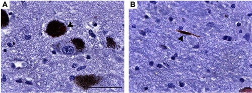
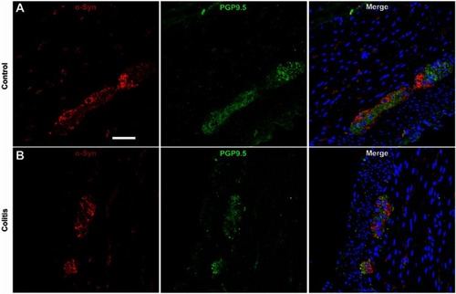
Figure S2 α-Syn co-labeling with the panneuronal marker PGP9.5 in myenteric ganglia is similar in control and colitis animals. (A and B) Representative photomicrographs at 40× showing immunofluorescent co-labeling of α-syn and PGP9.5 in myenteric ganglia and nerve fibers in (A) control and (B) colitis animals. Note that small punctate spots in the PGP9.5 immunofluorescence are technical artifact/background. Scale bar =50 μm.
Abbreviations: α-syn, alpha-synuclein; PGP9.5, protein gene product 9.5.