
Abstract
Every element or cell in the human body produces substances that communicate and respond in an autocrine or paracrine mode, consequently affecting organs and structures that are seemingly far from each other. The same also applies to the skin. In fact, when the integrity of the skin has been altered, or when its healing process is disturbed, it becomes a source of symptoms that are not merely cutaneous. The skin is an organ, and similar to any other structure, it has different functions in addition to connections with the central and peripheral nervous system. This article examines pathological responses produced by scars, analyzing definitions and differences. At the same time, it considers the subcutaneous fascias, as this connective structure is altered when there is a discontinuous cutaneous surface. The consequence is an ample symptomatology, which is not limited to the body area where the scar is located, such as a postural or trigeminal disorder.
Keywords:
Introduction: definition of skin and fascia
The integument is composed of the epidermis and the dermis, which derive from different embryological sheets. The epidermis is the surface epithelium that originates in the ectoderm, meaning it is part of the structures that are in contact with the outside world.Citation1,Citation2 Among the other structures derived from the ectoderm, we identify the central and peripheral nervous system, the pituitary gland, the dental epithelium, and the mammary gland.Citation3–Citation6 Here we can find not only keratinocytes (which represent the first immune barrier, as they act as sentinels) but also melanocytes and immune cells (such as the Langerhans cells and the T-lymphocytes).Citation7,Citation8 There are also tactile cells, which are differentiated for selective touch.Citation9
Figure 1 Transverse section at the level of the upper third of the leg. The fascia is the philosophy of the body, meaning each body region is connected to another, whereas osteopathy is the philosophy of medicine: the entire human body must work in harmony. Reproduced with permission anastasi et al. AA VV, anatomia dell’uomo, 4 ed, Edi.ermes, Milano [Human anatomy].Citation114
![Figure 1 Transverse section at the level of the upper third of the leg. The fascia is the philosophy of the body, meaning each body region is connected to another, whereas osteopathy is the philosophy of medicine: the entire human body must work in harmony. Reproduced with permission anastasi et al. AA VV, anatomia dell’uomo, 4 ed, Edi.ermes, Milano [Human anatomy].Citation114](/cms/asset/6a79ac0b-bbb1-49e1-b18e-61e5bb06012b/djmd_a_52870_f0001_c.jpg)
Figure 2 The photo shows atrophic scar on the shoulder.
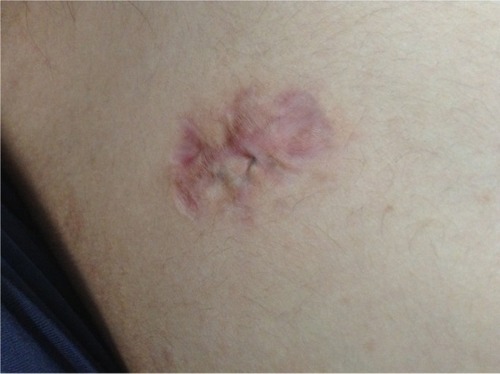
Figure 3 Intervention of sternotomy after cardiac surgery. There is a high percentage of risk that the scar could develop into a pathological scar.
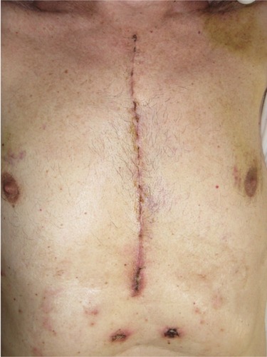
Figure 4 General organization and territories of innervation of the sympathetic nervous system (blue) and parasympathetic nervous system (red). We can logically assume that in the presence of a scar, these receptors may experience an alteration, resulting in transmitting nonphysiological signals and creating a pathological reflex arc. Reproduced with permission Anastasi et al. AA VV, Anatomia dell’uomo, 4 ed, Edi.ermes, Milano [Human Anatomy].Citation114
![Figure 4 General organization and territories of innervation of the sympathetic nervous system (blue) and parasympathetic nervous system (red). We can logically assume that in the presence of a scar, these receptors may experience an alteration, resulting in transmitting nonphysiological signals and creating a pathological reflex arc. Reproduced with permission Anastasi et al. AA VV, Anatomia dell’uomo, 4 ed, Edi.ermes, Milano [Human Anatomy].Citation114](/cms/asset/493deede-9c1f-43c8-826e-d9b3b6e17aa7/djmd_a_52870_f0004_c.jpg)
Figure 5 The structures of the spinal cord and blood vessels, rear projection, are made up of various layers but are continuous. However, it is usually ignored when the tissue where the tension is observable is in an unbalanced condition, as in the case when a scar is present, the cells cannot properly interpret the message, giving consequent anomalous responses. Reproduced with permission Anastasi et al. AA VV, Anatomia dell’uomo, 4 ed, Edi.ermes, Milano [Human anatomy].Citation114
![Figure 5 The structures of the spinal cord and blood vessels, rear projection, are made up of various layers but are continuous. However, it is usually ignored when the tissue where the tension is observable is in an unbalanced condition, as in the case when a scar is present, the cells cannot properly interpret the message, giving consequent anomalous responses. Reproduced with permission Anastasi et al. AA VV, Anatomia dell’uomo, 4 ed, Edi.ermes, Milano [Human anatomy].Citation114](/cms/asset/2a827ec7-502d-4885-a2aa-d9bb280346d4/djmd_a_52870_f0005_c.jpg)
Figure 6 Fascias of the neck. Deep traumas can also affect the fascia and the viscera, which then go through an identical healing process. Reproduced with permission Anastasi et al. AA VV, Anatomia dell’uomo, 4 ed, Edi.ermes, Milano [Human anatomy].Citation114
![Figure 6 Fascias of the neck. Deep traumas can also affect the fascia and the viscera, which then go through an identical healing process. Reproduced with permission Anastasi et al. AA VV, Anatomia dell’uomo, 4 ed, Edi.ermes, Milano [Human anatomy].Citation114](/cms/asset/f4944624-3c03-4feb-bab8-9606d4fccc22/djmd_a_52870_f0006_c.jpg)
Figure 7 A surgery to remove the radial artery. Furthermore, the fascial tissue is made of contractile fibers, which may possibly produce spasms and consequential dysfunction and pain.
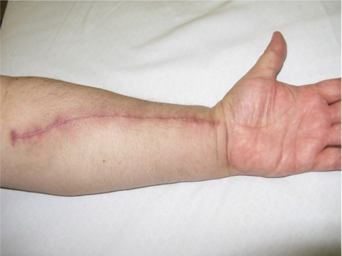
Figure 8 Continuity from the pelvis to the sternum. Muscles of the anterior wall of the trunk; view from inside. In addition, we can assume a malfunction of the respiratory diaphragm because of the stiffening of the lumbodorsal spine, to which the primary muscle of respiration is closely connected. Reproduced with permission Anastasi et al. AA VV, Anatomia dell’uomo, 4 ed, Edi.ermes, Milano [Human Anatomy].Citation114
![Figure 8 Continuity from the pelvis to the sternum. Muscles of the anterior wall of the trunk; view from inside. In addition, we can assume a malfunction of the respiratory diaphragm because of the stiffening of the lumbodorsal spine, to which the primary muscle of respiration is closely connected. Reproduced with permission Anastasi et al. AA VV, Anatomia dell’uomo, 4 ed, Edi.ermes, Milano [Human Anatomy].Citation114](/cms/asset/e1def55e-fd27-44db-ac13-a6eaafd2a85e/djmd_a_52870_f0008_c.jpg)
The dermis originates in the mesoderm, where, among other things, the connective tissue, bone, cartilage, blood, and their relevant organs are found.Citation10–Citation12 The dermis supports the epidermis and nourishes it.Citation13 Numerous lymphatic and vascular vessels go through it, as well as different nerve endings, which can be free, as in the epidermis, or encapsulated in complex structures.Citation9,Citation13,Citation14
The skin, with its 1.8 m2 surface, is one of the largest organs of the human body and is the one most exposed to the environment.Citation13 It is subject to a continual exchange of information, not just tactile but also concerning light and liquids. Therefore, we can reasonably affirm that it is always electrically active.Citation13–Citation15 The skin sends and receives information to the entire body, although with different intensities depending on the location.Citation16 For instance, the electrical activity of the dermis reflects its sympathetic sudomotor cholinergic function, which brings about continuous changes in the electrical conduction property of the skin, depending on external and internal stimuli.Citation17 To make another example, the mechanoreceptors of the skin always give information on posture, using low-threshold mechanosensitive afferents.Citation18,Citation19 Therefore, the skin is continuously developing. It is a mistake to think of the cutaneous surface as something unchangeable; on the contrary, it is always changing and directly participates in the homeostasis of the human body.
Beneath the dermis, there is an adipose layer, which also derives from the mesoderm.Citation20 Beneath the adipose layer, we find the so-called fascia. The fascia should be regarded as a connective sheet that covers various areas by perfectly adhering to them like velcro, composed of irregularly arranged collagen fibers that are markedly different from the regularly arranged collagen fibers recognizable in tendons, ligaments, or aponeurotic sheets.Citation21 The fascia surrounds and connects every muscle, even the tiniest myofibril, bones, nerves, and every single organ, forming a fascial system and bodily continuity.Citation22–Citation24 Embryologically, the fascia derives from the mesoderm, being the continuation of the connective tissue.Citation24 The fascial system consists of several layers, each characterized by different direction and thickness, which are constantly and jointly communicating and exchanging information. The fascia is the philosophy of the body, meaning each body region is connected to another, whereas osteopathy is the philosophy of medicine: the entire human body must work in harmony.
Definition of scar and endorsed hypotheses
There are four main stages in skin healing: hemostasis, inflammation, proliferation, and remodeling.Citation15 The healing process of the skin surface begins with the lesion, when the bleeding conveys the blood elements in the area of the injury (blood platelets, fibrin, fibronectin, glycoprotein), trying to produce a parallel vasoconstriction.Citation15 The blood platelets come in contact with the exposed collagen and with other elements of the extracellular matrix.Citation15 This contact induces the release of important growth factors (transforming growth factor-beta [TGF-β] and platelet-derived growth factor [PDGF]), whereas coagulants begin the reconstitution process.Citation25 Coagulation belongs to the first reconstitution stage of the injured tissue, hemostasis.Citation15 This process results in a deposit of fibrin and other similar substances, which represents a provisional matrix of successive healing events.Citation15 The growth factors, such as TGF-β and PDGF, are the most important cytokines that initiate the second stage of the skin healing process; namely, inflammation.Citation26,Citation27 PDGF produces the chemotaxis (ie, addressing movements toward a specific stimulus) of other elements of the process, such as neutrophils, macrophages, fibroblasts, and smooth muscle cells.Citation15,Citation27 The first cytokine, in contrast, attracts other macrophages and induces them to secrete further cytokines, controlling the action of the PDGF along with the production/secretion of collagen and of the relevant enzymes degrading the collagen.Citation15 Within 24 hours of the lesion, inflammation is increased by neutrophils, which enter the lesion and clean it of foreign matter.Citation15 This action can even last more than a week.Citation15 The mastocytes, which secrete various substances such as leukotrienes, interleukins, and other enzymes, along with the neutrophils, are responsible for the symptoms of inflammation and vasodilatation: redness, heat, swelling, and pain.Citation15 Other substances such as monocytes are activated within 2 days.Citation15 They indicate the beginning of a subsequent overlapping stage; namely, proliferation. Therefore, once the wound has been cleaned, within 8–14 days from the injury, the third healing stage begins, involving the migration of the fibroblasts toward the provisional extracellular matrix made of fibrin and collagen.Citation15 As this stage proceeds, the release of TGF-β produced by different groups of cells controls the numerous functions of the fibroblasts.Citation15,Citation26,Citation27
As the collagen increases (it is important to reinforce the wound), other substances such as the enzymes degrading the collagen decrease.Citation15 At the beginning, the rate of collagen production exceeds the decrease of other substances.Citation15 The fibroblasts, which are stimulated by the different growth factors, are divided into myofibroblasts comparable with smooth muscle cells (they hold a protein, which is the alpha smooth muscle actin).Citation15,Citation26 These cells can increase the traction force, and thus they contract and reduce the open area of the lesion.Citation15 After their contraction, they begin their apoptosis and gradually disappear.Citation15 The concurrent breaking of the provisional extracellular matrix stops the activity of the fibroblasts.Citation15 The last healing stage, remodeling, then begins. Remodeling can last for years and depends on the size and nature of the wound.Citation15,Citation27 In this phase, type 3 collagen is replaced by a stronger fiber such as type 1 collagen, but it is lined up without a specific order and is smaller than the collagen of an undamaged skin surface.Citation15,Citation27 This results in more strength but less elasticity. Gradually, as long as the healing process follows a physiological course of action, the scar will lose its erythematous appearance.Citation15 The healing process undergone by the internal organs, for example, after surgery, is the same as that observed for the skin.Citation26,Citation28
What happens, however, if these processes have been altered? The scar can shift the healing process toward a nonphysiological state, giving origin to a hypertrophic scar (HS), a keloid scar (KS), or an atrophic scar (AS), each one with a different etiology. Scars appear after traumas, surgery, or burns. The HS is, by definition, a healing process increasing in height but always confined to the area of the original wound.Citation25 A KS not only increases in height but also proliferates beyond the borders of the original lesion.Citation29 KSs and HSs may represent two different stages of the same disease.Citation30,Citation31 The AS appears as a cutaneous depression.Citation32 When the dermis and the fascia are affected by scars, these structures are altered, and their function and capacity of interaction with the external and internal environment are lacking.
Many are the reasons that provoke such events, from genetic predisposition, to age, but they are still not clearly understood.Citation29,Citation33 At this time, the scientific panorama offers several hypotheses, but the most endorsed is mainly the neuroinflammatory or neurogenic inflammation hypothesis.
According to some lines of thought, an excess of neuroinflammatory stimuli and a release of neuropeptides (substance P and peptides that release calcitonin) can be observed, which prolong the production of growth factors and cytokines, generating an extracellular matrix in excess.Citation30,Citation31 The neuroinflammatory overstimulation is probably a result of a reflex arc at the medullary level, which comes from the injury (sensory nerves with nonmyelinated fibers of type C and delta fibers) and then returns as a neuroinflammatory signal, with a consequent excess activity of neuropeptides.Citation34 Research has confirmed an increase of nerves in the region of scarring, particularly HSs, and an accumulation of neuropeptides.Citation34 This means a scar can present daily stimuli, enabling us to understand the symptoms we are going to examine later in this article. What generates this inflammatory overstimulation? According to some textbooks, skin-rubbing stimulates a neurogenic inflammatory response,Citation31 and a tight dress is enough to stimulate a neurogenic reflex arc.
According to another current thought, this neuroinflammatory response is mainly caused by stimuli of anomalous tension originated in the injury.Citation31,Citation33 This process provokes a release of neuropeptides from cells native to the extracellular matrix (therefore, not only those belonging to the nerve endings), stimulating an inflammatory reflex arc.Citation30,Citation31,Citation35 With reference to this idea, the tension on the injury depends on the direction of the injury itself; for example, in a vertical or horizontal direction compared with the axis of the leg.Citation36 In fact, in the case of a horizontal scar, there will be a tension three times greater.Citation37
Tension is certainly a factor predisposing the onset of these problems, but we still do not know whether it initiates a deviation from the physiological healing process of an injury or affects the scar once the healing process has concluded. The force lines recordable in KSs are addressed outside of the scar, with a consequent expansion of the tissue; recent studies have proven that their margins are pulled outward, with considerable peripheral tension, whereas the central area is subject to a mild tension.Citation30,Citation31 It is well-known that KSs and HSs frequently arise in specific sites; namely, the anterior chest, the shoulders (especially the scapular area), the lower abdomen, and the earlobes.Citation36
Mechanical forces may be perceived by two types of receptors: the mechanoreceptor (or mechanosensor) and the mechanosensitive nociceptor. The first deals with the information of mechanical modifications, whereas the second mostly controls the information of pain. However, both convey somatic information.Citation36 We can logically assume that in the presence of a scar, these receptors may experience an alteration, resulting in transmitting nonphysiological signals and creating a pathological reflex arc. It is worth remembering that the language of the cell is, precisely, the tension, which activates the mechanotransduction (ie, the chemicobiological and metabolic response to the tension).Citation35,Citation36,Citation38 This response will be different depending on how the tension emerges and depending on different parameters. However, it is usually ignored that if the tissue where the tension is observable is in an unbalanced condition, as in the case when a scar is present, the cells cannot properly interpret the message, giving consequent anomalous responses. At this time, one of the reasons explaining the finding of KSs is the constant stretching of injured tissue.Citation36,Citation39 However, the real causes that give rise to a nonphysiological scar are still obscure. With regard to the lymphatic system and its correlation with nonphysiological scars, there is little information found in the medical literature. A difficulty in the lymphatic drainage of the area concerned is presumed, but it is still unknown whether this may cause further damage or not.
Deep traumas can also affect the fascia and the viscera, which then go through an identical healing process. The fascia is rich in corpuscles (ie, the Golgi’s, the Pacini’s, and the Ruffini’s corpuscles), with proprioceptive properties and significant peripheral information, as well as with probable nociceptive function.Citation40 Furthermore, the fascial tissue is made of contractile fibers, which may possibly produce spasms and consequential dysfunction and pain.Citation40 Via the postsynaptic dorsal column and the spinothalamic tract, the viscera send signals to the spinal cord, which convey information related to pain and to interoceptive alterations. Visceral primary afferents are known to be rich in neuropeptides, such as substance P. This information will be useful in understanding the symptomatology described later.Citation41,Citation42 In conclusion, we emphasize that the percentage of adhesions as a result of surgery rises to 100%. An adhesion is a cicatricial event.Citation43
Connections
One of the most important connections between the skin and the body is that with the sympathetic nervous system. Its efferent fibers stem from the preoptical center of the hypothalamus and then descend to the brainstem and the spinal cord (intermediolateral fasciculus), where they finally join other neurons.Citation17,Citation44 The latter are known as preganglionic neurons, whose axons laterally derive from the medulla and finally reach the sympathetic ganglions.Citation17,Citation44
The preganglionic fibers emerge from the anterior roots and, through the white rami communicantes, reach the paravertebral sympathetic chain.Citation17,Citation44 Sympathetic postganglionic nonmyelinated fibers depart from here (gray rami communicantes) and then come in contact with different peripheral nervous fibers, finally arriving at the various sweat glands.Citation17,Citation44
The sympathetic preganglionic fibers housed between the second and the ninth thoracic segment concern the skin of the superior limbs, the preganglionic fibers located between the first and the fourth thoracic segment concern the face and the eyelids, and the fibers placed between the fourth and the twelfth thoracic segment concern the entire trunk, whereas those situated between the tenth thoracic segment and the third lumbar segment concern the inferior limbs.Citation17 The cervical tract is related to the acknowledged cervical ganglions, including the stellate ganglion of the last cervical vertebra and the first thoracic vertebra.Citation17 Therefore, there is a metameric overlap of the sympathetic innervations.
The hypothalamic area not only transmits efferents but also receives numerous sensitive afferents; for instance, those deriving from the thermoceptive system (hot and cold receptors of the skin) and the mechanoreceptive system (receptors that participate in the proprioceptive and motor control of the skin stretch pattern), the mechanosensitive nociceptor, or again, those stemming from the trigeminal nuclei (the trigeminal system is also connected to the cerebellum), and finally, from the thalamus.Citation14,Citation17,Citation45–Citation47 The information is then integrated so as to achieve an adequate thermoregulation (ie, the hematic variation of the skin) or a better endocrine response and postural change (the cerebellum has proven to be stimulated by information from the skin) and further functions.Citation17
The skin is connected, both in afferent and efferent mode, to the entire body and to all the systems, including emotions. Emotional sweating (also called mental sweating), which is mainly evident in the palms of the hands and feet or in the axillas, partly works independent of the sweating related to thermoregulation.Citation17 It is controlled at different levels and depends on the central nervous system. With reference to the cortex, the anterior cingulate cortex is chiefly involved in controlling emotional sweating, whereas its dorsal half receives visual sensory information and sends, in turn, information to the brainstem to control the relationship between the eyes and the movements of the head.Citation17 Recently, research has demonstrated that the skin performs an electrical sympathetic response, depending on the image watched and on the emotion it provokes.Citation48
Furthermore, it is important to remember that the skin also acts in response to the stimuli of light and that melanocytes directly originate from the central nervous system from an embryological perspective.Citation7,Citation49
However, the electrical response of the skin varies depending on the age of the patient. In fact, in people who are older than 60 years, reactions may have a lower value, even though the information received by the skin is always conveyed through the same means of connection.Citation17 The same variation also depends on the disease in progress. For example, people who suffer from diabetes or chronic renal failure, or people with disorders related to the central nervous system, have a reduced sympathetic response of the skin.Citation17
Deep breathing, as well as rapid eye movement, can stimulate a sympathetic response of the skin, which is electrically recordable.Citation17
The skin can stimulate the sympathetic nervous system, which is connected to the entire nervous system, both efferently and afferently.Citation50
The fascia has a high density of nerve endings belonging to the sympathetic system, and according to some studies on rats, it could also have a metameric innervation, corresponding to the underlying musculature.Citation21 However, scientific literature still offers little material on the innervation of the fascia.
The electrical activity of the nervous system is not restricted to the mere distribution of electrical efferent impulses toward a sole direction; in fact, nerves convey not only electrical impulses but also chemicobiological, neurotrophic, and at the same time, immune substances.Citation40 This process, which is not simply an electrical activity, can take place both in afferent and efferent mode, regardless of the nerve’s function.Citation40 For instance, depending on the nature of the muscular contraction, the contractile tissue synthesizes some neurotrophic molecules (such as neurotrophin 3, neurotrophin 4, brain-derived neurotrophic factor, and so on), which can move along the axis cylinder in retrograde motion until reaching the motoneuron structure to modify its form and, as a result, its function.Citation40 Therefore, a section of skin affected by a scar transmits, via the sympathetic nervous system, chemicobiological and metabolic information to the medullary neurons and interneurons of the related metameric level; this affects other motoneurons or sensitive neurons at the same level, whether ipsilaterally or controlaterally.Citation40 The transmission of an electrical impulse to a specific point is easier than the conveyance of a metabolic message to a single neuron. That is why there can be a general symptomatology that is different from the single symptoms observed in a restricted area.Citation40
When there is a fascial injury, there is a fascial dysfunction.Citation21,Citation40 A physiological alteration in any part of the body will affect, as a result, everything that is covered by the connective sheet: the symptom will arise in the area concerned with the alteration or, in contrast, in a distal area, when this is not capable of adapting to the new stressor.Citation40
With reference to the symptoms, what happens in the presence of a scar? We present here some clinical case-studies taken from the existing scientific literature and from our professional experience.
Symptoms and clinical scenarios: the ankle
A surgical operation or a burn can reduce the range of movement of an ankle or cause other problems to the patient as a result of the adhesions that, regardless of the surgical procedure adopted, are traceable in the underlying layers (from the skin to the bone, from the fascia to the nervous tissue).Citation51–Citation54 The information coming from the cicatricial area does not just originate from the skin but from all the tissues affected by the trauma. When the patient walks, not only will the ankle not work according to a physiological motor pattern but the afferents sent to the central nervous system will be altered.Citation9,Citation55 The same alteration will be observed in the efferents when they return to the ankle and to the whole motor system, as a consequence disturbing postural adjustments and posture in general.Citation18,Citation19 In the course of the variety of information, the symptom will arise in the area less capable of compliance.Citation40 For example, symptoms will be reported when touching an area of the spinal musculature that is more tonic and that coincides with the metameric innervation.Citation44 When stimulated in this way, the sympathetic nervous system can produce a local vasoconstriction of the ankle, again disturbing the postural balance.Citation56,Citation57 Frequently, the patient experiences pain, even in the case of a small or aesthetically acceptable scar, because the adhesions trap the nerve (the peroneal nerve).Citation52,Citation53,Citation58 As a result of the constant stimulation of pain, the central and peripheral nervous systems adapt themselves and change their structure (allodynia and hyperalgesia), creating a vicious circle.Citation59,Citation60 The same events occur in the lesions of the syndesmotic joints and the interosseous membranes. The mechanism of injuries to the tibiofibular syndesmosis includes isolated rupture and rupture in combination with fractures of the ankle. Current hypotheses of surgical treatment include fixation, by applying bioabsorbable screws, syndesmotic stapling, and syndesmotic hooks, and the widely used screw fixation.Citation61 Scars and adhesions at this level will alter the dynamics of the gait and the correct distribution of loads.Citation9,Citation62,Citation63 We can hypothesize the presence of a vertebral dysfunction at the metameric level, which results in a painful symptomatology, because of an abnormal increase in the muscular tone and in loads incorrectly distributed, with consequent postural alteration. Again, this dysfunction is attributable to a sympathetic return efferent, influenced by electrical and biochemical afferents.Citation64,Citation65 In addition, we can assume a malfunction of the respiratory diaphragm because of the stiffening of the lumbodorsal spine, to which the primary muscle of respiration is closely connected.Citation40 A scar located on the ankle may also be responsible for back pain for another reason: The entrapment of a nerve, for example, the peroneal nerve arising from the sciatic nerve, which cannot glide through the tissues, pulls out the root of the sciatic nerve.Citation66,Citation67 This will cause irritation, inflammation, and pain.Citation66
Consideration should be given to the fascial system of the lower limbs, communicating with the whole body, and in particular with the thoracolumbar fascia.Citation68 The gluteal fascia departs from the iliac crest and, through the sacrum and the coccygeal bone, runs to the femoral fascia; the latter will then become the tibial fascia, involving the tibia and fibula, finally enclosing the whole foot.Citation69–Citation72 The gluteus maximus is part of the thoracolumbar fascia, and the fascia of the lower limbs is its logical extension.Citation40 We can then assume that if a scar on the ankle creates an adhesion, this unusual tension will also be recorded by the thoracolumbar fascia, with the consequent appearance of back pain or dysfunctions in the shoulders. It is acknowledged that the thoracolumbar fascia can cause lower back pain or anomalies in the arthrokinematics of the shoulders.Citation21 Furthermore, when the fascial surface is not in its normal physiological condition, the normal fascial receptors can develop into nociceptors, with a more complex symptomatology.Citation68,Citation71
The sympathetic nervous system is connected to the emotions, and we can assume an emotional alteration resulting from nonphysiological information arising from the periphery.Citation48 We can still hypothesize the presence of thalamic and hypothalamic dysfunctions because of relations with the sympathetic system and skin.Citation17,Citation44
We can also logically theorize that in time, a scar on the ankle might generate a visceral dysfunction. The splanchnic nerves pass through the diaphragm and constitute the celiac ganglion (which originates in T4–T5–T9), the superior mesenteric plexus (which derives from T10–T11–T12), and the inferior mesenteric plexus (related to L1–L2).Citation40 Hence, because of a metameric relationship, in the case of chronic colitis, we may encounter some problems at the level of the superior mesenteric plexus or the inferior mesenteric plexus despite the absence of pain in the ankle itself. Continuing with other clinical hypotheses, we could find a patient whose trigeminal system has been involved in a case of cicatricial stimulation. The visceral afferents are connected to Lissauer’s tract (namely, the dorsolateral fasciculus or tract), which belongs to the trigeminal system.Citation40 Furthermore, the trigeminal system sends afferents to the hypothalamic preoptic region, creating a vicious cycle of pathological patterns.Citation17 The trigeminal system is connected to the entire medullary system; for instance, by means of the medial longitudinal fasciculus.Citation40 This is a composition of fibers that connect the mesencephalon and most cranial nerves, such as the trigeminal nerve (V), and the cranial nerves that innervate the eye (II, III, IV, the first branch of cranial nerve V, and VI), the tongue (the hypoglossal nerve, XII), and the cervical base (C1–C3).Citation40 Therefore, the medial longitudinal fasciculus is an important path of connection whose margins go from the mesencephalon-diencephalon to the lumbar spinal cord (L4) and further, at least according to some sources.Citation40 This pathway is essential to understanding the relationship between sight and posture. A cicatricial problem could send pathological information to the medulla, affecting the whole body system and expanding the symptomatology.
A symptom does not necessarily arise where the problem begins, but it can occur in distant sites because the body is a sole entity.Citation73
Symptoms and clinical scenarios: lumbar surgery
Let us now consider other examples. Surgery in the case of a herniated disk can produce many symptoms, whether local or distant from the scar, regardless of the surgical procedure adopted.Citation74,Citation75 The periradicular and epidural scar resulting from surgery prevents the nerve from sliding properly during the movement of the lower limbs; this, along with a decreased vascular supply recorded during the mechanical action, produces pain.Citation76,Citation77 We already know that when the dura mater exits from the skull, it contacts C2 before arriving at S2.Citation40,Citation78 We can logically presume the existence of a tension along the path of the dura mater because of an epidural scar, with symptoms that affect the nerve roots even if they are distant from the site of surgery. Furthermore, some anatomical structures are directly related to the dura mater, such as the nuchal ligament (innervated by C2) and the rectus capitis posterior minor (innervated by C1).Citation40,Citation79 The former is part of the thoracolumbar fascia, and the latter is part of the deep cervical fascia.Citation40
A branch of C2 innervates the lower area of the tentorium cerebelli. In this dermatomeric area, we find the spinal trigeminal ganglion and the transverse cervical nerve.Citation40 We can assume that a lumbar scar can produce symptoms of functional impairment in the shoulders or provoke trigeminal facial pain and temporomandibular joint pain, as well as cervical problems. Surgery in the lumbar area will necessarily involve the integrity of the thoracolumbar fascia. It develops posteriorly from the sacral region and, through the thoracic region, finally reaches the cervical region.Citation40 It involves muscles such as the latissimus dorsi, the trapezius, the serratus posterior inferior, the gluteus maximus, and the external oblique, as well as the ligaments that connect the ileum to the sacrum; this fascial system separates the paraspinal muscle from the muscles of the posterior abdominal wall.Citation21,Citation80 The thoracolumbar fascia is essential for the muscles that involve the spinal column, posture, transfer of loads, and respiration.Citation21 It is joined to the bicep femoris and the semimembranosus and semitendinosus muscles, in regard to the lower limbs, and represents the fascial continuation of the upper limbs precisely of the pectoral fascia (which is connected to the deltoid and brachial fascia and to the rectus abdominis fascia).Citation21,Citation81 The thoracolumbar fascia envelops the body posteriorly, continuing with other fascial structures up to the pubis.Citation81 Therefore, we can hypothesize a very wide scenario of symptoms, which can arise even far from the initial scar. The cluneal nerves arise from the first three lumbar vertebrae and the first three sacral roots, running through the surface of the muscular portion of the latissimus dorsi and gluteus maximus near the iliac crest.Citation82 Lower back pain or pelvic and perineal pain can be experienced in cases of tensional dysfunction related to the thoracolumbar fascia or because of the entrapment of the roots of the previously mentioned nerves.Citation82–Citation84 The serratus posterior inferior is covered by the latissimus dorsi, the trapezius, and the rhomboid muscles, and it acts in synergy with the quadratus lumborum.Citation85 According to some authors, its main function is to act as a proprioceptor muscle, in this way giving important information for correct spinal movement and breathing.Citation85 In connection with a scar involving the thoracolumbar fascia, we can also speculate an alteration of posture and of the distribution of the load during walking, in addition to some problems at the shoulders resulting from an altered tensional status of the serratus posterior muscle.Citation86 The same thoracolumbar fascia, in the presence of abnormal tensions, can convey information of pain to the spinal cord neurons, with consequent lower back pain.Citation87 When the pressure of the intervertebral disk is altered, the deepest muscles alter their contractile activation, increasing the possibility of spinal pain.Citation88 This event can be attributed to surgery or to the distribution of altered fascial loads, and again to different information coming from the skin itself.Citation86,Citation89 Finally, consideration should be given to the fact that the muscles of the pelvis are in a condition of preactivation and are more tonic if compared with other muscles at rest, which means that a further tensional change in the thoracolumbar fascia will give symptoms related to posture, walking, and visceral functionality of the pelvic area.Citation80 A lumbar scar could increase the tension of the deep fascia of the neck, pulling the stellate ganglion (which is located above the first rib and derives from the unification of the inferior cervical ganglion and the first thoracic ganglion of the sympathetic system). This event may cause symptoms at the level of the cervical and thoracic outlet and negatively affect all the neural structures related to it.Citation40,Citation90,Citation91 To exclusively treat the joint negatively involved will not solve the problem.
Symptoms and clinical scenarios: the elbow
In the case of an altered mechanical stimulation, the fascia might be constricted, with a consequent increase of its basic tone, creating in this way a pathological vicious cycle.Citation21,Citation23,Citation92 The fascia is the largest mechanosensitive organ, and with its muscle, skin, and periosteal connections, it can cause symptoms possibly far from the source of the problem.Citation71 In addition, the fascia, as any other tissue including the skin does, keeps in itself the memory of the trauma, which means a symptom may occur without any apparent cause of direct stimulation.Citation93 When there is a scar resulting from traumas or surgery in the elbow, the brachial fascia is involved, with adhesions of different subcutaneous layers.Citation68 Repetitive mechanical stress on a scar may result in an excessive collagen deposition, which enhances fibrosis.Citation94 Symptoms can arise later as the result of a scar.Citation66 The radial and ulnar nerves also may be trapped, thus causing pain.
According to our clinical experience, there is often rigidity in the vertebral body corresponding to the output of the nerves.Citation58 The arcade of Struthers, the Frohse’s arcade, and the sublime bridge are connective structures that correlate the brachial fascia anteriorly and posteriorly at the level of the elbow.Citation95–Citation97 The brachial fascia arrives at the deltoid fascia, which derives from the pectoral fascia.Citation81,Citation98,Citation99 The pectoral fascia, in its upper portion, is the continuation of the deep fascia of the neck, whereas in its lower portion, it is connected through the sternum to the abdominal fascia and the pubis; posteriorly, it is connected to the trapezius and the latissimus dorsi, and thus to the thoracolumbar fascia.Citation81,Citation100 A scar on the elbow may cause postural problems, which affect walking, and back and neck pain.Citation66,Citation81,Citation100 A fascial dysfunction that starts at the elbow can affect the pectoral fascia at the level of the clavicle and the subclavian muscle.Citation68 It can be assumed that the stellate ganglion and the ulnar nerve are negatively affected by this tension, developing the thoracic outlet syndrome. Overstimulation of the stellate ganglion, and thus of the sympathetic system, could alter the trigeminal function, particularly affecting the vasomotor control of the face.Citation45
Symptoms and clinical scenarios: abdominal surgery
Finally, we analyze the problems that can arise from abdominal visceral surgery. Communication between the viscera and the brain is continuous. The brain receives (and responds to) continuous dynamic feedback of afferent visceral signals through neural and humoral pathways.Citation42 Spinal visceral afferents project themselves toward the dorsal horns of the spinal cord, into the spinothalamic tract; humoral information is processed through circumventricular organs and within other brain regions, including the hypothalamus.Citation42 The visceral efferents, especially those of the sympathetic system, are the solar ganglia, the superior mesenteric ganglion and the lower one, and the sacral ganglion.Citation40
The incidence of detection of adhesions after visceral surgery may rise to 100%, depending on the texts examined.Citation43,Citation101,Citation102 Necessarily, we identify symptoms resulting from the scar, such as meteorism, irregular bowel movements, chronic abdominal pain, digestive disorders, and intestinal obstruction.Citation43 Abdominal fascial connections with the sternum and the pubis will determine postural problems, back pain, and dysfunctions in walking.
The distal rectus abdominis sheath has a direct connection with the gracilis and adductor longus muscles through fascial connections; this relationship is important to distribute the load of the step between the trunk and the lower limbs.Citation103 Over time, an alteration of the loads may lead to pubalgy, and even to back pain. The scar resulting from a cesarean section may lead to infertility, menorrhalgia, lower abdominal pain, dyspareunia and dysmenorrhea, endometriosis, and pelvic pain.Citation104–Citation106 A pelvis that is not physiologically mobile will result in dysfunctions of the respiratory diaphragm, the thoracic outlet, the mouth floor, and the reciprocal tensional membrane.Citation40 Therefore, a visceral problem can alter the structure.
It is worth remembering that there is a convergence of viscerosomatic information in the spinal cord.Citation107,Citation108 An abdominal scar may draw the inferior mesenteric plexus, bringing about symptoms related to the sympathetic nervous system and to the correlated visceral and somatic spheres (T11–L2), as studies on cadavers have already proven.Citation109 Therefore, visceral adhesions can affect the sympathetic nervous system, with varied symptomatology such as vertebral stiffness corresponding to the metameric innervation.Citation110 The spinal trigeminal nucleus receives visceral information via the vagus nerve (and the glossopharyngeal nerve). This may lead to a malfunction with symptoms related to the trigeminal system, from a toothache to a headache, and from painful chewing to optical problems.Citation111
The general rule is to check scars, even when they seem to be normal. Research has already proven that even if a scar appears to be hypotonic to the touch, its electrical activity is higher in a patient who has been ordered to move actively than the electrical activity recorded, in the presence of undamaged skin, in the same person performing the same movements.Citation112 This applies to every part of the body.
To conclude, we underline that the fascias envelop the viscera and that the fascia would seem capable of conducting electrical activity under mechanical stimuli, giving rise to additional symptoms.Citation113
Conclusion
The skin is an organ that can influence body homeostasis. It is good practice, when evaluating the clinical picture of a patient, to always ascertain the presence of scars. Looking at the patient as a system and not just as a symptom or a symptomatological segment will help the clinician, and all those professionals who use manual techniques, to find a more effective treatment. A scar can produce different symptoms, which can affect the neurological sphere, the fascial, and the visceral area. The skin surface is a means to communicate with the nervous system, to understand it, and to give therapeutic information. We can conclude by affirming that the skin and the fascias represent the skeletal system of the nervous system.
Acknowledgments
We want to thank our families, who have always been supportive and patient with us. We also want to thank the friends of Advanced Osteopathy Institute.
Disclosure
The authors report no conflicts of interest in this work.
References
- FuchsEHorsleyVMore than one way to skin …Genes Dev200822897698518413712
- BenitahSAFryeMStem cells in ectodermal developmentJ Mol Med (Berl)201290778379022570240
- MaciasHHinckLMammary gland developmentWiley Interdiscip Rev Dev Biol20121453355722844349
- MiletichISharpePTNeural crest contribution to mammalian tooth formationBirth Defects Res C Embryo Today200472220021215269893
- MonahanPHimesADParfieniukARaetzmanLTp21, an important mediator of quiescence during pituitary tumor formation, is dispensable for normal pituitary development during embryogenesisMech Dev201212811–1264065222154697
- TapadiaMDCorderoDRHelmsJAIt’s all in your head: new insights into craniofacial development and deformationJ Anat2005207546147716313388
- AdameykoILallemendFAquinoJBSchwann cell precursors from nerve innervation are a cellular origin of melanocytes in skinCell2009139236637919837037
- SidgwickGPBayatAExtracellular matrix molecules implicated in hypertrophic and keloid scarringJ Eur Acad Dermatol Venereol201226214115221838832
- RoweMJTraceyDJMahnsDASahaiVIvanusicJJMechanosensory perception: are there contributions from bone-associated receptors?Clin Exp Pharmacol Physiol2005321–210010815730443
- BrendTHolleySABalancing segmentation and laterality during vertebrate developmentSemin Cell Dev Biol200920447247819084074
- KardonGDevelopment of the musculoskeletal system: meeting the neighborsDevelopment2011138142855285921693508
- SlukvinIIDeciphering the hierarchy of angiohematopoietic progenitors from human pluripotent stem cellsCell Cycle201312572072723388453
- Di MeglioPPereraGKNestleFOThe multitasking organ: recent insights into skin immune functionImmunity201135685786922195743
- O’BrienGSRiegerSWangFCoordinate development of skin cells and cutaneous sensory axons in zebrafishJ Comp Neurol2012520481683122020759
- BranGMGoesslerURHormannKRiedelFSadickHKeloids: current concepts of pathogenesis (review)Int J Mol Med200924328329319639219
- JangSHSeoJPAhnSHLeeMYComparison of cortical activation patterns by somatosensory stimulation on the palm and dorsum of the handSomatosens Mot Res201330310911323593982
- VetrugnoRLiguoriRCortelliPMontagnaPSympathetic skin response: basic mechanisms and clinical applicationsClin Auton Res200313425627012955550
- MouchninoLBlouinJWhen standing on a moving support, cutaneous inputs provide sufficient information to plan the anticipatory postural adjustments for gait initiationPLoS One201382e5508123390513
- MacefieldVGPhysiological characteristics of low-threshold mechanoreceptors in joints, muscle and skin in human subjectsClin Exp Pharmacol Physiol2005321–213514415730450
- HanJLeeJEJinJThe spatiotemporal development of adipose tissueDevelopment2011138225027503722028034
- WillardFHVleemingASchuenkeMDDanneelsLSchleipRThe thoracolumbar fascia: anatomy, function and clinical considerationsJ Anat2012221650753622630613
- van der WalJThe architecture of the connective tissue in the musculoskeletal system-an often overlooked functional parameter as to proprioception in the locomotor apparatusInt J Ther Massage Bodywork20092492321589740
- ChaudhryHSchleipRJiZBukietBManeyMFindleyTThree-dimensional mathematical model for deformation of human fasciae in manual therapyJ Am Osteopath Assoc2008108837933790
- GrinnellFFibroblast mechanics in three-dimensional collagen matricesJ Bodyw Mov Ther200812319119319083673
- ZhuZDingJShankowskyHATredgetEEThe molecular mechanism of hypertrophic scarJ Cell Commun Signal Epub3182013
- SarrazyVBilletFMicallefLCoulombBDesmoulièreAMechanisms of pathological scarring: role of myofibroblasts and current developmentsWound Repair Regen201119Suppl 1s10S1521793960
- ProfyrisCTziotziosCDo ValeICutaneous scarring: Pathophysiology, molecular mechanisms, and scar reduction therapeutics Part I. The molecular basis of scar formationJ Am Acad Dermatol201266111022177631
- BondJEHoTQSelimMAHunterCLBowersEVLevinsonHTemporal spatial expression and function of non-muscle myosin II isoforms IIA and IIB in scar remodelingLab Invest201191449950821102503
- CarantinoIFlorescuIPCarantinoAOverview about the keloid scars and the elaboration of a non-invasive, unconventional treatmentJ Med Life20103212212720968196
- OgawaRMechanobiology of scarringWound Repair Regen201119Suppl 1s2s921793962
- AkaishiSOgawaRHyakusokuHKeloid and hypertrophic scar: neurogenic inflammation hypothesesMed Hypotheses2007113238
- WeissETChapasABrightmanLSuccessful treatment of atrophic postoperative and traumatic scarring with carbon dioxide ablative fractional resurfacing: quantitative volumetric scar improvementArch Dermatol2010146213314020157023
- WolframDTzankovAPülzlPPiza-KatzerHHypertrophic scars and keloids – a review of their pathophysiology, risk factors, and therapeutic managementDermatol Surg200935217118119215252
- ScottJRMuangmanPGibranNSMaking sense of hypertrophic scar: a role for nervesWound Repair Regen200715Suppl 1S27S3117727464
- ChinMSLancerottoLHelmDLAnalysis of neuropeptides in stretched skinPlast Reconstr Surg2009124110211319568049
- OgawaRKeloid and hypertrophic scarring may result from a mechanoreceptor or mechanosensitive nociceptor disorderMed Hypotheses200871449350018614294
- MiyamotoJNagasaoTMiyamotoSNakajimaTBiomechanical analysis of stresses occurring in vertical and transverse scars on the lower legPlast Reconstr Surg200912461974197919952653
- HuangCOgawaRRoles of lipid metabolism in keloid developmentLipids Health Dis2013126023634948
- ParkTHSeoSWKimJKChangCHManagement of chest keloidsJ Cardiothorac Surg201164921489249
- BordoniBZanierEAnatomic connections of the diaphragm: influence of respiration on the body systemJ Multidiscip Healthc2013628129123940419
- WangYWuJLinQNautaHYueYFangLEffects of general anesthetics on visceral pain transmission in the spinal cordMol Pain200845018973669
- CritchleyHDHarrisonNAVisceral influences on brain and behaviorNeuron201377462463823439117
- BrüggmannDTchartchianGWallwienerMMünstedtKTinnebergHRHackethalAIntra-abdominal adhesions: definition, origin, significance in surgical practice, and treatment optionsDtsch Arztebl Int20101074476977521116396
- LongmireDRAn electrophysiological approach to the evaluation of regional sympathetic dysfunction: a proposed classificationPain Physician200691698216700284
- NolanoMProviteraVCaporasoGCutaneous innervation of the human face as assessed by skin biopsyJ Anat2013222216116923078075
- HallSCFazalbhoyABirznieksIMacefieldVGBiphasic effects of tonic stimulation of muscle nociceptors on skin sympathetic nerve activity in human subjectsExp Brain Res2012221110711422766847
- UpadhyayJKnudsenJAndersonJBecerraLBorsookDNoninvasive mapping of human trigeminal brainstem pathwaysMagn Reson Med20086051037104618956455
- HendersonLAStathisAJamesCBrownRMcDonaldSMacefieldVGReal-time imaging of cortical areas involved in the generation of increases in skin sympathetic nerve activity when viewing emotionally charged imagesNeuroimage2012621304022580171
- AdameykoILallemendFGlial versus melanocyte cell fate choice: Schwann cell precursors as a cellular origin of melanocytesCell Mol Life Sci201067183037305520454996
- MondelliMAretiniABalleriniMVecchiarelliBRossiASympathetic skin response. Glabella stimulation may be more useful than peripheral nerve stimulation in clinical practiceAuton Neurosci20111641–210110421813339
- GrishkevichVMAnkle dorsiflexion postburn scar contractures: anatomy and reconstructive techniquesBurns201238688288822325850
- LuiTHExtensor tendons and deep peroneal nerve adhesion: Treated by complete anterior ankle arthroscopic capsulotomyFoot Ankle Surg2012181e1e322326011
- KimBSChoiWJKimJLeeJWResidual pain due to soft-tissue impingement after uncomplicated total ankle replacementBone Joint J201395-B337838323450024
- MelhamTJSevierTLMalnofskiMJWilsonJKHelfstRHJrChronic ankle pain and fibrosis successfully treated with a new noninvasive augmented soft tissue mobilization technique (ASTM): a case reportMed Sci Sports Exerc19983068018049624634
- GervasioSFarinaDSinkjærTMrachacz-KerstingNCrossed reflex reversal during human locomotionJ Neurophysiol201310992335234423427302
- GilbeyMPSympathetic rhythms and nervous integrationClin Exp Pharmacol Physiol200734435636117324150
- RajuSSanfordPHermanSOlivierJPostural and ambulatory changes in regional flow and skin perfusionEur J Vasc Endovasc Surg201243556757222326875
- ElliotDLloydMHazariASauerlandSAnandPRelief of the pain of neuromas-in-continuity and scarred median and ulnar nerves in the distal forearm and wrist by neurolysis, wrapping in vascularized forearm fascial flaps and adjunctive proceduresJ Hand Surg Eur Vol201035757558220494918
- ZwerverJKonopkaKHKeizerDDekkerRvan WilgenCPDoes sensitisation play a role in the pain of patients with chronic patellar tendinopathy?Br J Sports Med2013479e2
- DayJACopettiLRucliGFrom clinical experience to a model for the human fascial systemJ Bodyw Mov Ther201216337238022703750
- SteinGEichlerCEttmannLTibiofibular screw fixation for syndesmotic ruptures: a biomechanical analysisSurg Radiol Anat201234759359722415030
- SkrabaJSGreenwaldASThe role of the interosseous membrane on tibiofibular weightbearingFoot Ankle1984463013046735287
- HarrisonJWSiddiqueIPowellESShaabanHStanleyJKDoes the orientation of the distal radioulnar joint influence the force in the joint and the tension in the interosseous membrane?Clin Biomech (Bristol, Avon)20052015762
- BrumagneSJanssensLKnapenSClaeysKSuuden-JohansonEPersons with recurrent low back pain exhibit a rigid postural control strategyEur Spine J20081791177118418594876
- ShirzadiADrazinDJeswaniSLovelyLLiuJAtypical presentation of thoracic disc herniation: case series and review of the literatureCase Rep Orthop2013621476
- LeijnseJNRietveldABLeft shoulder pain in a violinist, related to extensor tendon adhesions in a small scar on the back of the wristClin Rheumatol201332450150623397144
- DrakeRLVoglAWMitchellAWMGray’s Anatomy for Students2nd edLondonElsevier-Churchill-Livingstone2009
- SteccoAMacchiVSteccoCAnatomical study of myofascial continuity in the anterior region of the upper limbJ Bodyw Mov Ther2009131536219118793
- PaolettiSThe Fasciae: Anatomy, Dysfunction and TreatmentSeattleEastland Press2006
- SteccoCPavanPGPorzionatoAMechanics of crural fascia: from anatomy to constitutive modellingSurg Radiol Anat200931752352919242635
- BenjaminMThe fascia of the limbs and back – a reviewJ Anat2009214111819166469
- Abu-HijlehMFHarrisPFDeep fascia on the dorsum of the ankle and foot: extensor retinacula revisitedClin Anat200720218619516944522
- BordoniBZanierECranial nerves XIII and XIV: nerves in the shadowsJ Multidiscip Healthc20136879123516138
- KimCHChungCKJahngTAYangHJSonYJSurgical outcome of percutaneous endoscopic interlaminar lumbar diskectomy for recurrent disk herniation after open diskectomyJ Spinal Disord Tech2012255E125E13322744610
- FransenPPrevention of scar tissue formation in spinal surgery: state of the art and review of the literatureJ Neurosurg Sci201155327728121968590
- KobayashiSTakenoKYayamaTPathomechanisms of sciatica in lumbar disc herniation: effect of periradicular adhesive tissue on electrophysiological values by an intraoperative straight leg raising testSpine (Phila Pa 1976)201035222004201420959779
- RönnbergKLindBZoegaBPeridural scar and its relation to clinical outcome: a randomised study on surgically treated lumbar disc herniation patientsEur Spine J200817121714172018946688
- GoodmanBSBayazitogluMMallempatiSNobleBRGeffenJFDural puncture and subdural injection: a complication of lumbar transforaminal epidural injectionsPain Physician200710569770517876368
- AndaryMTHallgrenRCGreenmanPERechtienJJNeurogenic atrophy of suboccipital muscles after a cervical injury: a case studyAm J Phys Med Rehabil19987765455499862543
- CarvalhaisVOOcarino JdeMAraújoVLSouzaTRSilvaPLFonsecaSTMyofascial force transmission between the latissimus dorsi and gluteus maximus muscles: an in vivo experimentJ Biomech20134651003100723394717
- SteccoAMasieroSMacchiVSteccoCPorzionatoADe CaroRThe pectoral fascia: anatomical and histological studyJ Bodyw Mov Ther200913325526119524850
- TubbsRSLevinMRLoukasMPottsEACohen-GadolAAAnatomy and landmarks for the superior and middle cluneal nerves: application to posterior iliac crest harvest and entrapment syndromesJ Neurosurg Spine201013335635920809730
- KuniyaHAotaYSaitoTAnatomical study of superior cluneal nerve entrapmentJ Neurosurg Spine2013191768023641672
- LabatJJRobertRDelavierreDSibertLRigaudJApproche symptomatique des douleurs neuropathiques somatiques pelvipérinéales chroniques [Symptomatic approach to chronic neuropathic somatic pelvic and perineal pain]Prog Urol20102012973981 French21056374
- VilenskyJABaltesMWeikelLFortinJDFourieLJSerratus posterior muscles: anatomy, clinical relevance, and functionClin Anat200114423724111424195
- LoukasMShojaMMThurstonTJonesVLLingannaSTubbsRSAnatomy and biomechanics of the vertebral aponeurosis part of the posterior layer of the thoracolumbar fasciaSurg Radiol Anat200830212512918087664
- HoheiselUTaguchiTTreedeRDMenseSNociceptive input from the rat thoracolumbar fascia to lumbar dorsal horn neuronesEur J Pain201115881081521330175
- KimYEChoiHWParaspinal muscle activation in accordance with mechanoreceptors in the intervertebral discsProc Inst Mech Eng H2013227213814723513985
- JeongYMShinMJLeeSHChungHWSagging posterior layer thoracolumbar fascia: can it be the cause or result of adjacent segment diseases?J Spinal Disord Tech2013264E124E12923096127
- MihalacheGIndreiATãranuTLes structures antérolatérale du cou et du tronc [The anterolateral structures of the neck and trunk]Rev Med Chir Soc Med Nat Iasi19961001–26974 Romanian9455400
- KitayamaTSaitoTIwabuchiTAnatomical study of the stellate ganglion position, the cervical sympathetic trunk, and the surrounding membrane structuresAnesth Prog20115814351
- VerjeeLSMidwoodKDavidsonDEastwoodMNanchahalJPost-transcriptional regulation of alpha-smooth muscle actin determines the contractile phenotype of Dupuytren’s nodular cellsJ Cell Physiol2010224368169020432463
- MinasnyBUnderstanding the process of fascial unwindingInt J Ther Massage Bodywork200923101721589734
- Martínez RodríguezRGalán del RíoFMechanistic basis of manual therapy in myofascial injuries. Sonoelastographic evolution controlJ Bodyw Mov Ther201317222123423561871
- TubbsRSMarshallTLoukasMShojaMMCohen-GadolAAThe sublime bridge: anatomy and implications in median nerve entrapmentJ Neurosurg2010113111011219895198
- ClavertPLutzJCAdamPWolfram-GabelRLiverneauxPKahnJLFrohse’s arcade is not the exclusive compression site of the radial nerve in its tunnelOrthop Traumatol Surg Res200995211411819297265
- TubbsRSDeepAShojaMMMortazaviMMLoukasMCohen-GadolAAThe arcade of Struthers: An anatomical study with potential neurosurgical significanceSurg Neurol Int2012184
- JindeLJianliangSXiaopingCAnatomy and clinical significance of pectoral fasciaPlast Reconstr Surg200611871557156017102728
- SteccoCGageyOMacchiVTendinous muscular insertions onto the deep fascia of the upper limb. First part: anatomical studyMorphologie200791292293717574470
- SteccoAMacchiVMasieroSPectoral and femoral fasciae: common aspects and regional specializationsSurg Radiol Anat2009311354218663404
- HedleyGNotes on visceral adhesions as fascial pathologyJ Bodyw Mov Ther201014325526120538223
- BoveGMChapelleSLVisceral mobilization can lyse and prevent peritoneal adhesions in a rat modelJ Bodyw Mov Ther2012161768222196431
- Norton-OldKJSchacheAGBarkerPJClarkRAHarrisonSMBriggsCAAnatomical and mechanical relationship between the proximal attachment of adductor longus and the distal rectus sheathClin Anat201326452253023553712
- MorrisHSurgical pathology of the lower uterine segment caesarean section scar: is the scar a source of clinical symptoms?Int J Gynecol Pathol199514116207883420
- MarsdenNJWilson-JonesNScar endometriosis: a rare skin lesion presenting to the plastic surgeonJ Plast Reconstr Aesthet Surg2013664e111e11323369738
- BiswasBKGuptaNMagonNIncisional endometriosis: A rare cause for a painful scar – A report and commentaryNiger Med J201253425725923661890
- MirandaAPelesSRudolphCShakerRSenguptaJNAltered visceral sensation in response to somatic pain in the ratGastroenterology200412641082108915057747
- KingHHJanigWPattersonMMThe Science and Clinical Application of Manual TherapyLondonChurchill Livingstone Elsevier2011
- JohnsonIPColorectal and uterine movement and tension of the inferior hypogastric plexus in cadaversChiropr Man Therap201220113
- McSweeneyTPThomsonOPJohnstonRThe immediate effects of sigmoid colon manipulation on pressure pain thresholds in the lumbar spineJ Bodyw Mov Ther201216441642323036875
- MaWLZhangWBXiongKHGuoFVisceral and orofacial somatic afferent fiber terminals converge onto the same neuron in paratrigeminal nucleus: An electron microscopic study in ratsAuton Neurosci20071311–2454916962830
- ValouchováPLewitKSurface electromyography of abdominal and back muscles in patients with active scarsJ Bodyw Mov Ther200913326226719524851
- FindleyTWShalwalaMFascia Research Congress Evidence from the 100 year perspective of Andrew Taylor StillJ Bodyw Mov Ther201317335636423768282
- AnastasiAA VV, Anatomia dell’uomo4 edEdi.ermes, Milano [Human Anatomy]www.eenet.it